▶️ Answer/Explanation
| Vibrio cholerae | D |
| Helicobacter pylori | B |
| Salmonella typhi | E |
| Treponema pallidum | C |
| Staphylococcus aureus | A |
| Streptobacillus moniliformis | G |
| Streptococcus pyogenes | F |
(a) Dichotomous Key Logic:
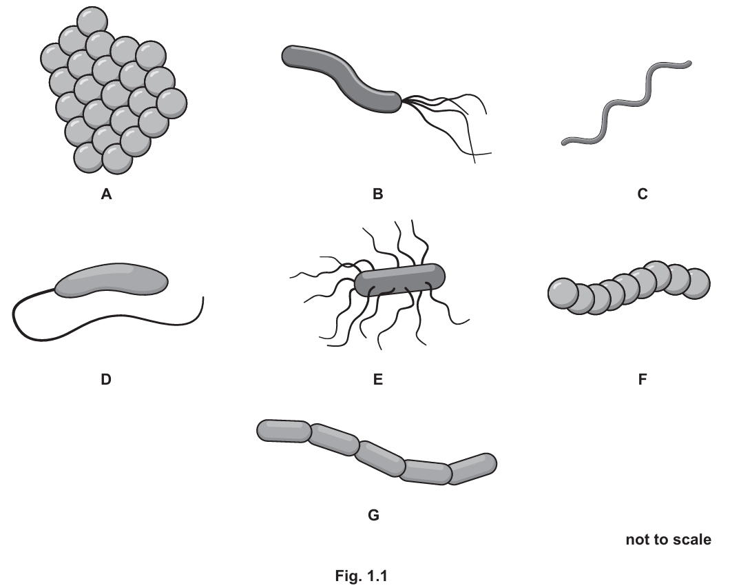
To identify the bacteria, observe the features in Fig 1.1 and follow the key:
- D: Has only one hair-like structure (flagellum) \(\rightarrow\) Vibrio cholerae.
- B: Has multiple flagella attached at only one end \(\rightarrow\) Helicobacter pylori.
- E: Has flagella attached all over the body \(\rightarrow\) Salmonella typhi.
- C: No flagella, but has a distinct spiral shape \(\rightarrow\) Treponema pallidum.
- A: No flagella, not spiral, does not form a chain (it is a cluster/clump) \(\rightarrow\) Staphylococcus aureus.
- G: No flagella, not spiral, forms a chain, and the individual bacteria are cylindrical (rod-shaped) \(\rightarrow\) Streptobacillus moniliformis.
- F: No flagella, not spiral, forms a chain, and bacteria are circular \(\rightarrow\) Streptococcus pyogenes.
(b)(i) Calculations:
Given: Length ($h$) = \(2\,\mu\text{m}\), Diameter ($d$) = \(0.5\,\mu\text{m}\).
Radius: \(r = \frac{d}{2} = \frac{0.5}{2} = \mathbf{0.25\,\mu\text{m}}\).
Volume: Using the formula for the volume of a cylinder \(V = \pi r^2 h\):
$$V = \pi \times (0.25)^2 \times 2$$ $$V = \pi \times 0.0625 \times 2$$ $$V = \pi \times 0.125 \approx 0.3927$$ Volume = \(\mathbf{0.39\,\mu\text{m}^3}\) (rounded to 2 significant figures/decimal places).
(b)(ii) Classification:
Bacteria belong to the kingdom Prokaryote (or Prokaryotae).
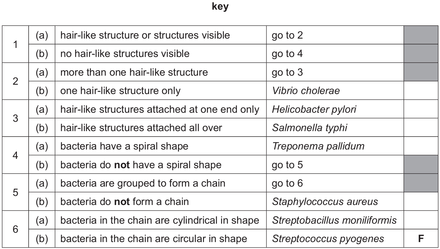
(c)(i) Virus Structure:
- J points to the outer shell of the virus, which is the Protein coat (or Capsid).
- K points to the internal coiled structure, which is the Genetic material (DNA or RNA).
(c)(ii) Bacteria vs. Viruses:
Bacteria are cellular organisms while viruses are not. Structures present in bacteria but absent in viruses include:
- Cell membrane
- Cytoplasm
- Ribosomes
- Cell wall
- Plasmids
▶️ Answer/Explanation
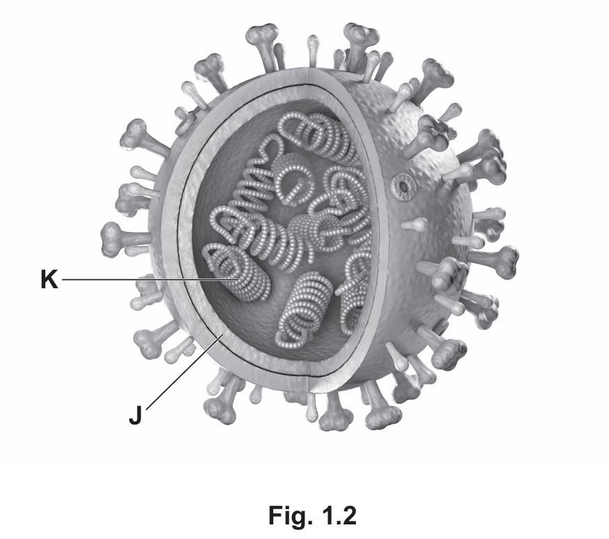
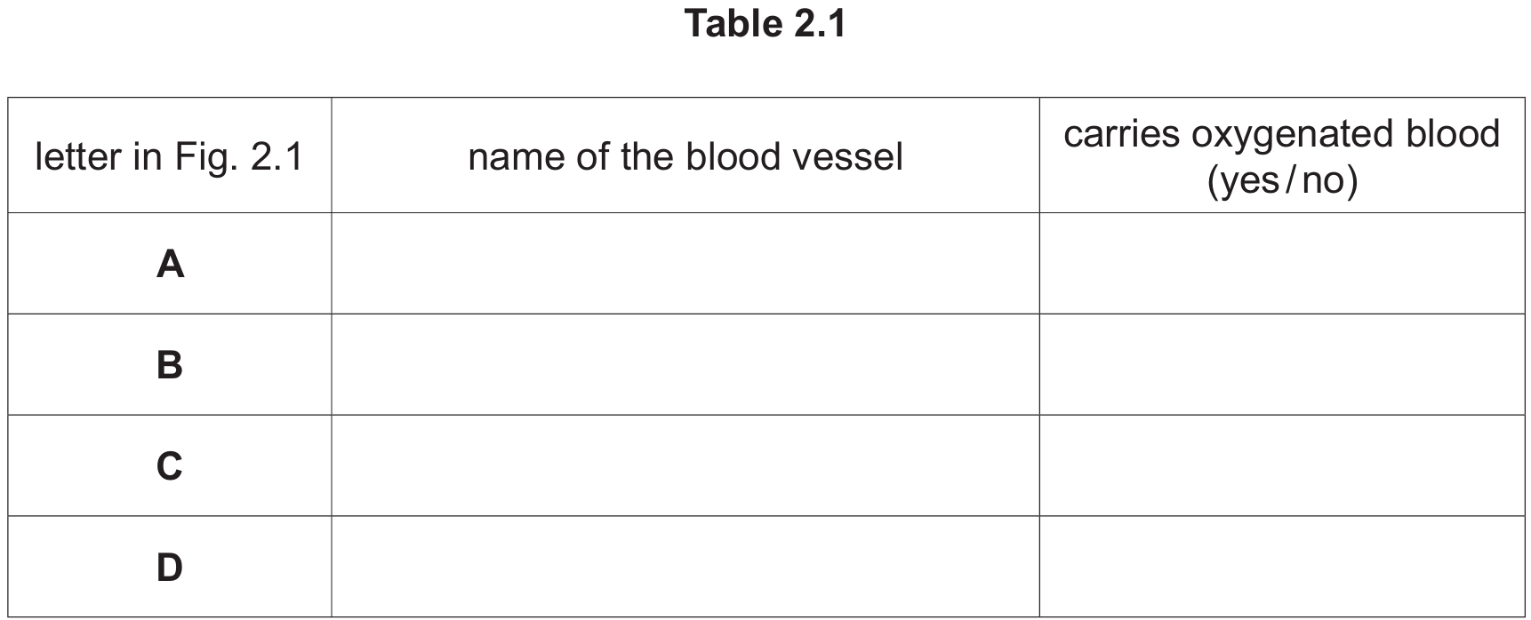
(a)(i) Table 2.1 Completion:
| Letter | Name of Blood Vessel | Carries Oxygenated Blood? |
| A | Pulmonary artery | No |
| B | Aorta | Yes |
| C | Hepatic portal vein | No |
| D | Renal vein | No |
Explanation:
• A (Pulmonary artery): Carries blood from the right ventricle of the heart to the lungs to be oxygenated. It is the only artery that carries deoxygenated blood.
• B (Aorta): The main artery carrying oxygenated blood from the left ventricle to the rest of the body.
• C (Hepatic portal vein): Connects the intestines to the liver. It carries nutrient-rich but deoxygenated blood.
• D (Renal vein): Carries deoxygenated blood away from the kidneys back toward the vena cava.
(a)(ii)
Name: Valve
Function: Prevents the back flow of blood.
Explanation: Veins (where X is located) operate under low pressure. Valves are essential to ensure blood flows in only one direction (towards the heart) and does not pool due to gravity.
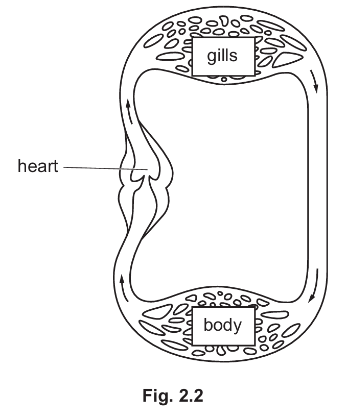
(b)(i) Differences in Heart Structure:
• The fish heart has only two chambers (one atrium and one ventricle), whereas the human heart has four.
• The fish heart lacks a septum separating left and right sides (since it is a single loop), while the human heart has a septum.
• The fish heart has fewer valves and fewer blood vessels attached compared to the human heart.
(b)(ii) Advantages of Human Circulatory System (Double Circulation):
• Separation of blood: Oxygenated and deoxygenated blood are kept separate by the septum, maintaining a steep concentration gradient for efficient gas exchange.
• High Pressure to Body: Blood loses pressure in the capillaries of the lungs. By returning to the heart (left side), it can be pumped out to the body at a much higher pressure, ensuring efficient delivery of oxygen and nutrients to tissues to support a high metabolic rate.
• Low Pressure to Lungs: The pulmonary circuit remains at lower pressure, preventing damage to the delicate capillaries in the lungs.
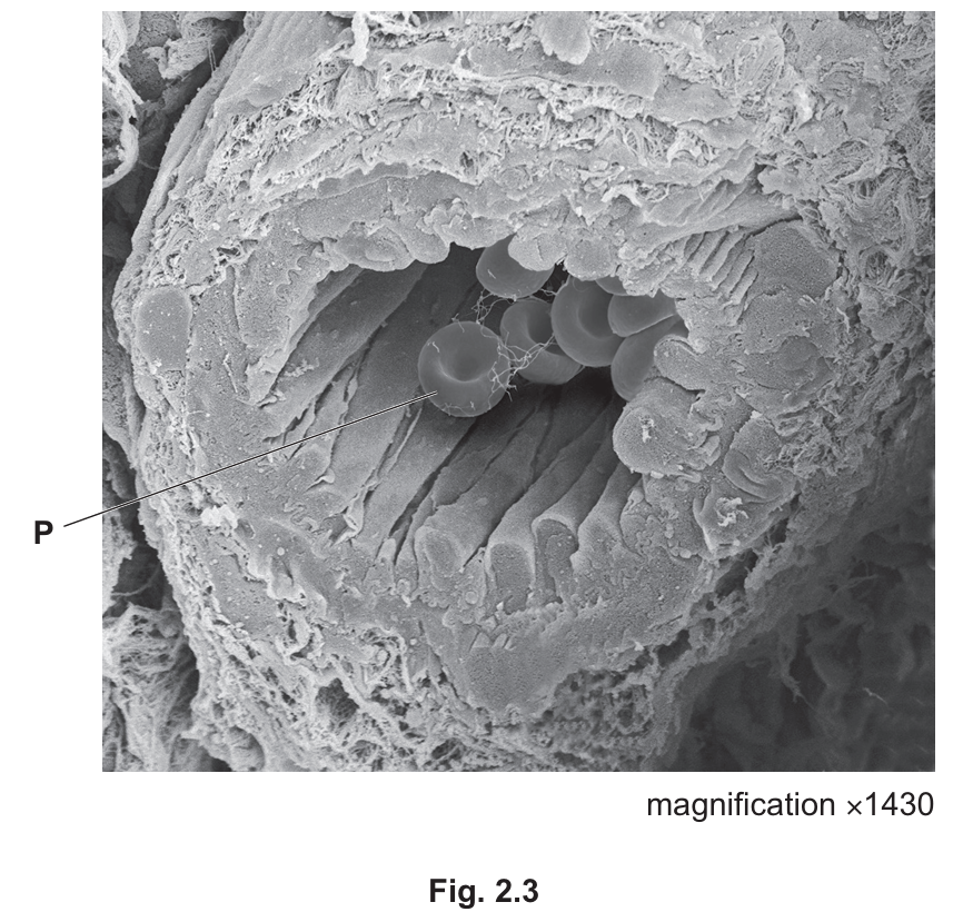
(c)(i)
Structure P: Red blood cell (Erythrocyte).
Note: These are recognizable by their biconcave disc shape and location inside the vessel lumen.
(c)(ii)
Formula: $$\text{Actual size} = \frac{\text{Image size}}{\text{Magnification}}$$
(c)(iii)
Answer: $31.5 \, \mu\text{m}$
Calculation: To convert millimeters (mm) to micrometers ($\mu\text{m}$), multiply by 1000.
$$0.0315 \, \text{mm} \times 1000 = 31.5 \, \mu\text{m}$$
▶️ Answer/Explanation
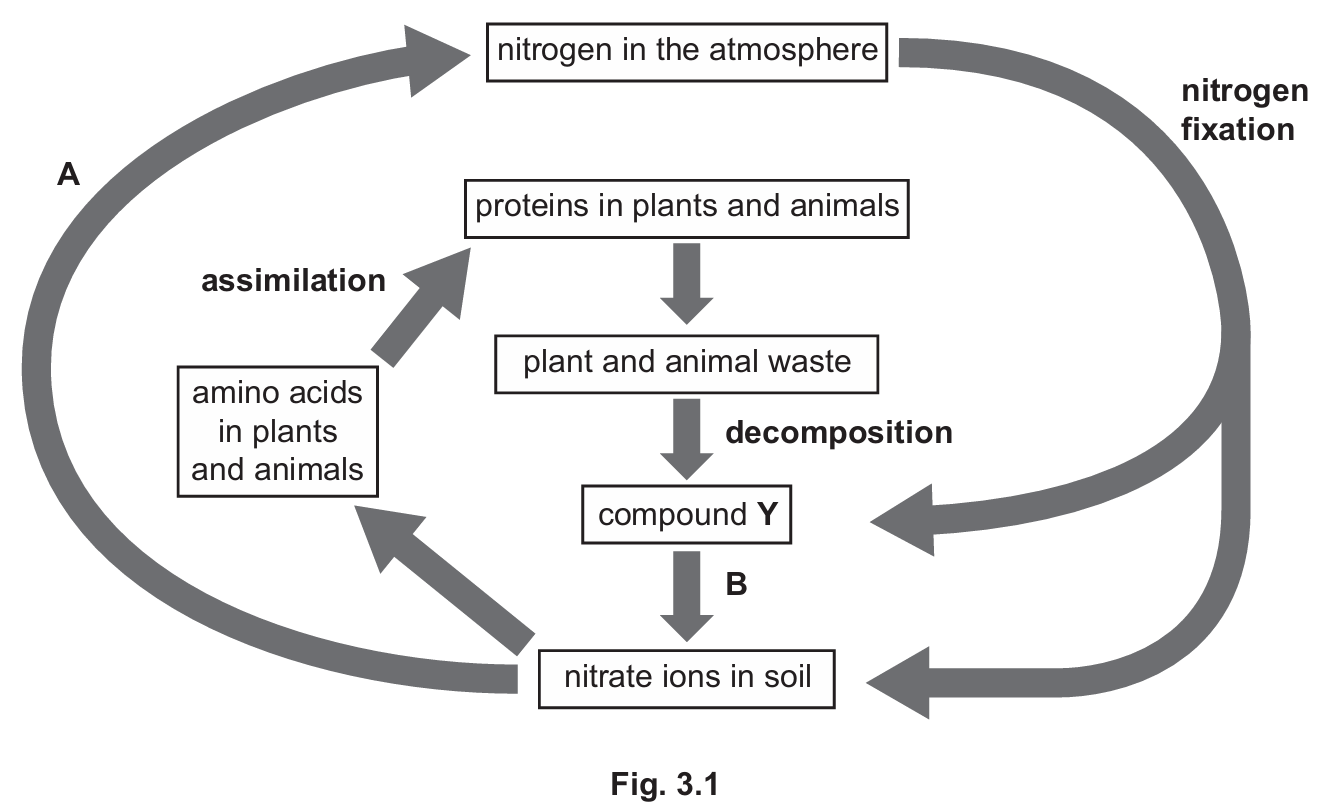
(a) (i)
A: Denitrification
B: Nitrification
(ii)
$1.$ Lightning
$2.$ Nitrogen-fixing bacteria (e.g., Rhizobium in root nodules)
(iii)
Ammonia / Ammonium ions
(iv)
Liver
(v)
Deamination is the removal of the nitrogen-containing part (the amino group) of amino acids. This process occurs when there is an excess of protein intake, as amino acids cannot be stored. The removed nitrogen is converted into urea to be excreted by the kidneys.
(b)
Methane is a pollutant gas that contributes to the enhanced greenhouse effect and climate change. Methane is released into the atmosphere from cattle and from the farming of rice crops.
Carbon dioxide is a gas that is absorbed by trees in the process of photosynthesis. This gas is released into the atmosphere by the burning / combustion of fossil fuels, where it also contributes to climate change.
(c)
Eutrophication occurs when untreated sewage or fertilizers (rich in nitrates/phosphates) leach into water bodies.
1. This leads to an increased availability of nutrients, causing an algal bloom.
2. The thick layer of algae blocks sunlight, preventing submerged plants from performing photosynthesis, leading to their death.
3. Decomposing bacteria feed on the dead organic matter and multiply rapidly.
4. These bacteria respire aerobically, using up the dissolved oxygen in the water.
5. This results in anoxic conditions where aquatic organisms like fish die due to suffocation.
Conceptual Explanation:
The nitrogen cycle is essential for converting inert atmospheric $N_2$ into forms usable by living organisms (like nitrates). Nitrification (Process B) turns ammonia into nitrates, which plants can absorb. Conversely, denitrification (Process A) converts nitrates back into atmospheric gas, usually in waterlogged soils. In animals, the liver is the metabolic hub where assimilation (incorporating nutrients into cells) and deamination (breaking down excess amino acids) occur to maintain chemical balance and remove toxic nitrogenous waste.
▶️ Answer/Explanation
(a)(i) Features attracting insects:
The three main features are: 1. Scent (perfume to attract insects from a distance). 2. Nectar (a sugary food reward). 3. Large / colourful petals (visual attraction/landing platform).
Other accepted answers: Pollen (as a food source).
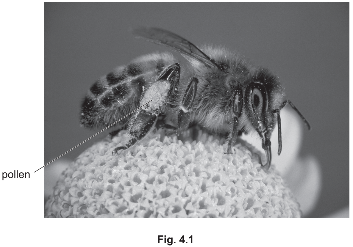
(a)(ii) Pollen adaptation:
Pollen grains in insect-pollinated flowers are sticky or spiky. This texture allows them to easily attach to the hairs on the insect’s legs and body (as seen in Fig 4.1) to be transported to the next flower.
(b) Monocots vs. Dicots:
Any two of the following differences:
• Leaves: Monocots have narrow/strap-like leaves with parallel veins, whereas dicots generally have broad leaves with a branching (reticulate) network of veins.
• Flower parts: Monocot petals usually appear in multiples of \(3\).
• Cotyledons: Monocots have \(1\) cotyledon (seed leaf) inside the seed, dicots have \(2\).
• Roots: Monocots have fibrous root systems.
• Stem: Vascular bundles are scattered in monocots (dicots have them in a ring).
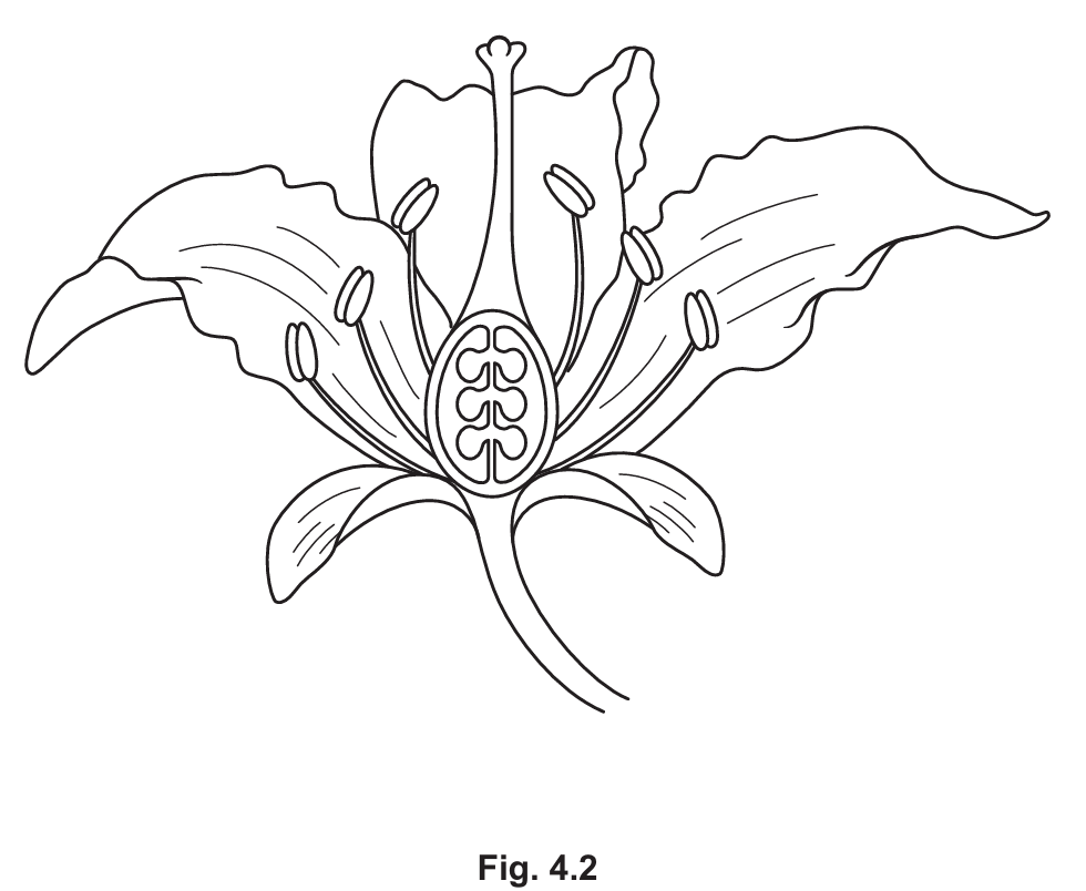
(c) Labeling Fig 4.2:
Based on the flower diagram:
• Structure supporting anther: The Filament (the stalk holding up the anther).
• Structure protecting bud: The Sepal (the outermost leaves, often green).
• An ovule: The small structures inside the ovary at the base of the carpel.
(d) Sexual reproduction in a flower:
This process follows a specific sequence :
1. Pollination: Pollen is transferred from the anther to the stigma.
2. Tube Growth: A pollen tube grows out of the pollen grain.
3. Journey: The tube grows down through the style towards the ovary.
4. Transport: The male nucleus (gamete) travels down the pollen tube.
5. Fertilisation: The pollen tube enters the ovule (via the micropyle), and the male nucleus fuses with the female nucleus (ovum).
6. Result: A zygote is formed.
(e) Human Reproduction:
(i) Male gamete: Sperm (cell).
(ii) Formation site: Testis (or testes).
(iii) Process: Meiosis (This is the specific type of cell division that reduces the chromosome number by half to create haploid gametes).
(iv) Prostate gland function: It secretes fluid (semen) which creates a medium for the sperm to swim and provides them with nutrients (sugar).
▶️ Answer/Explanation
(a)(i)
Transpiration (or diffusion).
Explanation: Transpiration is the biological term for the evaporation of water from plant leaves, typically through stomata.
(a)(ii)
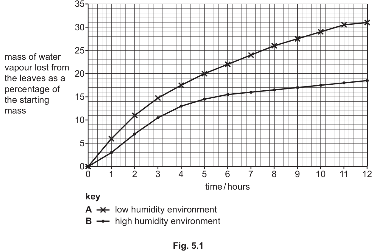
Comparison:
- Both groups lose mass over the 12-hour period.
- Group A (low humidity) loses significantly more water vapour (higher percentage mass loss) than Group B (high humidity). For example, A reaches ~31% while B reaches ~18%.
- The rate of loss is steeper initially for both groups but flattens out over time.
Explanation for high humidity effect:
In high humidity, there is a greater amount of water vapour in the atmosphere surrounding the leaf. This reduces the water potential gradient (or concentration gradient) between the moist air spaces inside the leaf and the outside air. Because diffusion relies on a concentration gradient, the reduced gradient results in a slower rate of water vapour loss through the stomata.
(a)(iii)
Prediction: The line should be drawn above the existing line for low humidity (Line A).
Explanation: Increasing the temperature to \(35^\circ\text{C}\) increases the kinetic energy of the water molecules, leading to a faster rate of evaporation and diffusion. Therefore, water loss would occur more rapidly and reach a higher percentage than at \(20^\circ\text{C}\).
(b)(i)
Sweat glands in the skin secrete sweat onto the skin surface. As the water in the sweat evaporates, it absorbs heat energy from the body (specifically, the latent heat of vaporisation). This removal of heat cools the blood flowing near the skin surface, helping to lower the internal body temperature back to the set point (homeostasis/negative feedback).
(b)(ii)
Expiration (breathing out).
Explanation: Air is warmed and moistened in the lungs; when humans exhale, this moisture is lost as water vapour.
▶️ Answer/Explanation
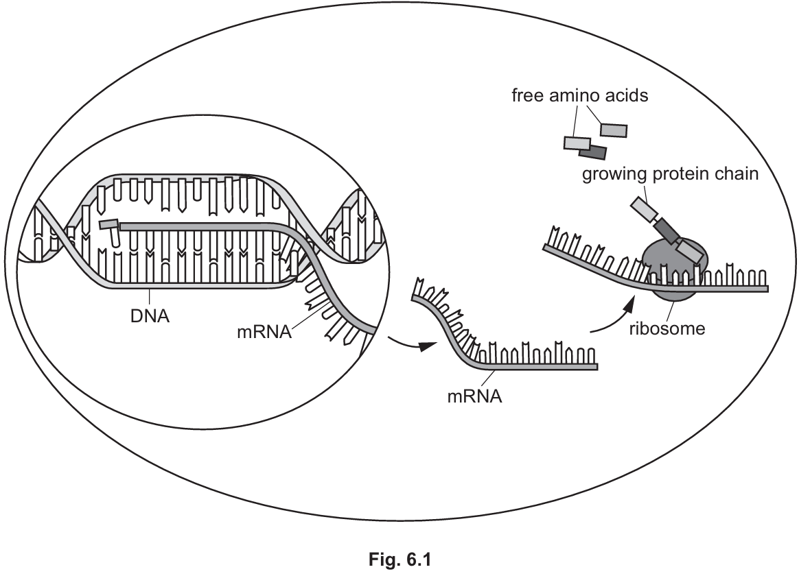
(a)
Nucleus
The DNA contains the genetic information and is located within the nucleus of the cell.
(b)
Enzymes (Other acceptable answers: membrane carriers, receptors for neurotransmitters, antibodies).
DNA controls cell function by controlling the production of proteins, such as enzymes which act as biological catalysts, or membrane carriers used in active transport.
(c)
Role of mRNA in protein synthesis:
Based on the diagram and biological principles:
- Copying the gene: mRNA is formed as a copy of a gene from the DNA found in the nucleus.
- Transport: The mRNA molecule moves out of the nucleus into the cytoplasm.
- Translation: The mRNA passes through a ribosome. The ribosome assembles amino acids into protein molecules based on the specific sequence of bases in the mRNA.