Digestion and Absorption Class 11 Notes Biology Chapter 16
Topics and Subtopics in for Class 11 Biology Chapter 16 Digestion and Absorption:
| Section Name | Topic Name |
| 16 | Digestion and Absorption |
| 16.1 | Digestive System |
| 16.2 | Digestion of Food |
| 16.3 | Absorption of Digested Products |
| 16.4 | Disorders of Digestive System |
| 16.5 | Summary |
Topic 1 Digestion: The System and Associated Glands
Animals depend on ready-made food for their nutritional requirements. Nutrition is a process by which animal obtain essential and non-essential substances called nutrients.
The way by which organisms derive their nutrients is called mode of nutrition.
It is mainly of two types
(a) Autotrophic or Holophytic The organisms having the capability to form their own food with the help of solar energy, e.g., Plants, Euglena, etc. It is of further two types, i.e., photoautotropic and chemoautotrophic.
(b) Heterotrophic The organisms which cannot use free energy of our atmosphere to synthesise necessary organic compounds as food. These normally obtain the nourishment from the autotrophs.
Based upon the mode of feeding the heterotrophs can be
(i) Holozoic (ii) Saprozoic (iii) Saprophytic
(iv) Osmotrophic (v) Parasitic (vi) Predatorship
The myxotrophic nutrition is the case in which more than one type of . nutritional modes are found within the single animal. The animals like Euglena show this kind of nutrition.
A balanced diet has various components (carbohydrates, hits, proteins, vitamins, water, mineral and roughagfi) in optimum proportion and quantity.
Human Digestive System (Structure)
Biomacromolecules in food cannot be utilised by our body in original form. So, they are subject to a process called digestion (complex food substances are converted to simple absorbable forms).
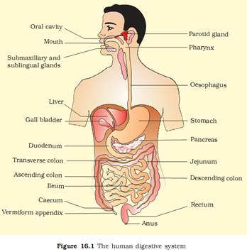
The system that helps in the complete process of digestion by mechanical and biochemical methods is called digestive system.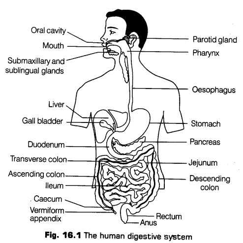
It is a long tube (about 8-10 m in length) with muscular walls and varying diameter. It begins with an anterior opening, i.e., mouth and opens out posteriorly through anus.
It is called canal (not tube), because it opens at both ends [i.e., mouth and anus).
Structure Wall of Alimentary Canal
If a transverse section of an alimentary canal is viewed, the wall of alimentary canal from oesophagus to rectum (large intestine) in general shows following four concentric layers
(a) Serosa It is the outermost layer made up of a thin mesothelium (epithelium of visceral organs) with some connective tissues.
(b) Muscularis It is the second coat present just below the serosa. It is a very thick and contains muscle fibre. It is formed by smooth muscles. It consists of outer longitudinal and inner circular muscle fibres (both are unstriped, i.e., smooth).
In some regions like stomach an additional layer of oblique muscle is found inner to the circular muscle fibres.
(c) Submucosa It lies below the muscular coat. Consist of loose connective tissues richly supplied with nerves, blood and lymph vessels and also with glands in some areas like duodenum. id) Mucosa It is the innermost layer lining the human gut or alimentary canal. It is named so, because it has its major role in secreting mucus in order to lubricate inner lining of gut.
This layer forms irregular folds (rugae) in the stomach and small finger-like folding called villi in the small intestine. It also forms gastric glands in the stomach.
All the four layer shows modification in different parts of the alimentary canal.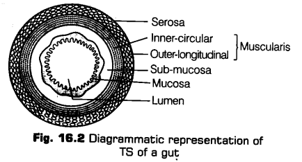
Various parts of alimentary canal are discussed below
i. Mouth and Buccal Cavity
Mouth is a slit like opening, bounded by two soft, movable lips. It opens into a small vestibule (space enclosed between lips and cheeks externally and gums and teeth internally), which inturn leads into the buccal or oral cavity.
Oral cavity further comprises of two main components a. Teeth
These are hard structures present in the mouth on both the jaws (i.e., upper and lower jaw). Each tooth is embedded in a socket of jaw bone.
Mammalian teeth are characterised by following three features
* Thecodont The teeth are fixed in sockets. They have very well-developed roots, which are implanted deeply in the jaw bone socket.
* Diphyodont Like other mammals, human beings also has two sets of teeth formed during lifetime. The first set of teeth is temporary and is known as milk or deciduous teeth.
Milk teeth are 20 in number.
* The milk set is replaced by the second set known as permanent teeth or adult teeth. Permanent teeth last for whole life, if lost, cannot be replaced.
* Heterodont An adult human has 32 permanent teeth, but they are of different size, shape and type.
They are of following four types
* Incisors (I) for cutting of food
• Canine (C) for tearing the food
* Premolars (Pm)
* Molars (M) for crushing, grinding and chewing the food.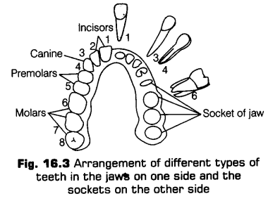
i. Dental Formula
The number of each type of teeth can be expressed by a dental formula, which is the arrangement of teeth in each half of the upper and the lower jaw in the order I, C, Pm, M.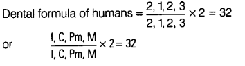
b. Tongue
It is a muscular organ, which is freely movable in the oral cavity. A fold called frenulum attaches the tongue to the floor of oral cavity. The upper surface of the tongue bears small projections (elevations) known as papillae. Some of the papillae bear taste buds.
Note:
* Papillae provides a characteristic roughness to the tongue.
* The hard visible chewing surface of tooth helps in the mastication of food and is covered by a thick, shiny and translucent substance called enamel (the hardest substance in the body).
* Taste buds present at the surface of the tongue contain chemosensory cells. Human taste buds are sensitive to four basic
* tastes, i.e., sweet, bitter, salty and sour. These four taste buds are present at different locations of the tongue.
ii. Pharynx
It is a small funnel-shaped chamber located behind the oral cavity. It serves as a common passage for both food and air, i.e., it communicates with both oesophagus (food pipe) and trachea (wind pipe).
The opening of trachea or wind pipe is called glottis, which is guarded by a cartilaginous flap or lid called epiglottis. The glottis normally remain open, but during swallowing of food it gets covered by epiglottis to prevent the entry of food in trachea.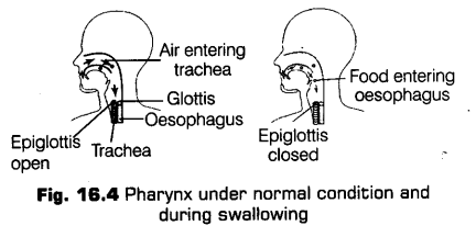
iii. Oesophagus
It is the thin, long muscular tube that extend posteriorly passing through the neck, thorax and diaphragm and finally leads into a J-shaped bag- like structure called stomach. A muscular gastro-oesophageal sphincter regulates the opening of oesophagus into the stomach.
iv. Stomach
It is the most dilated- structure of alimentary canal situated between the oesophagus and small intestine. It lies below the diaphragm in the abdominal cavity towards the upper left side.
Pails of Stomach
Stomach has three major parts as given below
(a) Cardiac stomach, the upper portion into which the oesophagus opens.
(b) Fundic stomach, the middle portion.
(c) Pyrolic stomach, the lower portion, which opens into the first part of small intestine, i.e., duodenum.
The terminal pyrolus part of stomach (i.e., opening of stomach into duodenum) is guarded by a pyrolic sphincter.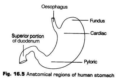
Functions of Stomach
Stomach serves the following Junctions
(a) Acts as a short term storage reservoir.
(h) The substantial chemical and enzymatic digestion is initiated here (especially of proteins).
(c) Gastric smooth muscles mix and grind the foodstuff by vigorous contractions with gastric secretions.
(d) Food become liquefied in the stomach and is released slowly in the small intestine.
Note:
* The lymphatic tissues of the pharynx and oral cavity are arranged in a ring like manner, that are collectively called Waldeyer’s ring. This ring consists of lingual tonsils and palatine tonsils.
The lower – part of oesophagus has only involuntary muscles.
v. Small Intestine
It is the longest part of the alimentary canal, which is about 6 m long in human beings.
It is divisible into three main reports
(a) Duodenum It is U-shaped, widest and shortest part of small intestine.
(b) Jejunum It is the middle part of small intestine, which is about 2.5 m long and coiled.
(c) Ileum It is highly coiled and the longest portion of the small intestine, which and enormously increases the surface area of the intestine.
Absorptive Surface Area of Small Intestine
The structure of small intestine is similar to all other regions of the alimentary canal, but it incorporates three important features, which account for its huge absorptive surface area.
These are as follows
(a) Mucosal folds Inner surface of small intestine is thrown into circular folds, i.e., it is not flat.
(b) Villi The inner mucosa layer of small intestine has, villi (about 1 mm in height), covered with columnar epithelial cells.
(c) Microvilli Numerous microscopic projections of microvilli are produced by the cells lining the villi. These microvilli gives it a brush border appearance showing villi Functions of Small Intestine.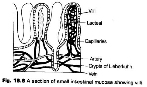
Following purposes are served by small intestine
(а) It acts as a major site for the digestion of food as it secretes most of the digestive enzymes and gastro-intestinal hormones.
(b) Maximum absorption of the end products of digestion takes place here, because it contains many villi that increases the surface area of absorption.
(c) It also helps in absorption of fats.
Although it is shorter but, is called large intestine, because it is wider in diameter than small intestine.
Large intestine lacks villi and microvilli
It is distinguishable into three main parts
(a) Caecum It is a small pouch-like structure connected to the terminal part of small intestine. It is a blind sac that functions as a host for various symbiotic microorganisms. Vermiform appendix, a narrow finger-like projection which is a vestigial organ arises from caecum.
Both the structures are not well-developed in human beings, but in herbivores it is developed very well in order to digest cellulose whose digestion is difficult.
finally opens into large intestine on the right side of the abdominal cavity.
(b) Colon It is the longest part of the large intestine. The caecum opens into colon, which is further divisible into three main parts, i.e., an ascending colon, transverse colon and descending colon.
(c) Rectum It is the last part of the large intestine. The descending colon finally opens into rectum, which serves to store the faecal matter temporarily. It further leads to a short anal canal, which opens to outside through anus.
The anal canal is guarded by another sphincter, i.e., internal and external sphincter,.
At the ileocaecal junction is an ileocaecal value is present, that regulates the passage of materials from small to the large intestines.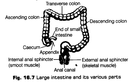
Digestive Glands
To bring about the chemical simplification of food, digestive juices are secreted by the different glands. The digestive glands associated with the alimentary canal include majorly salivary glands, the liver and pancreas.
i. Salivary Glands
These are exocrine glands that secretes saliva. There are three pairs of salivary glands in man. All three glands are situated just outside the buccal cavity and secrete salivary juice into the buccal cavity. These are as follows:
(a) Parotid Glands These are largest of the three glands present one on either side of the cheek on the upper palate.
(b) Sub-maxillary or Sub-mandibular Glands These are present at the angle of the lower jaw.
(c) Sub-lingual Gland These are situated beneath the tongue.
Each sublingual gland has about ten small duets called sub-lingual ducts or ducts of Rivinus, which open into the floor of mouth.
ii. Liver
It is the largest gland of the body, an exocrine gland. In adults, it weighs about 1.2-1.5 kg and lies in the abdominal cavity just below the diaphragm and has two lobes, i.e., left and right lobes.
It is a large organ and occupies most of the right side of abdominal cavity.
Liver is a double membrane structure. Interiorly, it is divided into many small units called hepatic or liver lobules (structural and functional units of liver) consisting of many hepatic cells (hepatocytes) that are arranged in the form of cords.
Each lobule is also covered by a thin connective tissue sheath called the Glisson’s capsule. Hepatic cells secretes the bile juice, which passes through the hepatic duct into the gall bladder.
Functions of Liver
Liver serves the following functions
(a) It helps in producing RBCs in embryo.
(b) Bile secreted by the liver helps in emulsification of fats, i.e., breaking down of fats into very small micelles.
(c) Bile also activates lipases.
(d) It also produces heparin for preventing clotting of blood inside the blood vessels.
Gall Bladder
It is a small pear-shaped, thin muscular sac-like organ situated just below the liver. It is attached by connective tissues to liver. The duct of gall bladder, i.e., cystic’duct along with the hepatic duct form a common bile duct, which regulates the amount of bile to be discharged into the duodenum.
After certain distance, the bile duct and pancreatic duct (duct of pancreas) form common hepato-pancreatic duct, which open into duodenum. It is guarded by a sphincter called the sphincter of oddi.
The common hepato-pancreatic duct carries both the bile (from liver) as well as pancreatic juice (from pancreas) into the duodenum.
iii. Pancreas
It is a compound elongated organ situated partly behind the stomach between the limbs of the U-shaped duodenum. As it is a mixed gland, it has both exocrine as well as endocrine activity.
An alkaline pancreatic juice containing enzymes is secreted by its exocrine portion and the endocrine portion is responsible for the secretion of hormones, insulin and glucagon.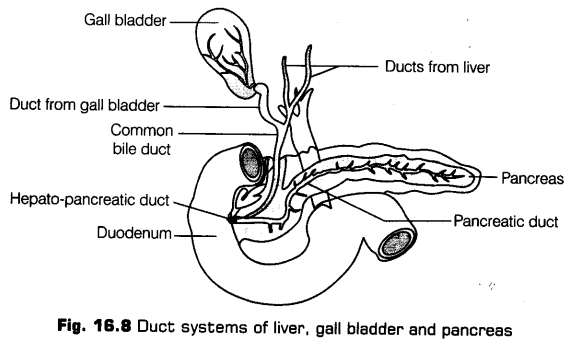
Other Glands
Apart from the above mentioned major glands, other glands also plays an important role in completion of the process of digestion.
These are mentioned below as
Gastric Glands
The glands of stomach are called gastric glands. These are present in the mucosa of the stomach.
The gastric gland contains the following three types of secretory cells
(a) Mucus or goblet cells, secretes alkaline mucus.
(b) Peptic o( chief or zymogenic cells, secretes inactive precursors of gastric enzymes.
(c) Parietal or oxyntic cells, secretes HCl and CIF (Castles Intrinsic Factor).
Intestinal Glands
The epithelium of intestine bears a large number of glands. Most of these glands are formed by the modification of the surface epithelial cells and are located on villi.
(a) Brunners glands are present only in the submiicosa of duodenum (not in ileum and jejunum).
(b) The lamina propria of small intestine contains large masses of lymphocyte cells called lymph nodules or Peyers patches. These help in destroying harmful bacteria of the region.
(c) The mucus portion contain simple, tubular intestinal glands or crypts of Leiberkuhn.
These are pit-like glands with three types of cell, i.e.,
* Undifferentiated epithelial cells
* Zymogenic cells or cells of Paneth
* Argentaffin or enterochromaffin cells.
In general intestinal juice is called succus entericus (secretion of cells of crypts of Lieberkuhn mainly,
i. e„ goblet cells and brush-bordered epithelial cells).
Note:
* The estimated number of gastric glands in humans is about 35 millions (3.5 crore).
* The unicellular goblet cells are also present in small intestine. Infact, these glands are present throughout the alimentary canal and secrete mucus.
Topic 2 Digestion : The Process and Control
The process of digestion involves the conversion of large, complex and non-diffusible substances into their respective simpler forms. The complete process of digestion is accomplished by mechanical and chemical processes.
Mobility of Gut
Alimentary canal being so long, does not allow the food to get jammed along its length. This is due to the mobility of gut, which helps the food to move forward.
The alimentary canal or gut shows following movements
i. In Buccal Cavity
The buccal cavity shows two major functions
(а) Mastication of Food It is the very first movement of the alimentary canal seen in buccal cavity. It involves the movement of teeth, which helps in chewing the food and the tongue, which help the food to mix thoroughly in the saliva, with the help of mucus.
The mucus lubricates and adhere the masticated particles of food into a bolus (mass of food that has been chewed before swallowing) and push it backward towards the pharynx for deglutition.
The mastication of food is a voluntary process (in human being).
(b) Swallowing (Deglutition) It is the process of passing bolus or mass of food in the oesophagus from the buccal cavity through pharynx.
The food is pushed back against the epiglottis, at the same time epiglottis covers the glottis (as already discussed in 1st topic). Due to this the oesophagus opening becomes wider and food enters it.
The bolus further passes down through the oesophagus by a successive wave (peristalsis) as a reflex along with the constriction of the oesophageal opening, which takes the food down towards the stomach.
ii. Peristalsis
It is the reflex wave that comprises of a series of muscle contraction that occurs in the complete digestive tract.
It pushes the food in the forward direction (away from the mouth).
Mechanism of Digestion
In human being, the digestion of food starts in the buccal cavity and continues till the anus of large intestine.
The mechanism of digestion continues in the following steps
i. Digestion in Buccal or Oral Cavity
Digestion starts in the oral cavity by the chemical hydrolytic action of the carbohydrate splitting enzyme, salivary amylase.
The saliva secreted into oral cavity contains electrolytes (Na+,K+, Cl–, HCO3, etc.) and enzymes, i.e., salivary
amylase and lysozyme (acts as an antibacterial agent that prevents infections). About 30% of the starch gets hydrolysed in the oral cavity by the action of salivary amylase (at optimum pH 6.8) into a disaccharide,
i. e., maltose.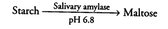
ii. Digestion in Stomach
The stomach stores the food for around 4-5 hours. Internal mucosa of stomach contains gastric glands, which mainly comprises of three types ofcells
(а) Mucus or neck cells for secreting mucus.
(b) Peptic or chief or zymogenic cells for secreting proenzyme pepsinogen.
(c) Parietal or oxyntic cells for secreting HCl and intrinsic factor (essential for vitamin-B12 absorption).
The gastro-oesophageal sphincter controls the passage of food into the stomach.
Food is slowly released from the stomach in small quantities into the small intestine, so that a slow process of digestion and absorption can occur.
The food mixes thoroughly with the acidic gastric juice secreted in the stomach by the churning movements of its muscles and becomes semi-digested, acidic, pulpy mass called chyme. The HCl and the enzymes of the gastric juice now helps in the chemical simplification of food.
The enzymes of stomach and their actions are given below
a. Pepsin
On exposure to HCl, the proenzyme pepsinogen gets converted into pepsin (proteolytic enzyme of the stomach) that further converts proteins into proteases and peptones (peptides).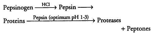
Pepsin usually attacks the peptide bonds between amino acids. It can attack all proteins except keratins, protamines, histones, etc.
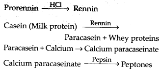
b. Rennin
It is a proteolytic enzyme found in gastric juice of only infants, in its inactive form. Its secretion takes place in order to digest the milk proteins.![]()
c. Gastric Lipases
Small amounts of lipases are also secreted by the gastric glands. Activity of this enzyme is inhibited in the stomach by the acidic condition.It act on emulsified fats and also help in digesting around 25% of milk fat (in infants).
It is mainly the digestion of proteins that occurs in the stomach.
Apart from all these enzymes, the amount of mucus and bicarbonates present in the gastric juice plays an important role in the lubrication and protection of mucosal epithelium from excoriation by the highly acidic pH.
iii. Digestion in Small Intestine
To further facilitiate the digestion of food, muscularis layer of small intestine shows various types of movements which allows a thorough mixing up of food with various secretions in the intestine.
These contractions of muscles in the small intestine allows the further churning and kneading of the chyme and finally pushing it into the large intestine.
The respective digestive juices from the liver (bile), pancreas (pancreatic juice) and srtiall intestine (intestinal juices) are released into the small intestine to bring out the further chemical simplification of food. The , pancreatic juice from the pancreas and the bile from the liver are released through the hepato-pancreatic duct.
Enzymes from Pancreas
The pancreatic juice secreted from the pancreas contains the various inactive enzymes.
These are as follows
(a) trypsinogen (b) chymotrypsinogen
(c) procarboxypeptidases (d) amylases (e) lipases (f) nucleases
Trypsinogen is activated by an enzyme enterokinase secreted by intestinal mucosa into active trypsin which in turn activates the other enzymes of pancreatic juice.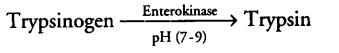
* The proteins, proteases and peptones (partially hydrolysed form of proteins) present in the chyme (reaching the intestine) are acted upon by the proteolytic enzymes of pancreatic juice. These are given below as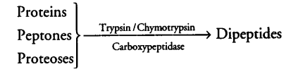
* Carbohydrates in the chyme are hydrolysed by pancreatic amylase into disaccharides.![]()
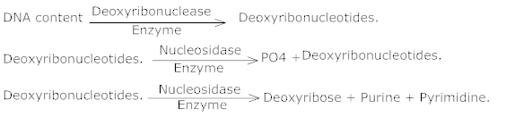
* Nucleases in the pancreatic juice acts on nucleic acids to form nucleotides and nucleosides.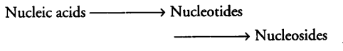
Enzymes from Liver
The bile secreted from the liver is released into duodenum of small intestine. Bile contains the bile pigments, i.e., bilurubin and biliverdin, bile salts, cholesterol and phospholipids.
Thus, fats are broken down into di and monoglycerides by the action of lipases.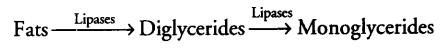
Note:
* Bile does not contain any enzymes as gastric juice. It helps in emulsifying fats, i.e., in breakdown of fats into very small micelles which are kept suspended in an aqueous medium.
* The process of emulsification is basically carried out by the salts of bile. This increases the surface area of fat available for digestion by the lipase (as bile also activates lipases).
Enzymes from Intestine
Intestinal mucosal epithelium has goblet cells (secretes mucus). Thus, the secretions of the brush border cells of mucosa together with the goblet cell secretions forms the intestinal juice (also known as succus entericus).
The succus entericus thus, contains various enzymes
(a) Disaccharidases, e.g., Maltase for digestion of maltose into glucose.
(b) Dipeptidases
(c) Lipases
(d) Nucleosidases
Pancreatic and intestinal lipases together helps in the emulsification of fats.
The mucus along with the bicarbonates from the pancreas helps in protecting the mucosal layer of intestine from the action of acid and also provides an alkaline medium (pH-7.8) for enzymatic activities.
Glands (Brunner’s gland) from sub-mucosal layer of intestine also helps in this. Hence, all the enzymes in the succus entericus acts on the end products of the above mentioned reactions in order to form their respective simpler forms.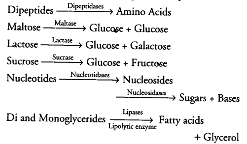
All these final steps takes place very close to mucosal epithelial cells of the intestine.
All the biomacromolecules mentioned above breakdown in the duodenum region of small intestine, while the simpler forms are absorbed in the other two regions of small intestine, i.e., jejunum and ileum.
Digestion in Large Intestine
The last stage of chemical simplification of food occurs in the last part of the alimentary canal, i.e., large intestine. This is carried out by bacterial action. Glands of this region tends to secrete mucus, i.e., enzymes are not secreted into this part of the digestive system.
The undigested and the unabsorbed substances are finally passed on to the large intestine.
Note:
* There are more than 500 species of bacteria found in the colon region of the large intestine which are not usually harmful as long as they remain in the large intestine.
* Infact, all these bacteria synthesise vitamin-K and B12 , also helps in absorption of calcium, magnesium and zinc (by increasing the acidity of colon region).
Followingfunctions areperformed by the large intestine
(i) Absorption of some water, minerals and certain drugs.
(ii) Secretion of mucus which helps in adhering the waste (undigested) particles together and lubricating it for an easy passage.
No significant digestive activity occurs in this region of digestive tract. .
The undigested, unabsorbed substances called faeces, enters into the caecum region of the large intestine (through the ileo-caecal valve, which prevents the back flow of faecel matter). It is temporarily stored in the rectum till defecation (egestion) through the anus.
Apart from absorbing vitamins secreted by various types of bacteria, large intestine also helps in absorbing water and electrolytes such as Na+, Cl–.
Neural and Hormonal Control of Digestion
For proper coordination- and functioning of different parts of the gastrointestinal tract, it should be under a proper, neural and hormonal control.
Neural Control
Secretion of saliva is stimulated by the sight, smell and the presence of food in the oral cavity. Similarly, the gastric and intestinal secretions are also under the control of neural signals.
The muscular activities of different parts of the alimentary canal are also moderated by neural mechanisms (both local and through CNS).
Hormonal Control
The major hormones that control the functions of digestive system are produced and released by the cells in the mucosa of the stomach and the large intestine.
CBSE Class 11 Biology Chapter-16 Important Questions
1 Marks Questions
1.Name the secretions of Goblet cell & parietal cells.
Ans. Goblet cells secrete mucus. Parietal cells secrete hydrochloric acid (HCl) and intrinsic factor.
2.Name the three parts of small intestine of man.
Ans. Duodenum, Jejunum and ileum.
3.Which is the largest gland in our body?
Ans. Liver.
4.What is the main function of bile salt?
Ans. It reduces the surface tension of fat droplets causing their break down into many small ones.
5.Name the watery fluid secreted from Bruner’s gland in duodenum.
Ans. Mucoid fluid is secreted from Bruner’s gland in duodenum.
6.What is atheroma?
Ans. Deposition of cholesterol on the walls of arteries.
7.What is egestion?
Ans. Passing out of undigested food from the body.
8.What are micelles?
Ans. Monoglycerids, long chain fatty acids and digested fats unite with bile salts and form small spherical droplets known as micelles.
9.What are crypts of lieberkuhn?
Ans. Pits into the sub mucosa of gastrointestinal tract wall.
10.What do you mean by the term malnutrition?
Ans. The stale of health due to improper intake of food or nutrients. It covers both under nutrition as well as over nutrition.
11.Name the hardest substance in the body.
Ans. Enamel
12.What is a lacteal?
Ans. Lymph vessel found in villi.
13.Name the small projections, found on the upper surface of tongue.
Ans. Papillac
14.Mention the function of epiglottis.
Ans. Prevention the entry of food in to the glottis.
15.Write the name of major parts of siornach.
Ans. Cardiac, fundic, pyloric.
16.Nane the enzyme that digest fast. Mention the end products of fat digestion.
Ans. Lipase, fatty acids and glycerol.
17.In which part of alimentariy canal does absorption of water, simple sugars and alcohol lakes place?
Ans. Stomach
18.Why are proteases generally released in inactive form?
Ans. If released in active form, they will start digesting the membranes and muscular of the alimentary canal.
19.Trypsinogen is an inactive enzyme of pancreatic juice. An enzyme, enterokinase, activates it. Which tissue/cell secrete the enzyme 7 How is it activatc4i ?
Ans. Intestinal mucosa.

20.What is the role of insulin?
Ans. Metabolis of sugar
2 Marks Questions
1.What is the role of micelles in the fat absorption?
Ans. During digestion, the fat in the intestine is converted to monoglycerides diglycerides and fatty acids which are insoluble in water. They cannot be directly absorbed from the intestinal contents. They are first incorporated into small, spherical and water soluble droplets called micelles by bile salts. It is from these micelles that fatty acids, glycosides, sterols and fat soluble vitamins are absorbed into the intestinal cells.
2.Give two functions of trypsin?
Ans. 1) Trypsin converts chymotrypsinogen into chymotrypsin.

2) Trypsin acts on proteoses and peptones and convert them into peptides.
Trypsin + Peptones + Proteoses → Peptides.
3.What are the specific functions of food?
Ans. Specific functions of food are –
(i) Food on oxidation inside the body supplies energy to perform various functions.
(ii) It serves to supply the material for growth and development of the body.
(iii) It also serves as a reserve material mainly as fat and glycogen. These can be utilized at the time of emergency.
(iv)It protects the body from diseases.
4.How does fat absorption takes place?
Ans. Fat absorption – It occurs as monoglycerides and fatty acids. These are resynthesized into triglycerides which in turn, combine with cholesterol. They form chylonicrons chylomicrons pass into the lymphatic system for circulation.
5.How is food absorbed?
Ans. The food eaten up by individuals is in complex form. The digestive glands secrete enzymes in different parts of alimentary canal and digest it into simpler form, mainly soluble form. The digested food consist of fatty acid and glycerol are absorbed through intestinal wall through lacteals. The sugars, amino acids, salts and water passed into blood circulation, water absorption takes place in colon (large intestine).
6.What are enzymes?
Ans. Enzymes are defined as “an organic catalyst found in a living organism, which alters the fate unchanged at the end of the reaction; but itself remain unchanged at the end of the reaction; and is produced by the living organism but is not itself alive.
7.If a major part of the small intestine of a mammal be removed, will this affect absorption of food?
Ans. The major part of the food is absorbed only in the small intestine, only some part of water is absorbed in the stomach. So, if the major part of the small intestine is removed it would seriously affect the absorption of digested food.
8.What is the role of micelles in the fat absorption?
Ans. Fats are digested into monoglycerides, diglycerides and fatty acids, which are insoluble in water. These are first incorporated into small, spherical and water soluble droplets called micelles. Micelles help in the absorption of fatty acids, glycerols, sterols and fat soluble vitamins into the intestinal cells.
9.Differentiate chylomicron & micelles on the basis of their structural components.
Ans.
| Chylomicrons | Micelles | |
| 1. | Protein coated water soluble fat droplets of about 150 mm released into the lymph. | It is formed by combination of fatty acids, mono acylglycerols and the bile salts. |
| 2. | In this form fats / lipids are put into circulation. | In this form, digested fats are absorbed in intestinal cells in alimentary canal. |
10. What is emulsification? Where and how does it occur?
Ans. The process of breakdown of large fat droplets into smaller ones. It occurs in small intestine. It is brought about by bile salts through reduction of surface tension of large fat droplets.
11. Name three part of large intestine. Which vestigial organ arises from the first part of it ?
Ans. Cancun, colon and rectum, Vermiform appendix.
12. Name the stand which perform/acts as exocrine and endocrine. Also name the products which am secreted by it.
Ans. Pancreas. Exocrine secretion is pancreatic juice containing enzymes and exocrine secretions are hormones : insulin and glucagon.
13. The wall of alimentary canal is made up of four layers. Give the names of these four layers.
Ans. Serosa, muscularis, submucosa and mucosa.
14. In which part of the digestive system the absorption of following substances takes place?
(a) Certain drugs
(b) Glucose, fructose and fatty acids
(c) Water, some minerals and drugs
(d) Simple sugar and alcohol
Ans. (a) Mouth(b) Small intestine (c) Large intestine (d) Stomach
3 Marks Questions
1.How is DNA content in our food digested in the body?
Ans. DNA content is digested in the intestinal part of our alimentary canal by the enzymes present in pancreatic juice & sucous entericus.
2.How would it affect the digestion of proteins if there is blockade in the pancreatic duct?
Ans. Pancreatic duct in addition to pancreatic juice brings bile juice also. The pancreatic juice contains many enzymes which are as fallows-
a) Trypsin – It acts on protein, proteases and peptones and converts them into amino acids.
b) Amylopsin – It acts on starch and converts it into soluble sugars.
c) steapsin or lipase – It emulsify the fats and convert them into fatty acids and glycerol.
Hence, if there is a blockade in the pancreatic duct then there will be no digestion of proteins, fats and starch because the digestive enzymes will be absent.
3.What is the action of salivary amylase? Differentiate between lipases and peptidases?
Ans. Salivary amylase digest starch into sugars.
Difference between lipases and Peptidases.
| Lipases | Peptidases | |
| 1. | They are insoluble in water. | They are soluble in water. |
| 2. | These hydrolyse fats & oils. | These hydrolyse proteins, |
4.It is absolutely not necessary to produce amylase in an active form in our body. But it is not in the case of trypsin. Given reasons.
Ans. Salivary amylase is secreted in buccal cavity and it digests starch and sugar (carbohydrates). Since amylase does not act on protein of which animal tissues (buccal cavity) is made from, it is secreted in its original form.
Trypis – It acts on proteins. The wall of the alimentary canal is also made of protein. Hence it is very essential that it is secreted in an inactive form and it should become active when food protein is available in the alimentary canal. Thus to prevent damage (digestion of body) it is secreted in an inactive form.
5.Describe coagulation of milk in alimentary canal.
Ans. When the food or milk reaches the stomach, the protein digestion starts. Pepsin stimulates the digestion of proteins in milk (casein) HCl activates pepsinogn into pepsin. It hydrolyses soluble casein into paracasein which precipitated as calcium paracaseinate to make solid curd i.e., coagulation of milk. There is a milk – coagulating enzyme called rennin which is found in calf gastric juice. Rennin is secreted as pro-rennin (inactive form) but in the presence of HCl, it is hydrolyses casein into paracasein leading to milk coagulation.
6.Name three enzymes secreted by pancreas specify the substance and the product of each.
Ans. Pancreas is a composite gland. It has exocrine and endocrine parts. The exocrine parts secretes pancreatic juice. It contain trypsin, amylopsin and steapsin.
a) Trypin – It converts proteins, peptones and proteoses into amino acids.
b) Amylopsin – It acts upon starch and converts them into soluble sugars.
c) Steapsin or lipase – It emulsifies fats and converts them into fatty acid & glycerol.
5 Marks Questions
1.Draw a labeled diagram of human alimentary canal & Describe its different parts.
Ans. The alimentary canal of man is a long coiled tube of varying diameter. It measures from 8 to 10 meters in length. It is divisible into the following parts –
a)Oral cavity – It is the initial enlarged part of the alimentary canal. It opens by mouth and consists of lips, cheeks, gums, teeth and the palate and its muscles. The salivary glands open into the oral cavity.
b)Pharynx – The oral cavity passes into pharynx.
c)Oesophagus – It is a muscular tube about 10 inches long through which food passes into the stomach where it joins the cardiac stomach.
d)The stomach is a sac – like structure and situated below the diaphragm. The wall of the stomach contains many small gastric pits into which ducts of gastric glands open.
e)Small intestine – It is a long tube – like structure measuring about 5-7 meters. It is divisible into 3 parts – duodenum, the jejunum and the ileum. The duodenum is the first part and u – shaped. In this open the opening of pancreatic duct and bile duct.
f)Large intestine – The large intestine is about 1.5m long. It consists of caecum with vermiform appendix, colon and rectum. The rectum opens to the exterior by anus.
2. Name the enzymes for protein digestion in the gastric, pancreatic and intestinal, the substrate they digest and products of their action.
Ans.
| Juices | Enzymes | Substrates | Products | |
| 1 | Gastric Juice | Pepsin Renin | Proteins, casein (milk) casein | Peptones, Paracasein (curd) Para casein |
| 2 | Pancreatic Juice | Trypsin
Chymotrypsin Carboxypeptidase | i) Protein ii) Chymotrypsinogen (inactive) iii)Procarboxypeptidases (inactive) iv)Protelactase (inactive) v) Fibrinogen (blood) casein Peptides | Peptides Chymotrypsin (active) Carboxy peptidases (active) Elactase (active) Fibrin (clot) Paracasein Small peptides, amino acids |
| 3 | Intestinal Juice | Enterokinase Amino peptidases Dipeptidases | Trypsinogen (inactive) Peptides Dipeptides | Trysin (active) small peptides, amino acid Amino acids. |
3. Explain the absorption of digested products.
Ans. Absorption of Digested products – The absorption is defined “as the process by which end products of digestion pass through the intestinal mucosa onto the blood or lymph” . The process of absorption is carried out by 3 mechanisms: by passive, active or transport mechanisms. The monosaceharides such as glucose, amino acids and certain electrolytes e.g. chloride are mostly absorbed by the process of simple diffusion against the concentration gradient some substances e.g., fructose and some amino acids are absorbed the help of carries ions like Na+. It is known as facilitated transport.
The transport of water – It depends upon osmotic gradient. Active transport takes place against the concentration gradient and it needs energy. The amino acids, monosaccharides like Glucose, electrolytes like Na+ are absorbed into the blood by active transport.
The fatty acids and glycerol – These are insoluble and so cannot be absorbed into blood. They are incorporated into small droplets termed as micelles. They move into the intestinal mucosa. They again form very small protein – coated fat globules or the chylomicrons. The chylomicrons are transported into lymph vessel or lacteals found in the villi. They ultimately release absorbed substances into the blood.
The absorption of various substances occurs in various parts of alimentary canal, mouth, stomach, small intestine and large intestine. Maximum absorption takes place in small intestine. The small intestine contains villi for it.
NCERT TEXTBOOK QUESTIONS FROM SOLVED
1. Choose the correct answer among the following:
(a) Gastric juice contains
(i) pepsin, lipase and rennin
(ii) trypsin, lipase and rennin
(iii) trypsin, pepsin and lipase
(iv) trypsin, pepsin and rennin.
(b) Succus entericus is the name given to
(i) a junction between ileum and large intestine
(ii) intestinal juice
(iii) swelling in the gut
(iv) appendix.
Solution: (a) (i) Pepsin, lipase and rennin
(b) (ii) Intestinal juice
2. Match column I with column II.
Column I Column II
(a) Bilirubin and (i)Parotid biliverdin
(b) Hydrolysis of (ii)Bile starch
(c) Digestion of fat (iii)Lipases
(d) Salivary gland (iv) Amylases
Solution: (a), – (ii),- (b) – (iv), (c) – (iii),- (d) – (i)
3. Answer briefly:
(a) Why are villi present in the intestine and not in the stomach?
(b) How does pepsinogen change into its active form ?
(c) What are the basic layers of the wall of alimentary canal?
(d) How does bile help in the digestion of fats ?
Solution: (a) The absorptive surface area of small intestine is enormously increased by microvilli and as maximum absorption
of digested food takes place in small intestine as compared to other organs, therefore, villi are present in small intestine and not in stomach. Moreover, stomach is primarily associated with temporary storage of food.
(b) The proenzyme pepsinogen, on exposure to hydrochloric acid, secreted by oxyntic cells of gastric glands gets converted into the active enzyme pepsin, the proteolytic enzyme of the stomach.
(c) The wall of alimentary canal from oesophagus to rectum possesses four layers, namely serosa, muscularis, sub-mucosa and mucosa. Serosa is the outermost layer and is made up of a thin mesothelium with some connective tissues. Muscularis is formed by smooth muscles. The sub-mucosal layer is formed of loose connective tissues containing nerves, blood and lymph vessels. In duodenum, glands are also present in sub-mucosa. The innermost layer lining the lumen of the alimentary canal is the mucosa. This layer forms irregular folds (rugae) in the stomach and small finger¬like foldings called villi in the small intestine.
(d) Bile has no enzymes but contains bile salts, namely, sodium bicarbonate, sodium glycocholate and sodium taurocholate that reduce the surface tension of large fat droplets and break them into many small droplets by a process known as emulsification. These small fat droplets present large surface area for lipase (fat digesting enzyme) to act upon them. Moreover, bile also activates lipases.
4. State the role of pancreatic juice in digestion of proteins.
Solution: The pancreatic juice contains inactive enzymes – trypsinogen, chymotrypsinogen, procarboxypeptidases. Trypsinogen is acti¬vated by an enzyme enterokinase, (secreted by the intestinal mucosa) into active trypsin, which in turn activates the other enzymes of the pancreatic juice. Proteins, proteoses and peptones (partially hydrolysed proteins) in the chyme reaching the intestine are acted upon by these proteolytic enzymes of pancre¬atic juice.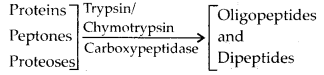
5. Describe the process of digestion of protein in stomach.
Solution: The gastric glands of the stomach secrete gastric juice that contains HCl and proenzymes – pepsinogen and prorennin. The proenzyme pepsinogen, on exposure to HCl gets converted into the active enzyme pepsin, the proteolytic enzyme of stomach. The pepsin converts proteins into proteoses and peptones (peptides). Prorennin is found in gastric juice of infants and is activated by pepsin into active rennin. It helps in digestion of milk protein casein.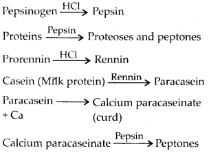
6. Give the dental formula of human beings.
Solution: The dental formula of human beings is![]()
It shows arrangement of teeth in each half of
the upper and lower jaw.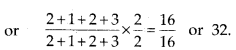
7. Bile juice contains no digestive enzymes, yet it is important for digestion. Why ?
Solution: Bile has no enzymes but contains bile salts, namely, sodium bicarbonate, sodium glycocholate and sodium taurocholate that reduce the surface tension of large fat drop¬lets and break them into many small droplets by a process known as emulsification. These small fat droplets present large surface area for lipase (fat digesting enzyme) to act upon them. Moreover, bile also activates lipases.![]()
8. Describe the digestive role of chymotrypsin. Which two other digestive enzymes of the same category are secreted by its source gland ?
Solution: Chymotrypsin is a proteolytic enzyme of pancreatic juice secreted by exocrine part of pancreas. It helps in digestion of proteins. It converts proteins, peptones and proteoses into oligopeptides and dipeptides. Two other proteolytic enzymes present in pancreatic juice are trypsinogen and procarboxypeptidase.
9. How are polysaccharides and disaccharides digested ?
Solution: Digestion of polysaccharides (starch and glycogen) starts from buccal cavity. In buccal cavity, polysaccharides are acted upon by salivary amylase or ptyalin which splits starch and glycogen into disaccharides and small dextrins called ‘a’ dextrin.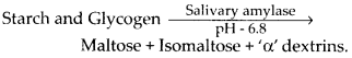
The digestion of carbohydrates does not occur in stomach because gastric juice itself has no carbohydrase.
In small intestine, the food mixes with two juices, pancreatic juice and intestinal juice. Pancreatic juice contains a carbohydrase named pancreatic amylase. This enzyme hydrolyses more starch and glycogen.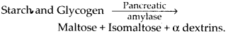
Intestinal juice contains carbohydrases; maltase, isomaltase, a-dextrinase, sucrase and lactase which act on disaccharides as follows: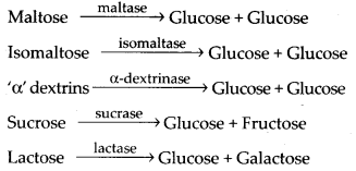
fructose and galactose are monomers of carbohydrates. These are absorbed by intestinal mucosa.
10. What would happen if HCl were not secreted in the stomach?
Solution: HCl is secreted by parietal or oxyntic cells of gastric glands. It serves the following functions:
- It activates the pepsinogen and prorennin into their active form pepsin and rennin.
- It provides the acidic pH (pH 1.8) optimal for pepsin.
- It kills the harmful bacteria present in the food.
- It stops the action of saliva on food. Pepsin and rennin are the principle proteolytic enzymes of stomach. If these enzymes are not activated by HCl then digestion of protein will not take place in stomach, and also the harmful bacteria can cause various diseases.
11. How does butter in your food get digested and absorbed in the body ?
Solution: Butter is a saturated fat. Fats and oils of the ingested food are triglycerides.
They are digested by lipases. Small intestine is the principal organ for fat digestion.
In the small intestine food meets three secretions, bile, pancreatic juice and intestinal juice, all alkaline in nature.
Bile contains no enzyme but it contains bile salts which reduces the surface tension of large fat droplets and breaks them into smaller ones (emulsification).![]()
Emulsified triglycerides Pancreatic juice contains pancreatic lipase, which is the principal fat digesting enzyme. It is activated by bile.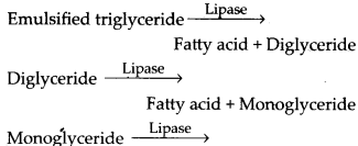
Fatty acid + Glycerol Intestinal lipase found in intestinal juice hydrolyses some triglycerides, diglycerides and monoglycerides to fatty acids and glycerol like pancreatic lipase.
Fatty acids, glycerol and monoglycerides are the end products of fat digestion and being insoluble in water cannot be directly absorbed from the intestinal contents. So they combine with the bile salts and phospholipids to form micelles (water soluble). From the micelles fatty acids, glycerides, sterols and fat soluble vitamins are absorbed into the intestinal cells by diffusion where they are resynthesised in the ER and are converted into very small protein coated fat molecules (droplets) called chylomicrons. The latter are released from the intestinal cells into the lymph present in the lymphatic capillaries, the lacteals. These lacteals ultimately release the absorbed substances into the blood stream.
12. Discuss the main steps in the digestion of proteins as the food passes through different parts of the alimentary canal.
Solution: Proteins of ingested food are broken down into amino acids by proteases (peptidases). Proteases are secreted in inactive forms called proenzymes which are converted into active forms at site of their action. Protein digestion starts in the stomach and is completed in the small intestine. Saliva contains no protease.
Digestion of proteins in stomach : Chief cells of gastric gland secrete pepsinogen and prorennin, which act as follows: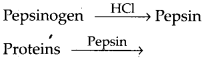
Digestion of proteins in small intestine: In small intestine, peptones and proteoses are acted upon by enzymes of pancreatic juice and intestinal juice.
Pancreatic juice contains 3 inactive proteases; trypsinogen, chymotrypsinogen and pro-carboxypeptidase. Their action is as follows: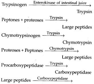
Dipeptides + Amino acids Intestinal juice contains two digestive pro-teases; aminopeptidases and dipeptidases and a nondigestive enterokinase (enteropep- tidase).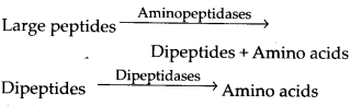
Amino acids are the end products of protein digestion which are absorbed by intestinal cells.
13. Explain the term thecodont and diphyodont.
Solution: Thecodont: In human, each tooth is embedded in a socket of jaw bone. Such teeth are described as thecodont.
Diphyodont: Majority of mammals including human beings form two sets of teeth during their life, a set of temporary milk or deciduous teeth replaced by a set of permanent or adult teeth. This type of dentition is called diphyodont.
14. Name different types of teeth and their number in an adult human.
Solution: Adult human has 32 teeth with the![]()
Human has heterodont dentition i.e., having four different types of teeth. The number of different types of teeth in human are as follows:incisors = 8, canines = 4, premolars = 8, molars = 12
15. What are the functions of liver?
Solution: Liver is the largest gland of the body and consists of hepatic cells. Besides being a digestive gland, the liver performs a number of functions for the welfare of body. Its varied functions are as follows
- Secretion of bile.
- Glycogenesis, gluconeogenesis and glycogenolysis.
- Storage of fat, glycogen, vitamins like A, D, E, K and B12, blood, water, etc.
- Deamination of amino acids.
- Synthesis of urea.
- Elimination of excretory substances.
- Detoxification of harmful substances.
- Formation and breakdown of blood
corpuscles, i.e., in embryos, liver is haemopoietic (produces red blood corpuscles) and in adults its Kupffer cells phagocytise and destroy worn out and dead RBCs. - Secretion of blood proteins, i.e., prothrombin and fibrinogen.
- Secretion of anticoagulant heparin.
- Production of heat.
- Secretion of enzymes.