Breathing and Exchange of Gases Class 11 Notes Biology Chapter 17
Topics and Subtopics in NCERT Solutions for Class 11 Biology Chapter 17 Breathing and Exchange of Gases:
| Section Name | Topic Name |
| 17 | Breathing and Exchange of Gases |
| 17.1 | Respiratory Organs |
| 17.2 | Mechanism of Breathing |
| 17.3 | Exchange of Gases |
| 17.4 | Transport of Gases |
| 17.5 | Regulation of Respiration |
| 17.6 | Disorders of Respiratory System |
| 17.7 | Summary |
Cells continuously use oxygen (O2) for the catabolic reactions that releases energy from molecules, e.g., breakdown of nutrient molecules like glucose. Thus, O2 has to be provided to the cells constantly. Simultaneously, these reactions releases carbon dioxide (CO2), which is harmful, so it must be removed quickly and efficiently.
The process that helps in the exchange of O2 from the atmosphere with CO2 produced by the cells is called breathing, commonly known as respiration.
Topic 1 Respiration : Types and Respiratory Organs
Respiration is a biochemical process that exchanges environmental oxygen with the CO2 produced in the cells. It include stepwise oxidation of food in cells with intake of oxygen and elimination of CO2 produced in oxidation, release of energy during oxidation and storing it in form of ATP.
Types of Respiration
Mechanism of respiration vary, depending mainly on an animal’s habitat and levels of organisation.
The different types of respiration are
1. Aerobic Respiration
When oxygen is used for respiration, its called aerobic respiration. The organism undergoing the process are termed as aerobes.
The term breathing and respiration are not same. Breathing is the first step of respiration and a physical process, while respiration is a biochemical process involving exchange of gases and oxidation of food.
Inspiration is an active process, while expiration is a passive process.
2. Anaerobic Respiration
It occurs in the absence of molecular oxygen in the cytoplasm (also called as fermentation). It yields only about 5% of the food’s energy. The organisms undergoing the process are called anaerobes, e.g., yeasts oxidise glucose to ethanol and CO2.
* Anaerobic respiration appeared first in primitive atmosphere due to absence of oxygen there.
* In frog 100% cutaneous respiration occurs during hibernation and in all marine snakes, 20% respiration is by skin.
* Respiration is a catabolic process. It breaks organic molecules and release their bond energy. It occurs in all organisms, at all times.
Respiratory Organs
Different animal groups have evolved different mechanism of breathing for the exchange of gases.
Lower animal like sponges, cnidarians, Platyhelminthes and free-living roundworms exhange 02 by simple diffusion through body surface.
Special vascularised structures called gills are used by most of the aquatic arthropods and molluscs whereas, vascularised bags called lungs are used by the terrestrial forms for the exchange of gases.
Parasitic flatworms (e.g.. Tapeworms) and roundworms (e.g., Ascaris) have anaerobic mode of respiration.
Human Respiratory System
Human respiratory system may be divided into two major components, i. e., conducting portion and respiratory or exchanging portion.
Conducting Portion
It is the passage for the air (transports the atmospheric air to alveoli and return from lungs to the exterior). This portion clear air from foreign particles, humidifies it and also brings it to body temperature. In this part, gaseous exchange does not take place. It is also called dead air space. It starts with the external nostrils upto the terminal bronchioles.
The various parts are as follows
(i) External Nares (Nostrils) These are a pair of slits at the lower end of the nose, which opens into the the nasal chamber through the nasal passage.
(ii) Nasal Chambers Pair of passage located at the back of nostrils just above the mouth cavity. Nasal septum is a median partition that separates the two chambers.
Each chamber has three regions, i.e., Vestibular, respiratory and olfactory. The chambers has special pseudostratified ciliated epithelium by which air is filtered (by hairs) and moistened (by mucus).
(iii) Internal Nares These are the posterior openings of the nasal chambers that leads into the nasopharynx.
(iv) Nasopharynx It is the upper part of pharynx, into which internal nares open.
(v) Larynx It is the upper part of trachea. It allows the air to pass into lungs. Nasopharynx opens through glottis of the larynx into trachea. Glottis is a slit-like aperture that remains open except during swallowing.
The glottis bears a leaf-like cartilaginous flap, the epiglottis at its anterior region. It closes the glottis to check the entry of food during swallowing. Larynx helps in sound production and hence, called the sound box.
Larynx is often called the Adam’s apple and is more prominent in men than in women.
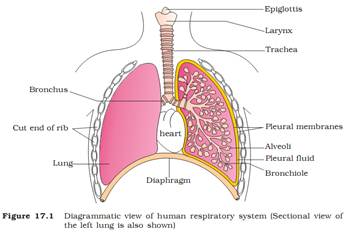
(vi) Trachea It is a thin-walled tube, about 11cm long and 2.5 cm wide. It extends up to the mid-thoracic cavity. It passes the air to the alveoli.
(vii) Primary and Secondary Bronchi At the level of 5th thoracic vertebra, the trachea divides into two tubes, right and left primary bronchi.
Each bronchi further divides into lobar or secondary bronchi. The secondary bronchi sub-divides into smaller tertiary bronchi, which divide into still smaller bronchioles. The small terminal bronchioles give off a number of thin, irregular walled, vascular bag-like structure called alveoli.
Note:
* Wall of trachea, bronchi and bronchioles is composed of fibromuscular tissue and is lined by pseudostratified ciliated columnar epithelium rich in mucus secreting cells.
* Cartilaginous rings support the walls of the trachea and the bronchi to prevent their collapsing.
Respiratory/Exchanging Portion
The alveoli and their ducts form this part of the respiratory system. It is the site of actual diffusion of O2 and CO2 between blood and atmospheric air. The branching network of bronchi, bronchioles and alveoli comprises the lungs, which provide the surface for exchange of gases in humans.
Lungs
These are the paired triangular bags that constitute the respiratory organ.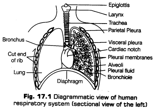
They lie in the thoracic cavity on the sides of the heart. The thoracic cavity is enclosed dorsally by thoracic vertebrae, laterally by the ribs, ventrally by the sternum and closed below by a dome-shaped diaphragm.
The arrangement of lungs is such that, any change in the volume of thoracic cavity will be manifested in the lung (pulmonary) cavity. This arrangement is necessary for breathing as the pulmonary volume cannot be altered directly.
Protective Coats (Pleurae)
Each lung is enclosed in two membranes called the pleura (layers of peritoneum of the thorax). The inner membrane, called the visceral pleuron, which is firmly bound to the surface of lungs. The outer membrane, called the parietal pleuron is held to the thoracic wall and diaphragm by connective tissue. Pleural cavity is a very narrow space that exists between the two pleura. It contains the pleural fluid secreted by the pleura, for reducing friction on the lung surface.
External Features
(a) The left lung has two lobes, i.e., superior lobe and inferior lobe separated by oblique fissure. It has a cardiac notch, a concavity where the heart lies. It is longer and narrower than right lung.
{b) The right lung is bigger and has three lobes, i.e., superior lobe, middle lobe and inferior lobe separated by horizontal fissure and oblique fissure.
Note:
* There are about 300 million alveoli in the two lungs with a combined surface area of about 70 m2.
* A film of lecithin lines the alveoli, that lowers the surface tension.
* Larynx is comprised of 9 cartilages (epiglottis, thyroid and cricoid are single, while arytenoid, comiculate and cuneiform are paired).
* If the chest wall is pierced (e.g., by a stab wound), atmospheric air rushes into the pleural cavity, eliminating the pressure difference across lung walls causing the lungs to collapse. The condition is called pneumothorax.
Topic 2 Respiration Processes : Breathing and Gaseous Exchange
The main mechanisms of respiration is categorised into following three steps
(i) Breathing (pulmonary ventilation) is the inflow of atmospheric air and release (outflow) of CO2 rich alveolar air.
(ii) Exchange of gases (O2 and CO2) across alveolar membrane as well as in tissues.
(iii) Transport of gases by the blood.
Breathing
Breathing is an extracellular, energy consuming and physical process. It involves movement of thorax, expansion (inflation) and deflation of lungs and flow of air into and from the lungs by creating a pressure gradient between the lungs and the atmosphere.
The diaphragm and a specialised set of muscles, i.e., external and internal intercostals between the ribs help in generation of such gradients.
Breathing mainly involves two steps
i. Inspiration
It is an active process by which fresh air enters the lungs. It can occur if the pressure within the lungs (intra-pulmonary pressure) is less than the atmospheric pressure, i.e., negative pressure in lungs with respect to atmospheric pressure.
Following muscles play an important role
(a) Diaphragm It is lowered by the contraction of its muscle fibres and becomes flat. This causes an increase in the volume of thoracic chamber in the antero-posterior axis.
(b) External intercostal muscles They occur between the ribs (internal intercostal muscles are related to expiration). The external intercostal muscles contract and pull the ribs and the sternum upward and outward thus, increasing the volume of thoracic chamber in dorso-ventral axis.
Accessory muscles of breathing, i.e. scaleni, sternomastoid and ala nasi come into action during forced inspiration.
Thus, the overall increase in the volume of thoracic cavity causes an increase in pulmonary volume. As a result, there is a decrease in the intra-pulmonary pressure. The , greater atmospheric pressure outside the body now causes air to flow rapidly into external nares, which sequentially leads to alveoli.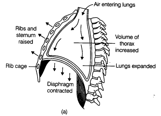
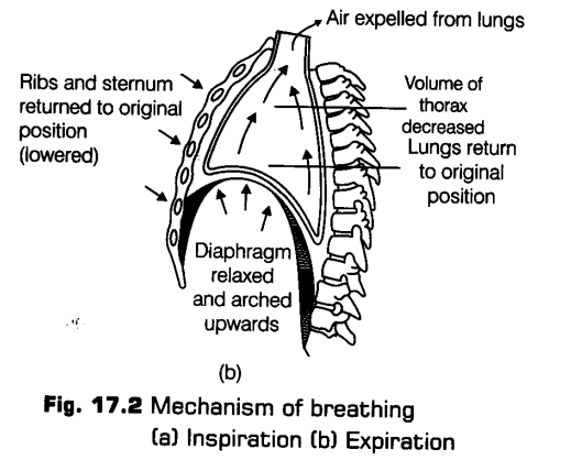
ii. Expiration
It is a passive process by which CO2 is expelled out from the lungs. It takes place when the intra-pulmonary pressure is higher than the atmospheric pressure.
The movement of muscles involved is as follows
(a) Diaphragm The muscle fibres of the diaphragm relax making it convex, decreasing volume of the thoracic cavity.
(b) Internal intercostal muscles These muscles contract thus, pulling the ribs downward and inward, decreasing the thoracic volume.
Abdominal muscles (i.e., external and internal oblique muscles) contracts, compressing the’ abdomen and pushing the diaphragm up.
The overall volume of the thoracic cavity thus, decreases thereby reducing the pulmonary volume.
As a result, the intra pulmonary pressure increases slightly above the atmospheric pressure. This inturn causes the expulsion of the air from the lungs. The process of expiration is simpler than inspiration.
Note:
* At rest, breathing occurs about 12-16 times/minute, being more in children.
* The volume of air involved in breathing movements can be estimated by using a spirometer. It helps in clinical assessment of pulmonary functions.
* Breathing in frog is considered as positive pressure.
Respiratory Volumes and Capacities
The quantity of air the lungs can receive, hold or expel under different conditions are called respiratory (or pulmonary) volumes.
Combination of two or more pulmonary volumes are called respiratory (pulmonary) capacities.
The different volumes and capacities are as follows
(i) Tidal Volume (TV) It Ms the volume of air inspired or expired during normal breathing in relaxed or resting position. It is about 500 mL. It consists of 150 mL of dead space volume and 350 mL of alveolar volume.
A healthy man can inspire or expire approximately 6000-8000 mL of air per minute.
(vi) Expiratory Capacity (EC) It is the total volume of air a person can expire after a normal inspiration. It includes tidal volume and expiratory reserve volume.
EC= TV+ ERV
(vii) Functional Residual Capacity (FRC) It is the volume of air that will remain in the lungs after a normal expiration. It includes residual volume and expiratory reserve volume.
FRC= RV+ ERV
(viii) Vital Capacity (VC) It is the maximum value of air a person can breathe in after a forced expiration or the maximum volume of air a person can breathe out after a forced inspiration. This includes TV+ IRV+ ERV.
It varies from 3400-4800 mL depending upon age, sex and height of individual.
(ix) Total Lung Capacity It is the total volume of air present in the lungs after a forced (maximum) inspiration. It includes (VC+RV) or (RV+ ERV+ TV+ IRV)
Note:
* The vital capacity is higher in athletes, mountain dwellers than in plain dwellers, in men than women and in the young ones than in the old persons.
* All pulmonary volumes and capacities are about 20-25% less in women than in men and they are greater in tall persons and atheletes than in small and asthenic people.
* During respiration, the lungs and the respiratory tract are never devoid of air. Instead, there is a tidal volume of air.
Exchange of Gases
The primary sites for exchange of gases are the alveoli and tissues.
It occurs by simple diffusion mainly based on pressure/ concentration gradient.
The factors that affect rate of diffusion are
(i) Thinness of the membrane.
(ii) Surface area of the membrane.
(iii) Permeability of the membrane.
(iv) Solubility of the gases.
(v) Partial pressure gradient (difference) of gases on the two sides of a membrane between them.
Partial pressure of a gas is the pressure it exerts in a mixture of gases. It is equal to the total pressure of the mixture divided by percentage of that gas in the , mixture.
1. Inspiratory Reserve Volume (IRV) It is the additional amount of air that can be inspired forcibly after a normal inspiration. It is about 2500-3000 mL of air.
2. Expiratory Reserve Volume (ERV) It is the additional volume of air that can be expired forcibly after a normal expiration. It is about 1000-1100 mL.
3. Residual Volume (RV) It is the volume of air remaining in the lungs even after a forcible expiration. It is about 1100-1200 mL. It can not be measured by spirometry.
4. Inspiratory Capacity (IC) It is the total volume of air a person can inspire after a normal expiration. It is about 2500-3000 mL. It includes tidal volume and inspiratory reserve volume.
IC= TV+ IRV
Partial Pressure (in mm Hg) of Oxygen and Carbon dioxide at different parts Involved in Diffusion in Comparison to those in Atmosphere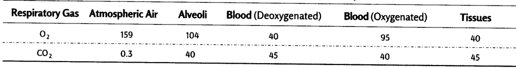
i. Exchange of Gases between Alveoli and Blood
This exchange between lung alveoli and pulmonary capillaries is called external respiration.
The alveolar wall is very thin with a rich network of blood capillaries. It is also called as respiratory membrane or diffusion membrane.
It consists of three major layers, i.e., the thin squamous epithelium of alveoli, the endothelium of alveolar capillaries and the basement substance in between them. All these layers form a membrane of about 0.2 mm thickness.
This membrane has a limit of gaseous exchange between alveoli and pulmonary blood, called the diffusing capacity.
At a particular pressure difference, the diffusion of CO2 is 20-25 times faster than oxygen. Thus, the amount of CO2 that can diffuse through the membrane per unit difference in partial pressure is much higher as corrtpared to oxygen.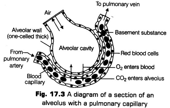
As seen in the above table, pO2 in the alveoli (104 mmHg) is higher than that in the deoxygenated blood in the capillaries of pulmonary arteries (95 mmHg). So, the movement of O2 is from alveoli to the blood.
Also, pCO2 is higher in deoxygenated blood (45 mmHg) than in alveoli, therefore, CO2 passes from blood to the alveoli.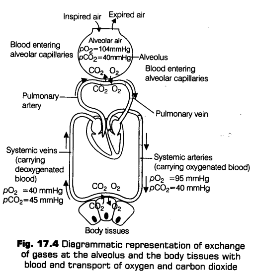
ii. Exchange of Gases between Blood and Tissue Cells
This exchange between tissue blood capillaries and tissue cells is called internal respiration. pO2 in oxygenated blood (95 mmHg) > pO2 in body cells (40 mmHg) and pCO2 in oxygenated blood (40 mmHg) < pCO2 in body cells (45 mmHg).
Due to this partial pressure differences, oxygen diffuses from the capillary blood to the body cells and CO2 diffuses from the body cells to the capillary blood via tissue fluid. Now, the blood will become deoxygenated, which is further carried to the heart and finally to the lungs.
CBSE Class 11 Biology Chapter-17 Important Questions
1 Marks Questions
1.Define partial pressure of a gas.
Ans. Pressure contributed by an individual gas in a mixture of gases is called partial pressure of gas and it is represented as PO2 for O2 and PCO2 for CO2.
2.Name the other pigments which are present in animals besides haemoglobin.
Ans. Haemocyanin and haemoerythrin.
3.What is the difference between alveolar air and inspired air?
Ans. Alveolar air – The air present in the alveoli.
Inspired air – The amount of air inspired at a time.
4.Define vital capacity.
Ans. Vital Capacity is the volume of air breathed out by a maximum forceful expiration.
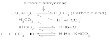
5.What is the role of carbonic anhydrase in RBC’s?
Ans. About 70% of CO2 reacts with water to form carbonic acid in RBCs in the presence of enzyme carbonic anhydrase. CO2 + H2O  H2CO3
H2CO3
6.What is carbamino haemoglobin?
Ans. Carbaminohaemoglobin is formed when CO2 combines with globin is reduced haemoglobin.
7.Name the place where actual exchange of gases takes place in insects.
Ans. Tracheoles.
8.What is the percentage of O2 in inspired & expired air?
Ans. Inspired air has 21% O2 and expired air has 16% O2.
9.What is the utility of chloride shift?
Ans. It maintains the ionic balance and electrochemical neutrality.
10.Name the organ in human respiratory system which produces sound.
Ans. Larynx (Sound box)
11.How many oxygen molecules can be carried out by one hemoglobin molecule.
Ans. Four molecules
12.Give the name and function of a fluid filled double membranous layer which surrounds the lungs.
Ans. Fleuron. It reduces the friction and keeps the two pleura together and the lungs inflated.
13.Which organ of our respiratory system acts as primary site of exchange of gases?
Ans. Alveoli of Lungs.
14.Cigarette smoking causes emphysema. Give reason.
Ans. Cigarette smoking damages alveolar walls due to alveolar sacs remaining filled with air leading to decreased respiratory surface fore
15.Name the principle of exchange of gases.
Ans. Diffusion.
16.What is the role of oxyhaernoglobin after releasing molecular oxygen in the
Ans. Amino group of refuced hoemoglobin combines with CO2 forming carbaminohaemoglobin to transport CO2.
17. Name the museles which facilitate breathing.
Ans. External and internal intercostals muscles, situated between ribs.
18.How is the entry of food pivoted in the respiratory tract?
Ans. Ans. During swallowing a cartilaginous flap like structure called epiglottis covers the glottis and prevents the entry of food into respiratory tract.
19. About 97% of O2 is transported by RBCs in the blood. How does the remaining 3% of O2 transported?
Ans. In simple solution form through plasma.
2 Marks Questions
1.Give role of intercoastals muscles in respiration.
Ans. The contraction of the external intercoastals muscles & diaphragm increases the volume of the thoracic cavity lowers the pressure in the lungs. To fill up the gap, the fresh air reaches to the lungs resulting in the inspiration.
The relaxation of the inspiratory muscles decreases the volume of the thoracic cavity and subsequently, pressure in the lungs increase. To equalize this pressure, the air from the lungs rushes out through the respiratory passage to bring about expiration.
2.Explain Erythrocytes can carry out anaerobic metabolism only.
Ans. Erythrocytes lack mitochondria and respiratory enzymes to perform the process of aerobic respiration. Therefore, they undergo aerobic respiration to carry out anaerobic metabolism only.
3.Describe how our brain gets a continuous supply of oxygen form the atmosphere.
Ans. Passage of air which contains oxygen:
Inhalation of fresh air → trachea → bronchi → lungs → alveoli → diffusion of O2 into blood (RBC) → formation of oxygemoglobin → some in plasma → pulmonary vein → carry it to heart → left auricle → to ventricle → Dorsal aorta → Carotid artery to the brain dissociation of oxyhaemoglobin, O2 supplied to the tissue, partial pressure of O2 facilitates diffusion.
4.What is chloride shift? Explain.
Ans. The diffusion of chloride ions from blood plasma into RBS’s is known as chloride shift.
a) Occurs from plasma to RBC’s in human body.
b) It maintains ionic balance and electrochemical neutrality.
5.Explain briefly the first step is respiration?
Ans. First step in respiration is called breathing. In breathing atmospheric air is taken in by inspiration and alveolar air is released out by expiration. The exchange of O2 and CO2 between deoxygenated blood and alveoli, transport of gases throughout body by blood, exchange of O2 and CO2 between the oxygenated blood and tissues and utilization of O2 by the cells are the other steps involved in it.
6.Write a note on bronchitis and its prevention.
Ans. It is “inflammation of the bronchi and is characterized by hypertrophy hyperplasia seromucous glands and goblet cells lining the bronchi”
Symptoms are coughing with thick greenish-yellow sputum. It shows infection, that excessive secretion of mucus. It is caused by pollutants as well as the cigarette smoking. Prevention of Bronchitis –
1) Avoiding exposure to allergens.
2) Treatment involves antibiotic theory & bronchodilator dugs, etc.
7.What is the difference between carbaminohaemoglobin and oxyhaemoglobin.
Ans.
| Oxyhaemoglobin | Carbominohaemoglobin | |
| 1. | It is formed by the combination of oxygen with the Fe2+ part of haemoglobin. | It is formed by the combination of carbon dioxide with the amine radical of haemoglobin. |
| 2. | It formation occurs on the alveolar surface. | Its formation occurs in the tissues. |
8.What is functional residual capacity?
Ans. When a person inhales and exhales in a normal way, the volume air that remains in the lungs is known as functional residual capacity (FRC). It includes the residual volume and expiratory reserve volume, i. e, FRC = RV + ERV.
9.Describe the transport of O2 and CO2?
Ans. O2 is transported as oxyhaemoglobin. In the alveoli of lungs (PO2 is higher), O2 gets bound to hemoglobin that dissociates at tissues where PO2 and H+ concentration are high. Approx 70% CO2 transported as bicarbonate (HCO3–) with the help of the enzyme carbonic anhydrase, 20-25% CO2 is carried by haemoglobin as carbaminohamoglobin. In tissues PCO2 is high its gets bound to blood but in alveoli where PCO2 is low and PO2 is high, this removed from blood.
10. Name the organs of respiration in the following organisms.
a) Flatworms b) Birds c) Frog d) Cockroach
Ans. a) Body surface b) lungs c) skin and lungs d) Network of trachea
3 Marks Questions
1.What is hypoxia, artificial hypoxia & Anaemic hypoxia?
Ans. Hypoxia – It is a condition of oxygen shortage in the tissues. It is 2 kinds – artificial hypoxia and anaemic hypoxia.
Artificial hypoxia – It is the result of shortage of air over 2400m altitudes. The mountain sickness is caused by headache, dizziness, nausea, vomiting, mental fatigue and breathless mess etc.
Anaemic hypoxia – results due to reduced O2 capacity of blood due to less content of Hb or carbon monoxide poisoning.
2.How is respiration regulated?
Ans. Respiratory centre located in floor of the medulla oblongata of the brain controls respiration. The centre is bilateral and its two halves which are connected together by commissural neurons. The sides of this centre are connected with motor respiratory neuron. The nerve cells of the centre are connected with the breathing apparatus forming a reflex arc. These nerve cells are secretive to chemical composition of blood. Half of the respiratory centre is an inspiratory centre and expiratory centre. It is believed that the inspiratory centre work in normal breathing and expiratory centre during other conditions like coughing, sneezing and laughing. These two centers control the entire breathing in man with his knowing about it. Dorsal respiratory group, ventral respiratory group and pneumoptaxic groups act as respiration centers in the brain.
Neumotaxic centre is located dorsally in upper pons. It transmits signals to inspiratory area. It controls the switch off point of inspiration.
3.Differentiate between vital lung capacity and total lung capacity.
Ans.
| Vital Capacity (VC) | Total lung Capacity. (CT2C) | |
| 1. | Sum total of tidal volume, expiratory reserve and inspiratory reserve volume. | Sum total of vital capacity and residual volume. |
| 2. | VC = Vt + ERV + IRV | TLC = VC + RV |
| 3. | Value is 3500-4500ml. | Value is 5000 – 6000ml |
| 4. | Represents maximum amount of air that a person can expel after filling the lungs to the maximum. | Represents maximum total amount of air which can be present in lungs after maximum inspiratory effort. |
4.Explain the mechanism of breathing in humans.
Ans. The mechanism of breathing in human involves breathing in of air in the lungs and breathing out of air from lungs thoracic cavity. The form is called inspiration and later expiration. The lungs are located in the closed thoracic cavity. A muscular partition called diaphragm separates the thoracic cavity from the abdominal cavity.
During inspiration the diaphragm is lowered due to contraction intercostals muscle. This result into the increase of volume of thorax causing fall of air pressure in the thoracic cavity lowers the pressure in the lungs and the air rushes from outside into lungs through external nares, trachea & bronchi.
During expiration the diaphragm move upward and the lateral thoracic walls move inwards due to the relaxation of muscles of diaphragm and the intercoastals muscles. This decrease the volume of thorax and the pressure inside the thorax and lungs is increased which results in the expulsion of some of air from the lungs to the atmosphere outside the body.
5.Define oxygen dissociation curve? Why it has sigmoidal pattern?
Ans. The relationship between O2 tension and its absorption by haemoglobin produces a graph called oxygen dissociation curve (O2 equilibrium curve). At about 100 mm Hg O2 tension Hb is 98% saturated (complete formation of haemoglobin). As it falls, the saturation of Hb decreases slowly. When O2 tension is about 40mm Hg, oxyhaemoglobin dissociates and O2 is available to the tissues.
The O2 gets bound to Hb in lung surface and it gets dissociated at tissues.
6.What is the role of carbonic anhydrase? Show by series of reactions how carbonic anhydrase starts the reactions leading to the formation of hemoglobinic acid?
Ans. Carbonic Anhydrate : CO2 reacts with water in presence of carbonic anhydrase in erythrocytes, Carbonic acid (H2CO3) is dissociated into hydrogen (H+) and bicarbonate (HCO3–) ions). Oxyhaemoglobin (HbO2) of RBC’s is weakly acidic and remain in association with K+ ions as KHbO2. H+ ions combine with haemoglobin. Bricarbonate ions diffuse out into plasma and combine with haemoglobin to from haemoglobinic acid (H. Hb)
5 Marks Questions
1.Describe transport mechanism of CO2.
Ans. Transport of CO2 in the blood.
(i) In the dissolved form
About 5 – 7 % of carbon – dioxide is transported in dissolved form in the plasma of blood.
(ii) In the form of bicarbonate.
The remaining part of carbon dioxide enters the erythrocytes, where it reacts with the water to form carbonic acid (H2 CO3) ; this reaction is catalysed by carbonic anhydrase.
O Carbonic acid immediately dissociates into hydrogen ions (H+) and bicarbonate (HCO3–)
O These H+ ions combine with haemoglobin, after the oxyglobin (KHbO2) dissociates to liberate the oxygen ; as a result haemoglobinic acid (H.Hb) is formed.
O The majority of bicarbonate ions (HCO–3) diffuses out of the erythrocytes into the plasma, following a concentration gradient.
O In response, chloride ions (Cl–) diffuse from the plasma into the erythrocytes (Chloride shift) and electrical neutrality is maintained.
O The chloride ions combine with potassium to form potassium chloride.
O In the plasma the bicarbonate ions combine with sodium and transported as sodium bicarbonate (NaHCO3)
O Nearly 70% of the carbon dioxide is transported in this form.
(iii) In the form of carbaminohaemoglobin
O Same amount of CO2 reacts with the amine radicals (NH2+) of haenoglobin and form carbaminohaemoglobin (HbCO2) molecule.
O About 23% of carbon – dioxide is transported in this form.
2. Describe in brief the respiratory organs of man.
Ans.The following are the main respiratory organs:-
1)Nostrils – These are the paired openings situated at the anterior and posterior ends of the nasal chambers. They are lined up with ciliated epithelium and mucous cells. These prevent the entrance of dust into lungs and help in warming and moistening the air. The nasal chamber opens interiorly by external nostril and posterior internal nostril into the pharynx.
2)Larynx – It is situated at the anterior part of trachea and communicates with the pharynx. The glottis is protected by a stiff cartilage called epiglottis. The larynx contains pairs of vocal cords which set into vibrations when the air enters into it and produces the sound.
3)Trachea – It is a long ringed tube. It is supported c – shaped elastic cartilaginou rings to prevent its collapsing. It is lined internally with mucous membrane to hold the dust particles, bacteria and other foreign bodies. It also warms the air.
4)Bronchi – Inside the thorax, the trachea bifurcates into two branchy and each of which enters into one lung. In each lung, the bronchus again redivides into numerous small branches known as bronchioles. These bronchioles further divide at its ends to form respiratory bronchioles.
5)Lungs – There are two large bag-like spongy structures which are the main respiratory organs. These are enclosed by double pleural membranes. The lungs are divided externally by lobes. The right lung consists of four lobes and left by two lobes. Inside the lungs, the respiratory bronchioles give rise to alveolar ducts, alveolar sac and finally alveoli. Each lung contains millions of alveoli. Each alveolus is exceptionally thin walled. Its walls are highly permeable and richly supplied with blood capillaries.
The blood is supplied to the lungs by a pair of pulmonary arteries. These bring blood which poor in oxygen & rich in CO2. The exchange of gases occurs in the alveoli of the lungs. The oxygenated blood from alveolar capillaries is called by pair of pulmonary vein to be conveyed to the heart.
3. Explain how our heart muscles get a continuous supply of atmospheric oxygen.
Ans. When inspiration occurs, the O2 is taken into lungs. O2 mixes with air already present in alveoli and becomes alveolar air, whose PO2 is 100 m Hg.
As PO2 of blood in the vessels is 40 mmHg oxygen differs into blood vessels from alveoli and the oxyhaemoglobin is formed when oxygen combines loosely with the Fe++ ions of haemoglobin.
Oxygenated blood from the lungs reaches the left auricle through pulmonary vein; to left ventricle is pumped at through aorta also.
The branch supplying blood to heart muscles is coronary artery. In heart muscles, as the PO2 is lower than that of the blood in the branches of coronary artery, oxyhaemoglobin dissociates and releases O2 to cardiac muscles.
NCERT TEXTBOOK QUESTIONS FROM SOLVED
1. Define vital capacity. What is its significance?
Solution: Vital capacity is defined as the maximum volume of air a person can breathe in after a forced expiration or the maximum volume of air a person can breathe out after a forced inspiration. It represents the maximum amount of air one can renew in the respiratory system in a single respiration. Thus, greater the vital capacity more is the energy available to the body.
2. State the volume of air remaining in the lungs after a normal breathing.
Solution: When a person breathes normally, the amount which remains in the lung after normal expiration, is called functional residual capacity. It is the sum of residual volume and the expiratory reserve volume (FRC = RV + ERV). It is about 2100 – 2300 mL of air.
3. Diffusion of gases occurs in the alveolar region only and not in the other parts of respiratory system. Why?
Solution: For efficient exchange of gases, respi: atory surface must have certain characteristics such as (i) it must be thin, me ist and permeable to respiratory gases (ii) it must have large surface area, (iii) it must be highly vascular. Only alveolar region has these characteristics. Thus, diffusion of gases occurs in this region only.
4. What are the major transport mechanisms for CO2? Explain.
Solution: Nearly 20-25 percent of CO2 is transported by haemoglobin of RBCs, 70 percent of it is carried as bicarbonate ion in
plasma and about 7 percent of CO2 is carried in a dissolved state through plasma. CO2 is carried by haemoglobin as carbamino- haemoglobin. This binding is related to the partial pressure of CO2.
5. What will be the p02 and pCO2 in the atmospheric air compared to those in the alveolar air?
(i) pO2 lesser, pCO2 higher
(ii) pO2 higher, pCO2 lesser
(iii) pO2 higher, pCO2 higher
(iv) pO2 lesser, pCO2 lesser
Solution: (ii) Air that has entered the alveoli through the bronchioles is called alveolar air. It has the same partial pressure of CO2 and 02 as is in the atmospheric air. Then, there occurs gaseous exchange between the adjacent blood capillaries and the alveoli. CO2 diffuses from blood into the alveolar air and O2 diffuses from alveolar air to the blood. As a result, new alveolar air has higher pCO2and lesser pO2, than the atmospheric air.
6. Explain the process of inspiration under normal conditions.
Solution: Inspiration is a process by which fresh air enters the lungs. The diaphragm, intercostal muscles and abdominal muscles play an important role. The muscles of the diaphragm and external intercostal muscles are principle muscles of inspiration. Volume of thoracic cavity increases by contraction of diaphragm and external intercostal muscles. During inspiration, relaxation of abdominal muscles also occurs which allows compression of the abdominal organs by diaphragm. Thus, overall volume of the thoracic cavity increases and as a result, there is a decrease of the air pressure in the lungs. The greater pressure outside the body now causes air to flow rapidly into the lungs. The sequence of air flow is.
7. How is respiration regulated?
Solution: Respiration is under both nervous and chemical regulation.
The respiratory centre in brain is composed of groups of neurons located in the medulla oblongata and pons varolii. The respiratory centre regulates the rate and depth of the breathing.
Dorsal respiratory group of neurons are located in the dorsal portion of the medulla oblongata. This group of neurons mainly causes inspiration.
Ventral group of neurons are located in the ventrolateral part of the medulla oblongata. These can cause either inspiration or expiration.
Pneumotaxic centre is located in the dorsal part of pons varolii. It sends signals to all the neurons of dorsal respiratory group and only to inspiratory neurons of ventral respiratory group. Its job is primarily to limit inspiration. Chemically, respiration is regulated by the large numbers of chemoreceptors located in the carotid bodies and in the aortic bodies. Excess carbon dioxide or hydrogen ions mainly stimulate the respiratory centre of the brain and increases the inspiratory and expiratory-signals to the respiratory muscles. Increased C02 lowers the pH resulting in acidosis. The role of oxygen in the regulation of respiratory rhythm is quite insignificant.
8. What is the effect of pCO2on oxygen transport?
Solution: Increase in pCO2 tension in blood brings rightward shift of the oxygen dissociation curve of haemoglobin thereby decreasing the affinity of haemoglobin for oxygen. This effect is called Bohr’s effect. It plays an important role in the release of oxygen in the tissues.
9. What happens to the respiratory process in a man going up a hill?
Solution: Rate of breathing will increase in order to supply sufficient oxygen to blood because air in mountainous region is deficient in oxygen.
10. What is the site of gaseous exchange in an insect?
Solution: Tracheae (Tracheal respiration) is the site of gaseous exchange in an insect. .
11. Define oxygen dissociation curve. Can you suggest any reason for its sigmoidal pattern?
Solution: The relationship between the partial pressure of oxygen (pO2) and percentage saturation of the haemoglobin with oxygen (O2)is graphically illustrated by a curve called oxygen haemoglobin dissociation curve (also called oxygen dissociation curve).
The sigmoidal pattern of oxygen haemoglobin dissociation curve is the result of two properties which play significant role in the transport of oxygen. These two properties are:
(i) Minimal loss of oxygen from haemoglobin occurs above p02 of 70-80 mm Hg despite significant changes in tension of oxygen beyond this. This is depicted by relatively flat portion of the curve.
(ii)Any further decline in p02 from 40 mm Hg causes a disproportionately greater release of oxygen from the haemoglobin. It results in the steeper portion of the curve and causes the curve to be sigmoid.
12. Have you heard about hypoxia? Try to gather information about it, and discuss with your friends.
Solution: Hypoxia is a condition of oxygen shortage in the tissues. It is of two types:
(i) Artificial hypoxia: It results from shortage of oxygen in the air as at high altitude. It causes mountain sickness characterised by breathelessnes, headache, dizziness and bluish tinge on skin.
(ii) Anaemic hypoxia: It results from the reduced oxygen carrying capacity of the blood due to anaemia or carbon monoxide poisoning. In both cases, less haemoglobin is available for carrying 02.
13. Distinguish between
(a) IRV and ERV
(b) Inspiratory capacity and expiratory capacity.
(c) Vital capacity and total lung capacity.
Solution:
(a) Differences between IRV and ERV are as follows: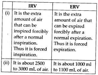
(b)Differences between inspiratory capacity and expiratory capacity are as follows: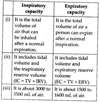
(c) Differences between vital capacity and total lung capacity are as follows: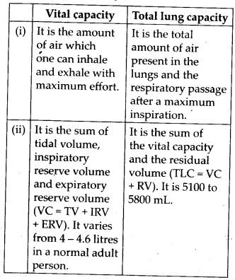
14. What is tidal volume? Find out the tidal volume (approximate value) for a healthy human in an hour.
Solution: Tidal volume is the volume of air inspired or expired with each normal breath. This is about 500 mL in an adult person. It is composed of about 350 mL of alveolar volume and about 150 mL of dead space volume. The alveolar volume consists of air that reaches the respiratory surfaces of the alveoli and engages in gas exchange. The dead space volume consists of air that does not reach the respiratory surfaces.
A healthy man can inspire or expire approximately 6000 to 8000 mL of air per minute. Therefore, tidal volume for a healthy human in an hour is 360 – 480 mL of air.