Structural Organization in Animals Class 11 Notes Biology Chapter 7
Topics and Subtopics in for Class 11 Biology Chapter 7 Structural Organisation in Animals:
| Section Name | Topic Name |
| 7 | Structural Organisation in Animals |
| 7.1 | Animal Tissues |
| 7.2 | Organ and Organ System |
| 7.3 | Earthworm |
| 7.4 | Cockroach |
| 7.5 | Frogs |
| 7.6 | Summary |
Topic 1: Animal Tissues
Tissue
All cells are well organised and coordinated to work in a group. A group of similar cells along with intercellular substances perform a specific function, such an organisation is called tissue. The term ‘Tissue’ was introduced by Bichat and he is known as the father of animal histology.
A tissue can also be defined as a group of one or more types of cells having a same origin and specialised for specific functions along with the intercellular materials.
The intercellular materials or fluid forms the environment of the cell. The cell receives almost all the materials it require from the intercellular fluid and transfer its waste materials again in this fluid.
Note:
* The microscopic study of the tissues and organs in relation to their function is called Histology.
* The term Histology was coined by Mayer, in 1819.
* The tissues arise from the undifferentiated cells of the primary germ layers (ectoderm, mesoderm and endoderm) in an embryo.Types of Animal Tissues
* The structure of the cells vary according to their function.
This variation in cells leads to the formation of following four types oftissues on the basis of their location andfunction
(i) Epithelial (ii) Connective
(iii) Muscular (iv) Neural
I.Epithelial Tissue
Epithelial tissue or epithelium (Epi – upon; thele – nipple) covers both external and internal surfaces of the animal body.
The epithelial tissue has a free surface, which faces either a body fluid or the outside environment and thus, provides a covering or a lining for some part of the body.
Characteristics
The characteristic features of epithelial tissue are as follows
(i) The cells are compactly arranged.
(ii) Intercellular spaces are narrow, 20-30 nm wide.
(iii) Adjacent cells are held together by intercellular junctions.
(iv) The epithelial tissue lies on a thin, non-cellular basement membrane.
(v) Blood vessels are not present in the epithelial tissue.
(vi) Materials are exchanged by diffusion between epithelial cells and the blood vessels of the connective tissues across the basement membrane.
(vii) Nerve endings may penetrate the epithelial tissues.
Junctions Between Epithelial Cells
The common intercellular junctions may include tight junctions, gap junctions, desmosomes, intercellular bridges and interdigitations.
Tight Junctions
The plasma membrane in the apical region of the adjacent epithelial cells become tightly packed together. These junctions check the flow of materials between the cells and are called occluding junctions.
Adhering Junctions
Facilitate the cementing process so as to keep the * neighbouring cells together. They include desmosomes and hemidesmosomes.
Desmosomes
These are thick and strong junctions. They serve, anchoring functions.
Gap Junctions
They are fine hydrophilic channels between adjacent cells formed with the help of protein cylinders called connexin. They help in chemical exchange between adjacent cells and hence are called communicating junctions.
Types of Epithelial Tissues
The epithelial tissues are broadly classified into two groups, i.e., simple and compound.
Simple Epithelia
Simple epithelium is made up of a single layer of compactly arranged cells which rest over a non-cellular basement membrane. It occurs over moist surfaces where a little wear and tear occurs by friction. The simple epithelium is generally related with absorption, secretion, diffusion and movement of materials.
It is further sub-divided into following types
i. Simple Squamous Epithelium
The squamous (squama-scale) is formed of a single layer of closely fitted, flattened, polygonal cells, which forms bulges on the cell surface. ,
The given cells are held together by various types of junctions, mainly tight junctions. The cells of squamous epithelium appear as tiles over a floor. They are also known as pavement epithelium.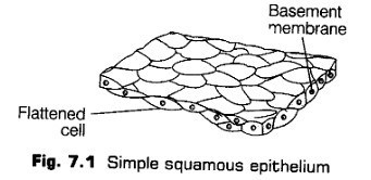
The squamous epithelium occurs in the alveoli of the lungs, Bowman’s capsule, Henle’s loop of uriniferous tubules, pericardial cavity, abdominal cavity, lining of various components of blood vascular system.
Functions Simple squamous epithelium performs the function of protection, excretion, gas exchange and secretion of coelomic fluid.
ii. Simple Cuboidal Epithelium
It is composed of a single layer of cube-like cells. The epithelium overlies on the basement membrane. Nucleus is rounded and placed centrally. The free surfaces of the cells may be smooth or bear microvilli. The microvilli increases the surface area of free ends of cells by many times.
The simple cuboidal epithelium is commonly found in the ducts of glands, tubular parts of nephrons in kidneys, ovaries seminiferous tubules of testes, etc.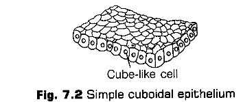
Functions The main function of this epithelium is protection, secretion, absorption, excretion and gamete formation.
iii. Simple Columnar Epithelium
It is composed of a single layer of tall and slender cells. A single oval or elongated nucleus is situated near the base of the cell. Some of its cells produce mucus, called goblet cells.
The simple columnar epithelium occurs in the lining of stomach, small and large intestine, digestive glands of stomach, intestine and pancreas, gall bladder, etc.
The brush border columnar epithelium occurs in the gall bladder. The mucus secreting goblet cells are found in the lining layer of stomach, intestine, respiratory tract, etc.
Functions The simple columnar epithelium helps in secretion, absorption and protection to the components of most glandular epithelia.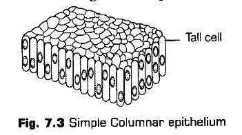
iv. Simple Ciliated Epithelium
If the columnar or cuboidal cells bear cilia on their free surface they are called ciliated epithelium. They move particles or mucus in a specific direction over the epithelium. The epithelium lies over a basement membrane. The number of cilia varies in different cellular forms.
In sensory cells of internal ear, a cilium accompanies number of stereocilia.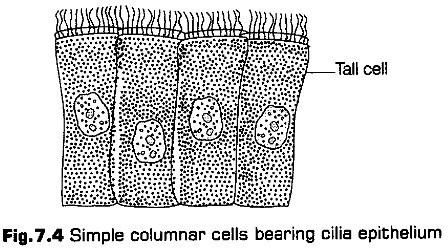
This epithelium is of two types, i.e., ciliated columnar and ciliated cuboidal.
(a) Simple Ciliated Columnar Epithelium It possess columnar cells that possess cilia over their free surface. It occurs in respiratory tract, fallopian tubes, parts of uterus and cervix, the different tubules of testes, etc.
(b) Simple Ciliated Cuboidal Epithelium It has cuboidal or cubical cells that bear cilia on their free surface. It occurs in many parts of ependyma of nervous system and parts of uriniferous tubules.
Functions The epithelium maintains a flow of mucus, liquid or suspended particles constantly in one direction. In the oviducts, cilia helps in the movement of egg towards the uterus. In respiratory tract, cilia helps to push the mucus towards the pharynx. In nephrons of kidney, cilia keep the urine moving.
In nervous system, cilia of the ventricles of the brain and central canal of the spinal cord helps in the circulation of cerebrospinal fluid.
Pseudostratified Epithelium
The epithelium is one-cell thick, but appears 2-layered because all the cells do not reach the free surface. The cells are attached to the basement membrance, hence they are called pseudostratified. The mucus secreting goblet cells also occur in this epithelium.
This epithelium is of two types
(a) Pseudostratified Columnar Epithelium It has columnar cells without cilia. It lines the large ducts of certain glands, like parotid salivary glands and the urethra of human male.
(b) Pseudostratified Ciliated Columnar Epithelium It has columnar cells. The tall cells bear cilia at the free surfaces and the short cells are without cilia. The epithelium lines the trachea and large bronchi. The movements of its cilia push the mucus laden with dust particles and bacteria towards the larynx.Functions The pseudostratified epithelium helps in protection, movement of secretions from glands, urine and semen in urethra and mucus loaded with dust particles and bacteria in trachea.
Compound Epithelia
The compound epithelium is made up of more than one layer of cells. They cover the surfaces where constant replacement of cells is required due to rapid wear and tear by friction.
The compound epithelia are of two types, i.e., stratified and transitional.
(a)Stratified Compound Epithelia
The stratified epithelia consist of many layer of cells. On the basis of the shape of the cells present in the superficial layers, the stratified epithelia are of four types (a) Stratified Squamous Epithelium The cells in the basal (deepest) layer are columnar or cuboidal with oval nuclei. It is called germinative layer. The cells in this region divide by mitosis to form new cells.
The stratified squamous epithelium is further sub-divided as two main types, i.e., keratinised and non-keratinised.
* Keratinised Stratified Squamous Epithelium The cells of the outer few layers replace their cytoplasm with a hard waterproof protein called keratin or horn. This is called keratinisation or cornification. These layers of flat, dead cells are called stratum corneum or homy layer.
The heavy deposits of keratin in the dead superficial cells makes the epithelium impervious to water and highly resistant to mechanical abrasions. This epithelium forms the epidermis of the skin in land vertebrates.
* Non-keratinised Stratified Squamous Epithelium This epithelium does not have keratin and is unable to check water loss. It provides moderate protection against abrasion. It lines the buccal cavity, pharynx, oesophagus,
canal, lower part of urethra, vocal cord, vagina, cervix (lower part of uterus), conjuctiva, cornea of eye and inner surface of eyelids.
(b) Stratified Cuboidal Epithelium It has outer layer of cuboidal cells and basal layer of columnar cells. It forms the epidermis of fishes and many urodeles (tailed amphibians like salamanders). It also lines the sweat gland ducts and larger salivary and pancreatic ducts.
(c) Stratified Columnar Ciliated Epithelium Its outer layer consists of ciliated columnar cells and basal layer of columnar cells. It lines the larynx and upper part of the soft palate.
(d) Stratified Columnar Epithelium It consists of columnar cells in both superficial and basal layers. It covers the epiglottis and lines mammary gland ducts and parts of urethra.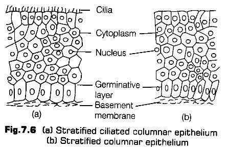
ii- Transitional Compound Epithelium
The epithelium consists of more than one layer of cells, but is much thinner and more stretchable than the stratified epithelium. It contains cuboidal cells at the base, two or three layers of large polygonal or pear-shaped cells in the middle and a superficial layer of large, broad, rectangular or oval cells.
The transitional epithelium lines the inner surface of urinary bladder, ureter and renal pelvis. They have thick membrane with thin regions that fold when the bladder contracts.
3. Glandular Epithelium
Some of the columnar or cuboidal cells get specialised for secretion and forms the glandular epithelium.
It is of two types
Unicellular Glandular Epithelium
It consists of isolated glandular epithelial cells called intraepithelial cells, e.g., Goblet cells of the alimentary canal are one such cell.u. Multicellular Glandular Epithelium
It consists of cluster of epithelial cells called extra epithelial cells. These cells unite to make up one gland, e.g., Salivary gland.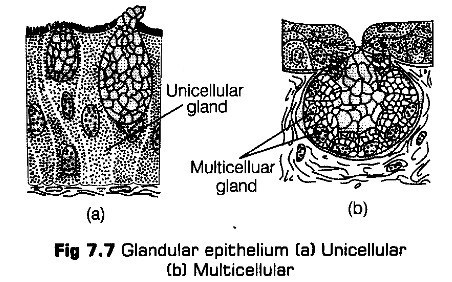
Gland
A cell, tissue or organ, which secretes some substance is called a gland. The secretions of glands may be protein (pancreas), lipids (adrenals), mixture of carbohydrates and proteins (salivary gland) or mixture of all the three materials (mammary glands).
The glands can be classified in different types based on site of secretion, mode ofsecretiori and involvement of single or many cells.
Based on Site of Secretion
The glands can be exocrine, endocrine or heterocrine based on the site where the secretion is released.
(a) Exocrine Glands These glands have ducts to pour their secretions to their site of action. They often secrete enzymes and its examples include salivary glands, intestinal glands, gastric glands, lacrimal or tear glands.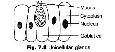
(b) Endocrine Glands These glands do not have ducts and pour their secretions directly into the blood or lymph. These glands are also called ductless glands and their secretions are known as hormones. Some examples of endocrine glands are pituitary, thyroid, parathyroid, adrenal, etc.
(c) Heterocrine Glands/Myxocrine Glands These glands are partly exocrine and partly, endocrine in function, e.g., Pancreas,
kidneys, stomach, gonads, intestine, placenta, etc.
Based on Number of Cells
According to the number of cells forming the glands, they are unicellular and multicellular.
(a) Unicellular Glands The mucus secreting goblet cells of the alimentary canal are called unicellular glands.
(b) Multicellular Glands These are composed of many cells and are formed by sinking of the gland into the underlying connective tissue. The multicellular glands may be simple or compound glands
* Simple Glands These may be simple tubular glands, e.g., Crypts of Lieberkuhn, simple coiled tubular glands (sweat glands) and simple alveolar glands having flask-shaped secretory units (mucus secreting glands in the skin of frog.)
* Compound Glands These have branch system of ducts. These may be compound tubular glands {e.g., Gastric glands of stomach, Brunner’s glands of intestine), compound alveolar glands {e.g., Some sebaceous glands and salivary glands) and compound tubuloalveolar glands having both tubular and alveolar secretory units {e.g., Pancreas, functional mammary glands).
Functions of Epithelial Tissue
The main functions of epithelial tissue are listed below
(i) The epithelial tissue protects the underlying tissues from mechanical injury, entry of germs, harmful chemicals and drying up.
(ii) It checks the absorption of harmful or unnecessary materials.
(iii) The epithelium of uriniferous tubules is specialised for urine excretion.
(iv) The sensory epithelia of sense organs help to receive various stimuli from the atmosphere and convey them to the brain.
(v) The epithelium of alveoli of the lungs brings about the exchange of gases between the blood and air.
(vi) The pigmented epithelium of the retina darkens the cavity of eyeball.
(vii) Epithelium also forms glands that secrete secretions such as mucus, gastric juice and intestinal juice.
(viii) The germinal epithelium of the ovaries and seminiferous tubules of the testes produce ova and sperms, respectively.(ix) Epithelium produces exoskeletal structures like scales, feathers, hair, nails, claws, horns and hoofs.
(x) Ciliated epithelia (e.g., of respiratory and genital tracts) serves to conduct the mucus and other fluids in the ducts they line.
Note:
* The term ‘Epithelium’ was coined by Ruysch.
* Transitional epithelium
ii. Connective Tissue
The connective tissues are most abundant and widely distributed in the body of complex animals. They are named as connective tissues because of their special function of linking and supporting other tissues/organs of the body.
Generally, connective tissue is made up of three components
1. Matrix
It is a clear and viscous substance. Its consistency may vary from liquid (e.g., blood) to semi-solid (e.g., cartilage) and solid (e.g, bone) form.
2. Cells Embedded in the Matrix
These are responsible for secreting the matrix and other substances.
The cells of connective tissue are of different types
(i) Fibroblasts produce fibres and matrix.
(ii) Adipose cells store fat.
(iii) Plasma cells synthesize antibodies. These are also called ‘cart wheel cells’ because thin chromatin in their nucleus forms four or five clumps giving the nucleus a resemblance of a cart wheel.
(iv) Mast cells produce histamine, heparin and serotonin. These are related to basophils of the blood.
(a) Histamine dilates the walls of blood vessels in inflammatory and allergic reactions.
(b) Heparin checks clotting of blood inside the blood vessels.
(c) Serotonin acts as a vasoconstrictor to check bleeding and to increase the blood pressure.
(v) Mesenchyme cells produce various types of connective tissue cells.
(vi) Macrophages ingest cell debris, bacteria and foreign matter.
(vii) Chromatophores (pigment cells) are found in the dermis of the skin which impart colour to the animals.
(viii) Reticular cells form reticular tissue and are phagocytic in nature.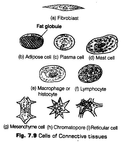
3. Fibres
These are non-living products of the cells.
These are of three types
(i) Collagen or Collagenous fibres (white fibres) are made up of collagen protein. When boiled in the water, collagen changes into gelatin.
(ii) Elastic fibres (yellow fibres) are formed of a protein called elastin. These fibres are branched and elastic.
(iii) Reticular fibres are delicate, branched and inelastic. They are made up of reticulin protein. They always form a network.
Types of Connective Tissues
The connective tissues are mainly of following three types
1. Loose Connective Tissue
Loose connective tissue has cells and fibres loosely arranged in a semi-fluid ground substance.
These tissues are of two types, i. e., areolar tissues and adipose tissue.
Areolar Tissue
It is found under the epithelial tissue of the skin, visceral organs like stomach, trachea and the walls of the blood vessels, etc. Its matrix is made up of glycoproteins. It contains two types of fibres, i.e., the white collagen fibres made up of collagen and the yellow elastic fibres made up of elastin.
The different cells of areolar tissue are fibrocytes, macrophages and mast cells.
Functions The tensile strength of collagen fibres and the elasticity of the yellow fibres protect the various organs from mechanical injuries.
This tissue also provides rapid diffusion of the materials and migration of wandering cells towards the areas of infection and repair.
Adipose Tissue
It is a modified type of areolar tissue. Its matrix contains large number of adipose cells along with fibrocytes and macrophages. White and yellow fibres are present in the matrix. The cells of this tissue are specialised to store fats.
The excess of nutrients which are not used immediately are converted into fats and are stored in this tissue. The adipose tissues are found in the subcutaneous region, around the heart, kidneys, eyeballs, etc.
e.g., It is also found in the blubber of whales and elephants, hump of camel, fat bodies of frog and yellow bone marrow.
Functions The adipose tissue is mainly a food reserve or fat depot for storage. It forms a shock-absorbing cushion around the eyeballs and kidneys. The tissue also helps in the production of blood corpuscles.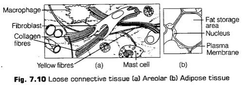
2. Dense Connective Tissue
Fibres and fibroblasts are found compactly packed in the dense connective tissues. This tissue is of two types i.e., dense regular and dense irregular connective tissue.
i. Dense Regular Connective Tissue
In this tissue, the collagen fibres are present in rows between many parallel bundles of fibres.
It is further of two types
It mainly consists of white fibres arranged in bundles. The fibroblasts are present in rows between the bundles.
It is of two types
* Tendons The white fibrous connective tissue forms the cords called tendons. These join the skeletal muscles with the bones.
* Sheets The white fibrous connective tissue also forms flat plates or sheets. It occurs in the dermis of the skin, periosteum of the bone,
perichondrium of Fi8-7’11 Dense re9ular cartilage, pericardium of heart etc .The white fibrous connective tissue has great strength however its flexibility is limited.
It mainly consists of yellow elastic fibres. The fibres are thicker. The fibroblasts and a few white fibres are found in between the yellow fibres.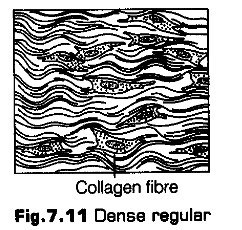
It is also of two types
* Ligaments The yellow elastic connective tissue forms the cords called ligaments. These join bones to bones.
* Sheets The yellow fibrous sheets formed by this tissue occur in the walls of blood vessels, lungs and bronchioles, true vocal cords, cartilage of larynx, trachea etc.
The yellow elastic connective tissue has considerable strength and remarkable elasticity. Thus, it allows the stretching of various organs.Dense Irregular Connective Tissue
It has fibroblasts and many fibres (mostly collagen) that are oriented in different pattern. This tissue is present in the skin. Collagen fibre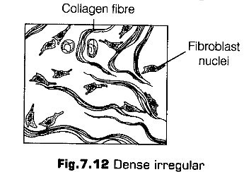
3 Specialised Connective Tissues
The specialised connective tissues are of following types
Skeletal Tissues
These tissues form the endoskeleton of the vertebrates. They form a rigid framework which supports the body, protects the vital organs and helps in locomotion.
The two types of skeletal tissues, i.e., cartilage and bone.
a.Cartilage
It is a tough, semitransparent, elastic and flexible tissue. The cartilage cells lie in groups of 2-3 in fluid filled spaces called lacunae. The cartilage is bounded externally by a stiff sheath called perichondrium containing white fibrous, tissue.
The cartilages are of three types, i.e., hyaline, fibrous and calcified.
* Hyaline Cartilage It has a clear, translucent, bluish green matrix. It is flexible and forms articular surfaces at the joints of long bones, where it is called articular cartilage.
* Fibrous Cartilage It has well-developed fibres in the matrix. It is of two types i.e., white fibrous cartilage and yellow elastic cartilage.
* It is a hard and rigid connective tissue. These are non-pliable ground substance rich in calcium salts and collagen fibres providing strength to the bone. The cells of bone are found in a calcified matrix made up of ossein. The bone cells known as osteocytes lodged in the spaces called lacunae..
They also interact with skeletal muscles attached to them to bring about movements.
The bone consists of four parts, i.e.,
* Periosteum It is a thick and tough sheath that forms an envelope around the bone. It is composed of collagen fibrous tissue. The periosteum contains blood vessels. It also contains bone-forming cells, the osteoblasts, which produce new bone material.
* Matrix It is composed of a protein called ossein. The Haversian canals, a characteristic feature of mammalian bones are present in the matrix. Each Haversian canal contains an artery, a vein, a lymph vessel, a nerve and some bone cells.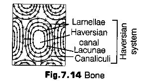
• Endosteum It is present outer to the bone marrow cavity. It comprises white fibrous tissue and the bone forming cells called osteoblast. The latter produces new bone material.
• Bone Marrow It is the vascular, soft pulpy connective tissue found in the bone marrow cavity of long bones like humerous, femur, etc.
Bone marrow is of two types
• Yellow marrow (rich in fat cells called adipocytes.) and
• Red marrow (Blood cells are formed in this marrow.)
In foetus, red marrow occurs in all bones. After birth, it restricts to limited places.
Calcified Cartilage When matrix of cartilage contains granules of calcium carbonate, the cartilage is called calcified cartilage.The bones can be spongy or compact on the basis of density and texture.
(a) Spongy (cancellate) Bone It contains a network of thin and irregularly longitudinal and transverse bony bars called trabeculae covered by the endosteum. It is found at the ends of long bones (epiphyses).
(b) Compact (Dense) Bone It is hard and compact and found in the shaft of long bones. It contains yellow bone marrow and has Haversian systems.
In a decalcified bone, the inorganic part of the matrix is removed. For deealcifieation, the bone is kept in dilute hydrochloric acid for long hours. This is to study living structures of the bone as it dissolves all the inorganic salts leaving behind only the organic matter.
Vascular Tissues
These are motile connective tissues consisting of fluid matrix and free cells. The matrix is without fibres. The vascular tissue helps in the transport of materials from one place to another.
Blood
It is a mobile, watery fluid with a slightly salty taste. It is composed of plasma (a fluid matrix) and the cells called blood corpuscles. It is bright red in colour when oxygenated and purple when deoxygenated. The volume of blood in an adult is about 5L.
It circulates within the blood vessels in higher animals. It is slightly alkaline (pH 7.4) in nature.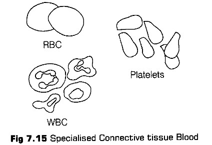
Plasma is a yellowish, straw-coloured liquid which is composed mainly of water (92%). About 55% of the total blood volume is plasma. The solid materials in plasma include plasma proteins, nutrients (glucose, amino acids, fatty acids and vitamins), hormones, antibodies, enzymes, lactic acid, cholesterol, dissolved gases (oxygen, carbon dioxide), mineral salts and waste products (urea; uric acid and creatinine). Functions It helps in transport of substances, provide body immunity, prevent the blood loses, retain fluid in blood, maintain blood pH and conduct heat to skin for dissipation.
Blood Cells
The blood cells or blood corpuscles forms about 45% of the blood volume. These cells are formed in the bone marrow of the long bones and the lymph nodes. The process of blood cells formation is called haemopoiesis and the tissues where these are formed are called haemopoietic tissues.
The blood cells are of following types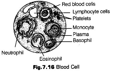
* Erythrocytes or Red Blood Corpuscles (RBCs) are the most abundant elements in blood. These carry red-coloured oxygen carrying pigment called haemoglobin. They are 7-8 |Xm in diameter.
* The human RBCs are smaller than the white blood corpuscles. In mammals, they are non-nucleated, biconcave and circular. The formation of erythrocytes is called erythropoiesis.
* Leucocytes or “White Blood Cells (WBCs) lack haemoglobin and are colourless. They are nucleated with rounded or irregular shape. They can change their shape and are capable of amoeboid movement.
* Thrombocytes (Blood Platelets) These are small, colourless, plate like discs having size of about 2-3 pm. Their number ranges between 0.15-0.4 million/mm3 of blood. Their normal life span is about a week. No nucleus is visible in these cells.
The blood performs following functions in the animal body.
* Blood transports oxygen from the respiratory organs to the tissues and carbon dioxide from the tissues to the respiratory organs.
* It transports nutrients to all parts of the body.
* Blood maintains the constant body temperature by distributing the heat throughout the body.
* Lymphocytes and eosinophils produce antitoxins to neutrilise the toxins, released by the microbes.
* Blood helps to maintain water balance to a constant level by bringing about constant exchange of water between circulating blood and tissue fluid.
* It helps to regulate the pH of the body fluids as it contains buffer materials such as proteins and mineral salts.
* Blood helps in healing of injuries by maintaining necessary supplies for the repair of damaged tissues.
It is a mobile connective tissue comprising lymph plasma (fluid) and lymph corpuscles (cells). It is pale yellow in colour and its composition is similar to plasma without the plasma proteins. It is present in the vessels called lymph vessels.
Lymph is formed of liquid components and formed elements or cells. It contains about 94% water and 6% of organic and inorganic substances. The organic part includes protein, fat droplets, carbohydrates, nitrogenous wastes and hormones.
Lymph performs the following functions in animal body
(i) It plays an important role in the defence of the body especially against invading organisms.
(ii) The digested products of fat digestion enter the lymph vessels present in the villus of the small intestine.
(iii) Lymph helps to maintain the blood volume by returning the interstitial fluid back to the blood during circulation.
(iv) The lymph nodes produce lymphocytes.
(v) It keeps the tissue cells moist.
4. Reticular Connective Tissues
Tissues consist of star-shaped reticular cells whose protoplasmic processes joins to form a cellular network. The reticular fibres are present on the reticular cells (composed of a protein called reticulin.)
The reticular connective tissue is present in the liver, spleen, lymph nodes, thymus, tonsils, bone marrow and lamina propria of the gut wall.
Function This tissue provides strength and support as it forms the supporting framework of many organs. It also helps to bind together the cells of smooth muscles. The reticular cells are phagocytic – and forms the defense mechanism of the body.
5. Pigmented Connective Tissue
The cells of pigmented connective tissue are irregular and are called pigment cells (chromatophores or melanophores). These cells contain yellowish brown, black or blue melanin pigment granules. Melanin is produced by other cells called melanocytes.
This tissue is present in the choroid, ciliary body and iris of the eye and dermis of the human skin.
Functions It gives colour to the structures.
6. Mucoid Connective Tissue
This tissue occurs as a foetal or embryonic connective tissue as it is present in the umbilical cord.
The mucoid tissue contains a jelly like substance called Whartson’s jelly and some delicate collagen fibres and primitive type of fibroblasts. It occurs as embryonic connective tissue in the foetus and vitreous humour of the eye.
Functions of Connective Tissue
The connective tissue performs fallowing main junctions
(i) The connective tissue mainly joins one tissue to another in the organs.
(ii) The adipose tissue stores fat.
(iii) The cartilage and bones form a supporting framework for the body.
(iv) Blood and lymph carry materials from one part to another in the body.
(v) The cells of connective tissues like macrophages, monocytes, neutrophils ingest bacteria, cell debris and foreign materials.
Thus, they protect and clean the body.
(vi) The adipose tissue acts as shock absorber around some organs, such as eye balls and kidneys. It also acts as packing material in various organs.
(vii) Bone marrow is the source of blood corpuscles.
(viii) The collagen fibres help in the repair of injured tissues.
Note:
* In old age, the bone marrow of the cranial bones undergo degeneration and is called gelatinous marrow.
* Bone marrow is a special kind of myeloid (myelogenous) tissue.
* Prothrombin and fibrinogen are the largest blood proteins and albumins.are the smallest one.
i. Muscular Tissue
The striated muscle fibres are multinucleated or syncytial in nature. The cytoplasm (sarcoplasm) of each fibre has a large number of myofibrils (actin and myosin myofibrils) which are tightly packed.
Each myofibril shows dark and light bands of stripes alternating with each other. Hence, they are called as striped muscle fibres.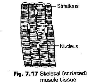
ii. Non-Striated (Smooth) Muscle
The non-striated muscles are found in the posterior part of oesophagus, stomach, intestine, lungs, urinogenital tract, urinary bladder, blood vessels, iris, ciliary body of eye, dermis of skin, etc.
The non-striated muscle consists of long, narrow, spindle-shaped fibres that are generally shorter than the striated muscle fibres. Their size may range from 20 |Xm (Small blood vessels) -500 (1m in pregnant uterus). Each non-striated muscle fibre contains a single oval nucleus in its thick middle part. In the cytoplasm, the myofibrils are arranged longitudinally. They are composed of myosin. There is no sarcolemma, however, the fibre is enclosed by the plasma membrane.
>
The smooth muscles help in the peristalsis which occurs in the tubular viscera. The autonomic nervous system controls these muscles. Hence, they are not under the control of animal’s will.
iii. Cardiac Muscles
The cardiac muscles are contractile tissues present only in the heart and in the wall of large veins which enter the heart. The cardiac muscle fibres show the characters of both unstriped and striped muscle fibres.
The myofibrils have transverse faint dark and light bands which alternate with each other.
The cardiac muscle fibres have some specialfeatures
(i) These muscle fibres are supplied with both central and autonomic nervous system and are not under the will of the animal.
(ii) These fibres never get fatigue.
(iii) Blood capillaries penetrate the cardiac muscle fibres, hence they have very rich blood supply.
(iv) These fibres have the property of contraction, even when they are isolated from the body temporarily.
Functions of Muscular Tissues
The muscle tissues perform following important junctions
(i) These are involved in the movement of body parts and locomotion of the organism.
(ii) Muscles are responsible for heart beat, production of sound and peristalsis in tubular viscera.
(iii) The muscles support the bones and other structures.
(iv) Muscles are essential during parturition.
The neural tissue is ectodermal in origin. It is specialised to receive stimuli and conducts impulses for controlling and coordinating body functions. It exerts the greatest control over the body’s responsiveness to changing conditions. The neural tissue consists of nerve cells and packing cells. The packing cells are called Schwann cells in the peripheral nervous system and neuroglia cells in the central nervous system.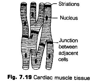
The muscle tissue consists of elongated and contractile cells called muscle cells or myocytes.
Due to their elongated nature, the muscle cells are also called muscle fibres. It develops from mesoderm. The muscle cells are surrounded by connective tissue. Each muscle cell is covered by a membranous sheath called sarcolemma.
It consists of plasma membrane and basement membrane. The cytoplasm of a myocyte is called sarcoplasm. The endoplasmic reticulum is called sarcoplasmic reticulum and the mitochondria are called sarcosomes.
The myoglobin keeps the reserve oxygen for immediate supply during muscle activity.
It also provides light pinkish colour to the muscles. The muscle cells may be uninucleate or multinucleate. The contractile structures of muscle cells are called myofibrils. The myofibrils are made of myofilaments. The myofilaments are of two types, i.e., thicker myosin and thinner actin. The contraction of muscles occurs due to sliding of actin filaments passing over the myosin filaments.
Types of Muscles
The muscles can be grouped into three types based on their structure, location andJunction.
(i) Striated or striped or skeletal or voluntary muscles.
(ii) Non-striated or unstriped or visceral or smooth or involuntary muscles. .
(iii) Cardiac muscles.
(iv) Striated Muscles
The striated or skeletal muscles form about 40% of total body weight. These muscles are attached and bring about the movement of the various bones of the skeleton, so are called skeletal muscles. The striated muscles give shape to the body and also release heat during contraction. These muscles have huge supply of nerves and blood vessels. Each striated muscle is a long, narrow, cylindrical, unbranched cell.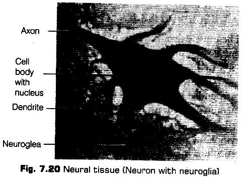
Nucleus
It is a long and cylindrical structure which has a definite sarcolemma. The fibres are uninucleate and the nuclei lie near the centre.
Neurons
Neurons are the functional unit of neural system. These are excitable cells. A neuron consists of a cell body (cyton) or soma and fine protoplasmic processes called neurites arising from the cell body.
i. Cyton
It contains neuroplasm (cytoplasm), a spherical nucleus, endoplasmic reticulum, mitochondria, Golgi bodies, ribosomes, lysosomes, fat globules, Nissl’s granules etc. The Nissl’s granules are probably involved in the synthesis of proteins.
Neurites
The processes arising from the neurons are called neurites. These are dendrites and an axon. Axon is single, but dendrites may vary from one to several. The dendrites are usually shorter and tapering processes. The axon is usually a long process of uniform thickness.
Nerve Fibres
The nerve fibres are elongated and slender processes of the neurons, which are formed by ensheathing of axons. A space of 15-20 nm occurs between axolemma and the covering sheath. It is called periaxonal space.
Depending upon the covering sheath, nerve fibres are of two types
(a) Myelinated Nerve Fibre
An axon covered by myelin sheath is called myelinated or medullated nerve fibre. The myelin contains lipids, proteins and water. The medullary sheath serves as an insulating layer, preventing loss of energy of the nerve impulse during its passage along the fibre./; Non-Myelinated Nerve Fibres
A non-medullated or non-myelinated fibre consists of an axis cylinder enclosed by neurilemma and connective tissue. It lacks medullary sheath and appears grey in the fresh state.
On the basis of function also, the nerve fibres are of two types (a) Afferent (Sensory) Nerve Fibres The afferent nerve fibres carry the nerve impulses from the sense organs to the central nervous system (brain and spinal cord).
(b) Efferent (Motor) Nerve Fibres
They carry nerve impulses from the central nervous system to the effector organs (muscles and glands).
Nerves
A nerve is a complex bundle of nerve fibres enclosed together by a common sheath of connective tissue along with the blood vessels. Each nerve fibre is covered by a thin sheath of connective tissue called endoneurium.
A number of nerve fibres, each covered by its own endoneurium are joined together to form a bundle called fasiculus or fascicle.
According to the nature of fibres, nerves can be of following three types
(i) Sensory (Afferent) nerves These nerves bring sensory impulses or excitation from different parts of the body and sense organs.
(ii) Motor (Efferent) nerves These nerves carry message from central nervous system to parts of the body and effector organs to perform their function.
(iii) Mixed nerves The nerves contain both sensory and motor fibres.
Neuroglia
The neuroglia or glia cells are supporting cells which form a packing around the neurons in the brain, spinal cord and ganglia. These cells have different shapes and many processes. The neuroglia cells have various roles like myelin formation, transport of materials to neurons, maintenance of ionic balance and phagocytosis.
Neurosecretory Cells
These are specialised neurons or neuron-like cells, which secrete biologically active substances that are effective in other structures, often at a different site. The neurosecretory cells occur in hypothalamus. They produce hormones called neurohormones.
Functions of Neural Tissue
Neural tissue perform the following functions
(i) The neural tissue coordinates and controls the functioning of different parts of the body.
(ii) The sensation of smell, vision, taste, hearing, pain, pleasure, etc., are performed through the nervous tissue.
(iii) The neural tissue helps in meditating conscious activities.
(iv) The information about the changes in various internal structures is provided by nerves.
(v) It makes us aware about the environment around us.
(vi) The nervous tissue brings about an appropriate response to each and every stimulus.
(vii) The tissue is also a seat of experiences, memories, etc.
Topic 2. Morphology and Anatomy of Animals
In this topic we will discuss morphology and anatomy of three organisms—Earthworm, Cockroach and Frog representing invertebrates and vertebrates at different evolutionary levels to show their organisation and functioning. Morphology refers to study of from or externally visible features. The word anatomy is conventionally used for the study of morphology of internal organs in the animals.
Earthworm
Earthworm is a reddish-brown terrestrial invertebrate that inhabits the upper layer of the moist soil. During day time, they live in the burrows made by boring and swallowing the soil.
In the gardens, they can be find out by their faecal deposits called as worm castings. The two common Indian species of earthworms are Pheretima and Lumbricus.
Systematic Position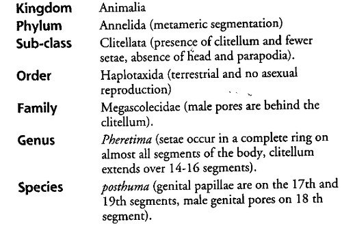
Earthworms inhabit almost all areas over the world, except the Arctic and Antarctic regions. There are about 500 species of Pheretima of which 13 species occur in India.
Habitat and Habit
Earthworm lives in moist soil rich in humus they are nocturnal animals (i.e., they come out at night for feeding and to mate and sleep during the day time).
Locomotion
The earthworm moves by crawling (creeping) in which its body remains on the ground. It moves by muscular contraction and relaxation of the body which is aided by chitinous setae or chaetae. It moves about 15 cm/min.
Food
The earthworm eats decaying organic matter found in the soil. It is omnivorous. The food is digested in the gut and undigested food along with the soil is passed out through the anus as small pills called worm castings.
Breeding
Earthworm is a hermaphrodite (i.e., bisexual or monoecious). It breeds in the rainy season. It is protandrous (i.e., male sex organs mature earlier than the female). Thus, self fertilisation is not possible, only cross fertilisation occurs in them.
The copulation occurs when two earthworms closely attach to each other by their ventral surfaces in a way that the head region of one is opposite to the tail region of the other. Then, the two worms separate after the exchange of spermatozoa.
Several eggs and spermatozoa are packed in an egg case, the ootheca (cocoon), which is deposited just beneath the surface of ground. About four baby earthworms develops in one cocoon.
Regeneration
The earthworm has great power of regeneration. If it is cut into two parts, its anterior half develops into tail, but in the posterior half, the head can be formed only if 4-6 anterior segments are removed.
Defence
Earthworm can defend itself only by ejecting the foul smelling coelomic fluid through the dorsal pores.
Morphology
Size, Shape and Colour
It has a long, cylindrical body. The anterior end is pointed, but there is no distinct head. The posterior end is rounded. The size of an adult worm is about 150 mm long and 3-5 mm wide. The dorsal surface is a bit darker than the ventral surface and bears a dark median line.
Perostomium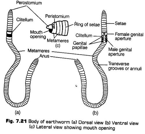
Segmentation
The body of earthworm is divided into more than hundred short segments, which are similar (metameres, about 100-120 in number). The segments are also divided internally by the septa. This is called true segmentation or metamerism.
The first segment is called peristomium. The mouth is present in this segment. A fleshy lobe called prostomium covers the mouth. The segmentation in the first few segments is visible externally without corresponding internal septa.
The clitellum is a prominent circular muscular band present in the 14th, 15th and 16th segments. It divides the body into pre-clitellar, clitellar and post-clitellar regions.
The female genital pore is a single aperture present mid-ventrally in the 14th segment. The male genital pores are a pair of openings found on the ventral surface of the 18th segment. Two pairs of genital papillae are present in 17th and 19th segments which helps the animal in copulation.
In the grooves of 5th-9th segments, four pairs of spermathecal pores are present ventro-laterally. These are connected to the sperm storing organ called spermathecae.
On the dorsal side minute pores are found called dorsal pores through which the coelomic fluid exudes out of the body. This fluid keeps the body surface moist.
There are numerous pores called nephridiopores present on the ventral surface of the body. These are the opening of the excretory organs called nephridia that expel out the nitrogenous waste from the body.
The last segment is called the anal segment and it bears the anus.
Anatomy
Body Wall
The body wall of earthworm has four layers, i.e., cuticle, epidermis, musculature and coelomic epithelium or parietal peritoneum.
a. It is a thin, transparent, non-cellular surface layer. The cuticle is secreted by the epidermis and is perforated by numerous minute pores.
b. It is the next layer after cuticle, made up of a single layer of columnar epithelium which contain secretory gland cells, i.e., basal cells, sensor or receptor cells, setigerous cells (seta forming cells), etc.c.
c.It is composed of an outer thin layer of circular muscle fibres and an inner thick layer of longitudinal muscle fibres. Contraction of circular muscles makes the body long and thin whereas, the contraction of longitudinal muscle fibres makes the body thick and short.
d. It is a thin, membrane like coelomic epithelium consisting of flattened squamous cells.It protects the internal organs and prevents excessive evaporation. The receptor cells play a vital sensory function. Setae and muscles help in locomotion. The excretory matter is passed out through the nephridiopores present in the body wall.
Digestive System
A complete alimentary canal is present in the body cavity of earthworm beginning with the mouth in the first segment and ends with the anal opening situated in the last segment. The earthworm swallows the soil and the organic content of the soil is digested.
The various regions of earthworm’s alimentary canal are following
(i) Buccal cavity l-3rd segments.
(ii) Pharynx 4th segment.
(iii) Oesophagus (food pipe) 5-7th segments.
(iv) Gizzard 8-9th segments. It helps in grinding the food.
(v) Stomach 10-14th segments. The stomach wall, contains calciferous glands to neutralise the humic acid in the soil.
(vi) Intestine 15th to the last segment where it opens out by anus.
(vii) Typhlosole Between 25-95th segments, there is a prominent infolding on the dorsal wall called the typhlosole. This enhances the area of absorption of the intestine of the digested food.
Respiration
The skin serves as the organ of respiration. It is thin, transparent and richly supplied with blood vessels. Respiration through the skin is called cutaneous respiration. Haemoglobin is found dissolved in the blood plasma.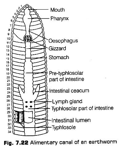
Circulatory System
Pheretima exhibits closed type of blood vascular system, consisting of blood vessels, capillaries and heart. Due to closed circulatory system, blood is confined to the heart and blood vessels.
There are three main median longitudinal blood vessels namely a dorsal vessel (above the alimentary canal), ventral vessel (below the alimentary canal) and a sub-neural vessel (lying on the ventral side below the nerve cord). In the blood vessel, the blood flows from the posterior to the anterior end. In the ventral and subneural vessel, the flow of blood is from anterior to the posterior end.
There are four pairs of hearts, a pair of each lying in 7th, 9th, 12th and 13th segments. All the hearts have muscles and pulsatile walls to pump the blood into the ventral vessel by rhythmical contractions. The backward flow of the blood is prevented by the valves present in the heart.
(viii) Anus The undigested food is sent out through it.
Excretory System
The excretory organs occur as segmentally arranged coiled tubules called nephridia (sing, nephridium). They are offollowing three types.
These are present on both the sides of inter segmental septa of segment 15 to the last that opens into intestine. They discharge the waste matter into the gut via septal excretory ducts and supraintestinal ducts.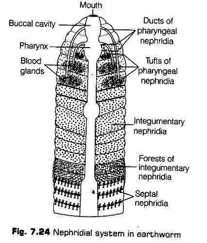
Nervous System
Nervous system is basically represented by the ganglia which are arranged segment-wise on the ventral paired nerve cord.
The nerve cord in the anterior region (3rd and 4th segments) bifurcates laterally, encircling the pharynx and joins the cerebral ganglia dorsally to form a nerve ring. The cerebral ganglia along with other nerves in the ring integrate sensory input as well as command muscular responses of the body.
Reproductive System
Earthworm is a hermaphrodite (bisexual), i.e., testes and ovaries are present in the same individual. ‘
Mule Reproductive System
It possess two pairs of testes present in the 10th and 11th segments. The ducts of testes (vas deferentia) run upto the 18th segment, where they join the prostate duct. Accessory glands are present, on the ventral side of the 17th and 19 th segments.
They open out by the fine ducts of genital papillae situated on the under surface of the segments 17th and 19th. Four pairs of sac-like structures called spermathecae are found one in each of the 6th to 9th segments. They receive and store spermatozoa during copulation.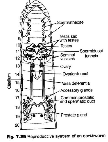
Femaie Reproductive System
One pair of ovaries is attached to the intersegmental septum between the 12th and 13th segments. Ovarian funnels are present beneath the ovaries which continue into oviduct. These ducts join together and open on the ventral side as a single female genital pore on the 14th segments.
Breeding and Fertilisation [An earthworm becomes sexually mature when it develops the clitellum. A mutual exchange of sperm occurs between the two worms during mating. Mature sperm, egg cells and nutritive fluid are deposited in the cocoons produced by the gland cells of the clitellum. Fertilisation and development occurs within the cocoons, which are deposited in the soil. The eggs are fertilised by the sperms within the cocoons, which then slips off the worm and is deposited in or on the soil.
The cocoons hold the worm’s embryo. After about three weeks, each cocoon produces two to twenty baby worms. Earthworms development is direct, i.e., there is ‘ no larval stage and all earthworms lay eggs.
Economic Importance of Earthworms
Merits
Earthworms are useful in several ways for humans
(i) Earthworms make the soil porous by digging burrows in the soil. Hence, they are called friends of the fanners.
(ii) The nitrogenous wastes and other waste products of the earthworms form food for plants. This process of increasing fertility of soil by earthworms is called
vermicomposting.
(iii) Earthworms are used as fish bait for catching fishes.
(iv) Some tribals in India use earthworms as medicine to cure jaundice, piles, diarrhoea, bladder stones, gout, etc.
(v) In some countries like China, Japan, Australia and Myanmar, earthworms are used as food.
(vi) The worms reduces both acidity and alkalinity of the soil and create optimum conditions for the plant growth.
(vii) Earthworms are eaten by frogs, birds, which are useful to man in some ways. Thus, they are an important part of food chain.
(viii) These are used in scientific studies and dissected in zoological laboratories for academic studies.
Demerits
Earthworms may also be harmful in many ways
(i) Earthworms may damage young and tender plants by eating them.
(ii) During rainy season, they make burrows and cause soil erosion.
(iii) They spoil the play grounds by digging burrows in them.
(iv) Some earthworms are intermediate hosts for some parasites such as tapeworm of chicken and lung nematode of pigs.
(v) The burrows of earthworms in the banks of irrigation channels sometimes cause leakage of the water.
Cockroaches are one of the common insects found in our house. They are brown or black bodied animals, although, bright yellow, red and green coloured cockroaches have also been reported in the tropical regions. In India, two species of cockroaches are found, i. e., Periplaneta americana and Blatta orientalis.
Locomotion
Cockroaches are cursorial insects, i.e., run very fast. They show double mode of locomotion-running and flying. The cockroach run on the tarsi of their legs. At a time, three legs are kept on the ground and the other three are carried forward. By repeating this step, the animal moves forward.
Cockroach flies by beating the hindlimbs with the help of special muscles. They are beaten up and down alternately.
Breeding
Cockroaches are unisexual. They show sexual dimorphism, i.e., male and female sexes can be seen externally. They are oviparous. The young cockroaches called nymphs resemble the adults in many features. The nymph undergo moulting or ecdysis in which the casting of older skin takes place. The nymph gradually become adults under the parental care.
The cockroaches are tropical and sub-tropical insects, but they have reached all parts of the world with trading ships. They are good enough to adapt to new habitats.
Habitat
Cockroaches inhabit the warm, dark and damp places. They are commonly found in underground drains, kitchens, restaurants, godowns, store houses, railway wagons, ships, etc., where food and moisture is available.
Habits
Cockroaches show some peculiar habits. They are nocturnal, i.e., come out of their hiding places at night to feed. These are omnivorous and eat all types of animals and vegetable v foods.
Morphology
The body of cockroaches is dorsoventrally flattened, elongated and bilaterally symmetrical. The adult cockroach is about 34-53 mm long with wings that extends beyond the tip of the abdomen in males.
The entire body of cockroach is covered by a hard chidnous exoskeleton (brown in colour) made of tough plates called sclerites. These are formed of chidn, a polysaccharide of acetoglucosamine molecule. ,
The exoskeleton protects the body and provides space for the attachment of muscles. The adjacent sclerites are joined together by thin, soft, flexible arthroidal membranes.
Head Antenna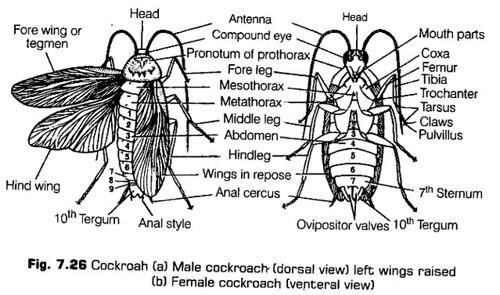
The body of cockroach is segmented and divisible into three parts, i.e., head, thorax and abdomen.
Head
The head of cockroach is triangular in shape and lies anteriorly at right angles to the longitudinal body axis. It is formed by the fusion of six embryonic segments. It is flattened anteroposteriorly and movably articulates with the thorax by a short neck. It is covered by sclereites and bear sense organs, mouth parts and mouth. The sclerites of head are fused to form a compact head capsule called vertex.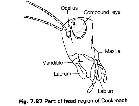
Sense Organs
The sense organs in cockroach includes compound eyes, antenna and fenestrae or ocellar spots.
(i) Compound eyes are a pair of large, black, kidneyshaped organs situated dorsoventrally on the head, one on either side. Their surface is marked by a large number
of hexagonal areas called facets. The eyes are the organs of sight.
(ii) Antenna are a pair of long, slender, jointed, tapering j filaments that articulates in the antennal sockets situated on the frons, close to the compound eyes. The antenna are organs of touch and smell. They can be moved in all directions to receive the stimuli. Antenna is made up of many segments called podomeres.
(iii) Fenestrae are a pair of small, whitish spots, each lying just above the inner to the antennal socket of its side, They are sensitive to light.
Mouth
It is a narrow opening that lies at the base of the pre-oral cavity. It is bounded by the mouth parts and leads into the pharynx. The mouth parts of cockroach are of biting and chewing type. They also help in swallowing. The mouth parts are attached to the head capsule.
The mouth parts include the following structures
(a) Labrum is also called upper lip that helps in holding food particles during feeding.
(b) Mandibles lie on the sides of the mouth just behind the labrum. The two mandibles work against each other in a horizontal plane to crush and cut the food into pieces.
(c) First maxillae are a pair of maxillae that lie beneath the mandibles, one on either side of the head.
(d) Second maxillae or labium is also called lower lip. It is a single structure, but it is formed by the fusion of a pair of second maxillae. It lies behind the mouth and forms a type of lower lip.
Neck
It is a slender, flexible (can move in all direction) tube articulating the head with the thorax. It is supported by a few ring-like sclerites.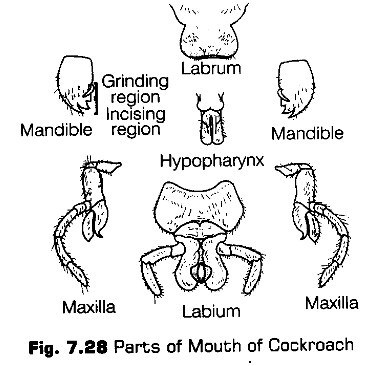
Thorax
The thorax forms the middle part of the body. It consists of three segments, the anterior prothorax, the middle mesothorax and the last metathorax. Each thoracic segment bears a pair of waiting legs. The thorax also contains spiracles for gas exchange.
Each thoracic segment is enclosed by four chitinous skeletal sclerites, a dorsal tergum, a ventral sternum and two lateral pleura. The tergum of the prothorax is called the protergum or pronotum.
The tergum of the mesothorax is called mesotergum or mesonotum. The tergum of metathorax is termed the metatergum or metanotum. The sterna of all the thoracic segments are largely covered by the legs.
The thorax contains three pairs of kgs and two pairs ofwings
(a) Legs are jointed and a pair is present in each thoracic segment which are three in number. Based on the segment that bears them, legs are prothoracic, mesothoracic and metathoracic or simply prolegs, mesolegs and metalegs. They articulate with their respective segment between the sternum and the pleura.
(b) Wings These are paired structures, one on the mesothorax (forewings) and another on the metathorax (hindwings). The wings are movable folds of the integument that grow out from the region between the tergum and the pleura near the anterior end of the segment.
Forewings called tegmina are opaque dark and leathery, used to cover the hindwings when they are at rest. While hindwing are transparent membranous and are used in flight.
They bear two thin sheets of cuticle with a framework of branching tubes, the veins or nervures.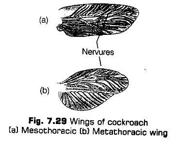
Abdomen
It is the posterior most and the largest part of the body. It is composed of ten segments in adults and eleven in the embryo. It is dorso-ventrally flattened, broader at the anterior end and narrow posteriorly.
Each abdominal segment bears sclerites. Certain segments have spiracles and stink glands. The terminal segments carry appendages and some apertures.
An abdominal sclerite is enclosed by four sclerites, a dorsal tergum, one ventral sternum and two lateral pleura.
There are ten terga. The 7th tergum covers the 8 th tergum in male and 8th and 9th terga in the female cockroach. The 10th tergum is large and notched in the middle. It projects backwards beyond the body. The abdomen bears 9th sterna, while the 10th one is absent. In male, all the nine sterna are visible and in female, only the first seven can be seen.
The abdomen in both male and female cockroaches comprises of 10th segments. In females, the 7th sternum is boat-shaped and together with 8th and 9th sterna forms a genital pouch.
In males, genital pouch lies at the hind end of the abdomen. It contains dorsal anus, ventral male genital pore and gonapophyses. The abdomen of female cockroach is broader than the male cockroach.
Male bears a pair of short thread like anal styles which are absent in females.
Abdominal Appendages
The abdomen bears small appendages at its hind end only. These appendages are a pair of anal cerci, (joined filamentous structures found in both sexes) a pair of anal styles and gonapophysis or external genitalia.
Apertures
The abdomen bears following three apertures
(a) Anus lies beneath the 10 th tergum between the two chitinous plates. These are called podical plates or paraprocts. They represent the remains of the eleventh segment.
(b) Genital aperture of the male cockroach lies just below the anus on one of the gonapophyses and that of female lies on the eighth sternum in the broad pouch.
(c) Abdominal spiracles are the eight pair structures. They are smaller than the thoracic spiracles.
(d) Stink glands A pair of stink gland is present between the 5th and 6th abdominal terga. These glands produce a secretion that gives a characteristic stinky smell.
Anatomy
The anatomical structure of different parts of cockroach body is described below
Body Wall
The body wall contains cuticle, epidermis and basement membrane. The cuticle is impermeable to water because of its thick, non-cellular surface layer. The epidermis consists of a single layer of columnar cells, enclosing some gland cells.Body Cavity
Cockroaches are coelomate. But, true coelom occurs only in embryonic stage. In adults, it is found in small cavities only around the gonads. The body cavity is filled with haemolymph and is called haemocoel.
Endoskeleton
Certain processes of exoskeleton extend into the body and forms endoskeletal elements. These provide attachment to the muscles and hence called apodemes.
Abdomen of cockroach does not have endoskeletal elements.
Digestive System
The alimentary canal of cockroach is 6-7 cm in length.
It is divisible into following three parts
a. It is the anterior part of the alimentary canal. It is surrounded by the mouth parts. Food is crushed initially by mandibles and mixed with saliva and passed to the short tubular pharynx.
The pharynx inturn bends to join a narrow tubular passage called oesophagus, whichs passes through the neck and opens into a sac like structure called crop (a large pear shaped sac that stores the food).
From crop, the food enters a conical and muscular part called gizzard or proventriculus having, outer layer of thick circular muscles.
The gizzard has six large chitinous teeth. (Formed by inner culide layer) and fine bristles in ks grooves. Therefore, it is efficient in grinding of food particles and straining apparatus.
Gizzard marks the end of foregut. The whole foregut is lined by cuticle protecting the alimentary canal from rough food particles. Its posterior end projects in the form of a narrow tube into the midgut, called stomodael valve.
b. It is a short, narrow tube of uniform diameter, lined by endodermal glandular epithelium. A ring of 6-8 blind tubules called gastric or hepatic caeca present at the junction of foregut and midgut secrete digestive juices. The midgut is the major organ of digestion and absorption of digested food.
The posterior part of the midgut has a sphinter that keeps it closed.
c. It is broader than the midgut and is divisible into ileum, colon and rectum. Ileum is short and narrow bearing short spines. A ring of about 100-150 fine yellow coloured thread like filaments. Malpighian tubules is joined to the beginning of the ileum. Colon is a wide coiled tube which do not contain spines while, rectum is the last part of the hindgut.
The papillae present in the rectum absorb water and salts from the undigested food. The rectum thus, opens outside by anus (which lies below the 10th tergum.)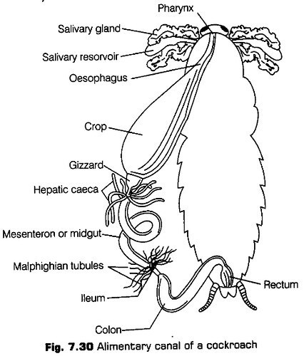
Respiratory System
The respiratory system is comprised of a network of blind, glistering white air tubes called tracheae. The tracheae are connected to outside by 10 pairs of lateral apertures called spiracles. Two pairs of spiracles occur in thorax and eighth pairs are present in the abdomen.
During rest, some of the spiracles are open so, that the oxygen can diffuse continuously reaching the body fluid present in the terminal region of the tracheoles. Exchange of gases occurs between the living cells and the body fluid. Expansion of abdomen draws fresh air into the tracheal system through stigmata and its contraction expels foul air.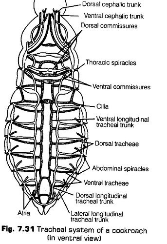
Circulatory System
The circulatory system of cockroach is of open type. The blood vessels are poorly developed and blood flows freely in the body cavity which is called haemocoel. Visceral organs located in the haemocoel are bathed in blood (haemolymph).
It contains colourless plasma and corpuscles called haemocytes.
Each heart chamber opens into next chamber through a ventricular valve. Each chamber has a pair of incurrent pores called ostia (which possess valvular mechanism to pass the blood only from haemocoel to the heart chambers).
The heart chambers contract one after the other rapidly. This pushes the blood into the anterior aorta as well as a few lateral or excurrent arteries. From aorta, blood passes into head sinuses and then into perivisceral and perineural sinuses.
Excretory System
Cockroach is urecotelic, i.e., main excretory product of it is, uric acid. The. main excretory organs of cockroach are Malpighian tubules.
The tubules are lined by cuboidal, brush bordered, glandular cells that extract the nitrogenous waste matter from the haemolymph and discharge it into the ileum as uric acid.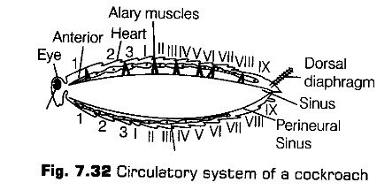
In rectum, the epithelial lining picks up most of the salts from urine and transport it into the haemolymph. Urine becomes nearly solidified and eliminates along with faces.
In addition to this, the fat body, nephrocytes and urecose glands also helps in excretion.
Nervous System
The nervous system of cockroach consists of a series of fused, segmentally arranged ganglia joined by paired longitudinal connectives.
Three ganglia lie in the thorax and six in the abdomen. The nervous system of cockroach is spread throughout the body. The head holds a bit of a nervous system while, the rest is situated along the ventral part of the body.
In the head region, the brain is represented by supra-oesophageal ganglion, which supplies nerves to antenna and compound eyes. Nerves arise from all the ganglia in the head, thorax and abdomen and innervate various parts in their respective regions.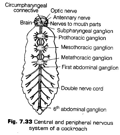
The sense organs in cockroach, are antenna, eyes, maxillary palps, labial palps, anal cerci, etc. The compound eyes are situated at the dorsal surface of the head. Each eye consists of about 2000 hexagonal facets or ommatidia (each capable of forming an image in it).
Reproductive System
Cockroaches are dioecious animals, i.e., both the sexes have well developed reproductive organs.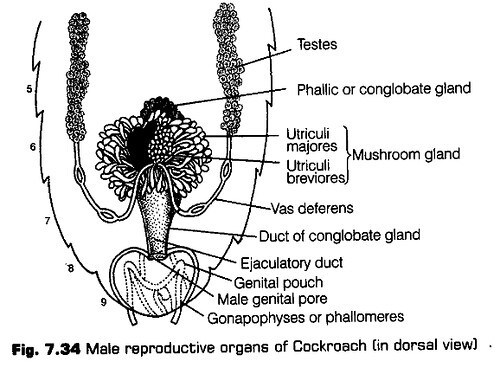
It consists of a pair of testes lying one on each lateral side in the 4th-6th abdominal segments. From each testis arises a thin vas deferens which opens into ejaculatory duct through seminal vesicle.
The ejaculatory duct opens into male genopore situated ventral to the anus. A characteristic mushroom shaped gland is present in the 6th-7th abdominal segments which functions as an accessory reproductive gland.
The external genitalia are represented by male gonapophysis or phallomere (chitinous asymmetrical structures surrounding the male gonopore). The sperms are surrounded in the seminal vesicles and are glued together in the form of bundles called spermatophores, which are discharged during copulation.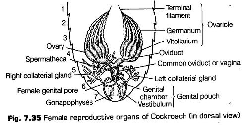
The male and female cockroaches come together by their posterior ends. The spermatophores are transferred to the genital chamber of the female. The sperms are liberated from the spermatophores and reaches the left spermatheca slowly.
The eggs come from both the ovaries alternately into the common oviduct and passes through the female genital pore into the genital chamber where they are fertilised by the sperms coming from the left spermatheca.
The secretion of collateral glands form the egg case of the ootheca (ploothecae). The fertilised eggs are encased in oothecae. Ootheca is a dark reddish to blackish-brown capsule, about 3/8″ (8 mm) long.
The oothecae are dropped to a suitable surface, usually in a crack or crevice of relatively high humidity near a food source. On an average, females produce 9-10 oothecae, each containing 14-16 eggs.
Economic Importance of Cockroaches
Many species of cockroaches are wild and are of no economic importance. A few species thrive in and around human habitat. They damage and destroy household objects such as eatables, clothes, shoes, etc. They also carry harmful germs of diseases like diarrhoea, cholera, typhoid, tuberculosis, etc.
The contaminate food items with their smelly excreta. The animals like frogs, toads, lizards, birds and snakes, etc., eat cockroaches. Thus, they form the part of food chain. They are used in laboratories as experimental animals.
Distribution
Frogs are widespread in tropical and temperate regions. There are about 2600 species of frogs all over – the world. In India, four species of frogs are mainly found. ”
These are as follows
(i) Rana tigrina Most widely distributed found all over the world except in countries like Australia, New Zealand and Southern South America and largest species of India.
(ii) Rana cyanophlyctis Found in Rajasthan, UP and MP.
(iii) Rana malabaricus Common in Maharashtra.
(iv) Rana temporaria Common British frog.
Habitat and Habit
Rana tigrina is the most common frog found in India. It is also called bull frog because, of its large and loud call. It is found in or near freshwater, marshes, ditches, ponds and shallow water bodies. It has various reasons to lead amphibious (Amphi — two; bios — life) life, such as
(i) It can respire both through skin and lungs.
(ii) Frog breeds in water and spends its early life in water.
(iii) It is unable to drink water and absorbs it through the skin. Hence, it lives near the water.
(iv) It gets its food, from live insects, worms and spiders mosdy near the water.
The important habits of frog are described below
Feeding
Frog feeds on insects, worms and spiders, etc. It is. carnivorous. The prey is captured with the help of tongue.
ii. Locomotion
The frog usually shows three types of locomotion such as
(a) Swimming The body of frog is boat-shaped. During swimming, the hindlimbs are alternately folded and strengthened quickly. The backward stroke of hindlimbs pushes the body forward and thus, the animal swims.
(b) Leaping The frog moves on land by leaping. In leaping, the hindlimbs are folded and strengthened alternately. When the hindlimbs are extended, the frog’s body is pushed forward and upward in the air.
(c) Walking During walking, each limb is lifted, swing forward and placed on-the ground again.
iii. Breeding
Frogs breed in rainy season. The male frog produce a high pitched croaking sound to attract the female. The male frog lacks copulatory organs. The sexual embrace in which the eggs and sperms are discharged in water is called amplexus or false copulation.
It is a characteristic of amphibians. The shedding of oocytes (eggs) by the female at the end of amplexus is called oviposition. The shedded oocytes and sperms remain embedded in a jelly like sac called as spawn. Fertilisation in frogs is exterrtal. The fertilised eggs develop into fish like tailed larvae, the tadpoles, which respire through gills. It feeds on plant matter (herbivore) and gradually develops into adult frog.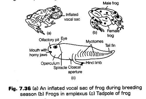
iv. Hibernation and Aestivation
Frog is a cold-blooded (poikilothermic) animal. Its body temperature fluctuates according to its surrounding temperature. To avoid the adverse conditions of environment, frog burrows inside the soil and lies in a ” state of rest or sleep during summers and winters.
The resting condition in winter is called hibernation or winter sleep and the condition of rest in summer is called aestivation or summer sleep.
v. Defences
Frogs escape from their enemies by several ways. If chased, they leap away to safe places or jump into water. They can also darken or lighten their green shade to blend with the background and thus prevent easy detection (camouflage). This protective colouration is called mimicry.
Morphology
The frog has an ovoid, streamlined and slighdy flattened body. It is about 10-15 cm long and shows bilateral symmetry, i.e., its right and left halves are mirror images of each other.
The skin of frog is naked, smooth, moist and slippery. A thin film of mucus is secreted by the cutaneous glands present in the skin. The body of frog is divisible into head and trunk. It is to be noted that neck and tail in frogs seem to be absent.
The head is triangular in shape with a blunt snout. It bears mouth, external nares, eyes, browspots and ear drums on the upper side and throat on the lower side.
The mouth extends along the entire border of the head. It is bounded by upper and lower jaws. (The lower jaw is toothless in frogs). The mouth gets open only during feeding.
At the top of anterior end, the head bears two small apertures called external nares. Air enters and leaves the body through the nares.
Eye
A little behind the nostrils, two large eyes are present, > situated along the sides. The eyes are spherical and protruded laterally.
Each eye has a thick upper and a thin lower eyelid. The upper part of the lower eyelid is modified into a transparent fold called nictitating membrane. This membrane protects the eye and is pulled over the eyeball when the frog is in water or under the mud and frog can see through it.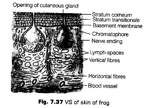
Coelom
The body cavity is a true coelom and large having two pans, a very small pericardial cavity around the heart which contains pericardial fluid and a very large pleuroperitoneal cavity around the other viscera that have coelomic fluid except the kidneys.
Both the fluids are watery, colourless and secreted by peritoneum.
Digestive System
The alimentary canal of frog is short because frogs are carnivores and hence, the length of intestine is reduced. It contains, mouth, buccopharyngeal cavity, oesophagus, stomach, small intestine, rectum and cloaca.
(a) Mouth It is present as a wide opening which opens into bucco-pharyngeal cavity. In frog teeth are not used for chewing but they prevent the escape of live food.
Mouth opens into the buccal cavity that leads to oesophagus through pharynx.
(b) Oesophagus It is a narrow, short tube, which continues in large and distended stomach.
(c) Stomach It helps in converting the food into chyme and secretes gastric juice containing HC1 and proteolytic enzymes.
(d) Intestine It is a coiled structure continued with the stomach. The intestinal wall has several finger-like folds called villi and microvilli, projecting into its lumen to enhance the surface area of absorption for digested food. The first part of small intestine lying parallel to stomach is called duodenum. The duodenum is followed by ileum. Small intestine continues into a wider rectum which opens into a cloaca.
A litde behind and below the eye on each side of head, a circular patch of tightly stretched, dark skin is present. This is called tympanic membrane or tympanum or eardrum. It receives sound waves.
The floor of the head is occupied by a soft throat. In a male frog, there is a pair of bluish-wrinkled patches of skin called vocal sacs. They help in intensifying the croaking sounds.
Trunk
The trunk contains an anterior portion called thorax and a posterior larger portion called abdomen. A pair of forelimb and hindlimb is attached to the trunk. Each forelimb has an upper arm, a forearm and a hand. The hand has a wrist, a palm and four digits.
Each hindlimb consists of an upper thigh, a middle shank and a lower foot. The foot has an ankle, a sole and five digits. The digits are joined together by a fold of skin called web.
At the end of the trunk, between the hind legs, there is a circular aperture called cloacal aperture through which faeces, urine and gametes passes out.
Anatomy
The body cavity of frogs accomodate different organ systems such as digestive, circulatory, respiratory, nervous, excretory and reproductive systems with well-developed structures and functions.
Skin
The skin of frog consists of an outer epidermis and inner dermis. Epidermis is the outermost, non-vascular layer made up of stratified epithelium. The innermost layer of epidermis consists of Malpighian layer or stratum germinativum.
Dermis layer contains mucous glands and poison glands. The poison glands secrete poisonous fluid which protects the frogs from their enemies.
(e) Pulmonary respiration occurs by the lungs and is less frequent than the cutaneous and buccopharyngeal respiration. It occurs when more oxygen is required.
The urinary bladder opens into cloacal chamber through ureter. The cloaca opens externally by a cloacal aperture. This aperture serves both as an anus and as urinogenital pore.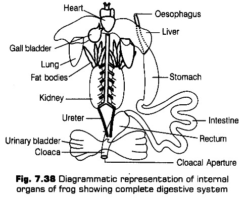
The lungs are a pair of elongated, pink coloured sac-like structures present in the upper part of the trunk region (thorax). Air enters through the nostrils into the buccal cavity and then to lungs.
The exchange of gases occurs by diffusion in all the three modes of respiration.
Circulatory System
The circulatory system of frog is well developed and is of closed type. It also has a lymphatic system. The blood vascular system includes the heart, blood and blood vessels. The lymphatic system consists of lymph, lymph channels and lymph nodes.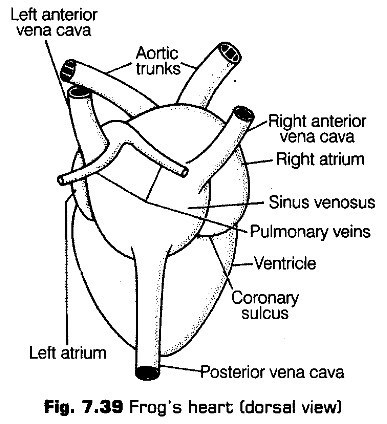
The digestive glands include gastric glands, intestinal glands, liver and pancreas. The gastric glands secrete gastric juice and the intestinal glands secrete intestinal juice, which contains a number of digestive enzymes.
The liver produces bile which is temporarily stored in the gall bladder before being released info duodenum. Pancreas is an irregular, elongated gland that produces pancreatic juice containing several digestive enzymes.
Digestion of Food
The digestion of food takes place by the action of HCI and gastric juices secreted from the walls of stomach. The partially digested food called chyme is passed from stomach to the first part of the intestine, the duodenum.
The duodenum receives bile from gall bladder and pancreatic juice from the pancreas through a common bile duct. Bile emulsifies fat and pancreatic juices digest carbohydrates and proteins.
Final digestion occurs in the intestine. Digested food is absorbed by the numerous finger-like folds in the inner wall of intestine called villi and microvilli. The undigested solid waste moves into rectum and passes out through cloaca.
Respiratory System
Frogs respire by three modes of respiration
(a) Cutaneous respiration occurs through moist skin of the frog. Frogs respires only by the skin when under water or deep under the mltd.
(b) Buccopharyngeal respiration occurs when the animal is on land or partially immersed in water. It occurs by
* thin, vascular, moist lining of buccopharyngeal cavity.
It is a muscular structure situated in the upper part of body cavity. It has three chambers, i.e., two atria (sing, atrium) and one ventricle. The heart is covered by a membrane called pericardium.
There are two accessory chambers in the frog’s heart, a tubular truncus arteriosus placed on the ventral side of the right auricle and a triangular sinus venosus present on the dorsal side of the heart.
Left anterior
Sinus venosus receives blood through the major veins called vena cava and opens into right auricle.
The left auricle receives oxygenated blood from the lungs by a common pulmonary vein. The auricles send their blood into ventricle where the blood gets mixed up. The ventricle opens into a sac-like conus arteriosus on the ventral side of the heart.
Nervous System
The system for control and coordination is highly evolved in the frog. It includes both nervous system and endocrine system. The nervous system of frogs consists of a central nervous system, a peripheral nervous system and an autonomic nervous system.
It includes brain and spinal cord. The brain is enclosed in a bony structure called cranium or brain box and the spinal cord lies inside the vertebral column. The brain is classified as forebrain, midbrain and hindbrain.
The forebrain consists of olfactory lobes, paired cerebral hemispheres and unpaired diencephalon.
The midbrain comprises paired optic lobes and the hindbrain comprises cerebellum and medulla oblongata. The medulla oblongata thus, passes out through the foramen magnum and continues into spinal cord present in the vertebral column.
It contain nerves arising from the central nervous system and extending into the organs of the body. Ten pairs of cranial nerves arises from the brain and nine pairs of spinal nerves supply the trunk and limb regions.
It consists of sympathetic nervous system of two ganglionic chains and parasympathetic nervous system of isolated ganglia in the viscera. The autonomic nervous system controls the functions of organs that are not under the voluntary control.
Sense Organs
The frogs have different types of sense organs such j as organs of touch (sensory papillae), taste (taste buds), smell (nasal epithelium), vision (eyes) and hearing (tympanum with internal ears), Out of these, eyes and ears are well organised structures and the rest are cellular aggregations around the nerve endings. Eyes in a frog are a pair of spherical structures situated in the orbit present in the skull. These are simple eyes.
External ear is absent in frogs and only tympanum can be seen externally. The ear is an organ of hearing as well as balancing (equilibrium).
Special venous connection between liver and intestine as well as the kidney and lower parts of the body are present in frogs. The former is called hepatic portal system and the latter is called renal portal system.
The blood vessels found in frogs are arteries, arterioles, veins, venules and blood capillaries.
Arteries carry blood from the heart to different body parts. Veins bring blood from different body parts to the heart. Arteries further divide to form arterioles. The arterioles branch out to form capillaries, which further unite to form venules. The venules thus, join to form veins (which have valves to prevent backflow of the blood).
The blood is composed of plasma and blood cells. The blood cells are RBCs (Red Blood Cells) or erythrocytes, WBCs (White Blood Cells) or leucocytes and platelets. RBCs are nucleated and contain red coloured pigment namely haemoglobin.
The lymphatic system comprises of lymph vessels, lymphatic channels and lymph. Lymph is a mobile connective tissue filtered out from the blood through capillaries. It contains plasma and white corpuscles and lacks red blood cells,
Lymph in the body always flows in following direction Lymph capillaries —> Lymph sinus —> Lymph hearts —> Veins
Excretory System
The elimination of nitrogenous wastes is carried out by a well-developed excretory system.
The excretory system consists of a pair of kidneys, ureters, cloaca and urinary bladder. The kidneys are compact, dark red and bean like structure, situated little posterior in the body cavity on both sides of vertebral column.
Each kidney is composed of several structural and functional units called uriniferous tubules or nephrons. Each nephron is the structural and functional unit of kidney. The Bowman’s capsule leads into a coiled, urinary tubule. The urinary tubule opens into transverse collecting tubules, which ultimately communicate with the ureter or urinogenital duct.
In females, the ureters and oviduct open separately into the cloaca. The thin-walled urinary bladder is present ventral to the rectum which also opens in the cloaca.
” The frog excretes urea and thus is a ureotelic animal. The excretory wastes are carried by blood into the kidney where these are separated and excreted.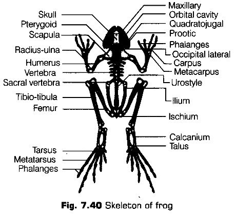
Endocrine System
The chemical coordination of various organs of the body is carried out by hormones, which are secreted by the endocrine glands. The prominent endocrine glands found in the frog are pituitary, thyroid, parathyroid, thymus, pineal body, pancreatic islets, adrenals and gonads.
Skeletal System
In frog exoskeleton is absent. The endoskeleton has two parts
(i) Axial skeleton that includes skull located in the head and vertebral column situated in the trunk. Appendicular skeleton includes limb bones in the arms and legs and girdles (pectoral and pelvic) that connects the limb bones with vertebral column. The skull consists of cranium, sense capsules and jaws. Vertebral column consists of 9 ring like vertebrae and a long urostyle. Each forelimb contains many bones namely, a humerus in the upper arm, a radioulna in the forearm, carpals in the wrist, metacarpals in the palm and phalanges in the fingers.
Each hindlimb consists of many bones namely, a femur in the thigh, a tibiofibula in the shank, tarsals in the ankle, metatarsals in the instep and phalanges in the toes.
Frogs have well organised male and female reproductive system. It shows sexual dimorphism.
Maxillary Orbital cavity Quadratojugal Prootic Phalanges Occipital lateral Carpus Metacarpus Urostyle
Hiium
Ischium
Calcanium
Talus
It includes a pair of yellowish, ovoid testes which are found adhered to the upper part of the kidneys by a double fold of peritoneum called mesorchium.
From testes, 10-12 vas efferentia arises. They enter the kidneys on their side and open into Bidder’s canal. The urinogenital duct comes out of the kidneys and opens into the cloaca. The cloaca is a small, median chamber that is used to pass faecal matter, urine and sperm to the exterior.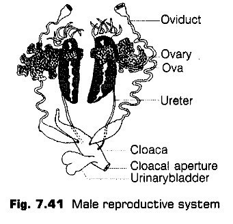
It consists of a pair of ovaries situated near the kidneys and there is no functional connection with kidneys. A pair of long oviducts from ovaries opens into the cloaca. Ovaries release about 2500-3000 eggs at a time.
The eggs are released in water, so, the fertilisation is external. Development is indirect. A fish-like tailed larva called tadpole is formed. The tadpole undergoes metamorphosis to form an adult in about three months.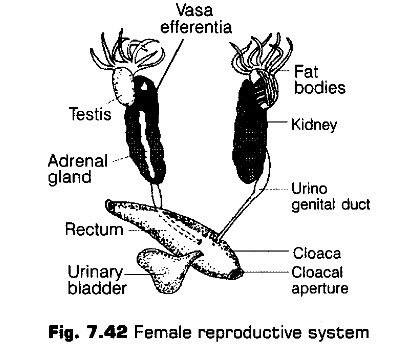
Frog is a useful animal for humans in following ways
(i) It feeds on insects which destroy crops and frogs eat up mosquitoes, which are the carriers of various diseases.
(ii) The muscles of legs of frogs are used as food in some parts of India and many other countries.
(iii) Baby frogs are used as fish baits, (any substance which is used to attract or catch fish).
(iv) Frogs are used for researches in medical science and pharmacology.
(v) They help to maintain ecological balance by forming an important part of food chain
CBSE Class 11 Biology Chapter-7 Important Questions
1 Marks Questions
1. Name the tissue which contains Have risen canals.
Ans. Mammalian bone.
2. Mention two special properties of nervous tissues.
Ans. Excitability and conductivity.
3. Name the large cells present in adipose tissue.
Ans. Adipocytes.
4. Name the cells responsible for clotting of blood.
Ans. Blood platelets.
5. What am exocrine glands?
Ans. Glands which discharge their secrctions into ducts.
6. Differentiate between tendon and ligament.
Ans. Tendon: Attach skeletal muscles to bones
Ligament: Attach bone to bone
7. Where are RBC s fornic4?
Ans. Bone narrow
8.Name the kind of tissue which forms the lining of blood vessels?
Ans. Squamous epithelium.
9.Name the chemical which helps in transmitting nerve impulse at the synapse?
Ans. Acetylcholine.
10.What is the main function of WBCs?
Ans. They protect the body from infection & diseases.
11.What are the organs of excretion in insects?
Ans. Malphigian tubules.
12.Which tissue has fat globules?
Ans. Adipose tissue.
13.Name two anticoagulants of blood of man?
Ans. Heparin & antiprothrombin.
14.Name the type of epithelium that lines the inner surface of stomach?
Ans. Columnar epithelium.
15.What causes fatigue of the muscle fibres?
Ans. Due to accumulation of lactic acid or due to prolonged contraction
16.Name the type of epithelium lines the buccal cavity?
Ans. Stratified squamous epithelium
17.Why muscle cells are usually called muscle fibres?
Ans. Muscle cells are usually called muscle fibres because muscle cells are thin & elongated into thread like structures.
18.Define glands.
Ans. Glands are secretary structures formed of epithelial tissues.
19.How many spermathecae are present in earthworms.
Ans. Four pairs of spermathecae are found in each of 6 to 9 segments of earthworm
20.Name the proteins which constitute muscle fibres.
Ans. Actin & myosin
21.Which type of epithelium is found in urinary bladder.
Ans. Transitional epithelium.
22.From which germ layers do the following organs originate
(a) kidney (b) urinary bladder.
Ans. (a) Mesoderm (b) Endoderm.
2 Marks Questions
1.What are the two typos of fibres of connective (issues? Distinguish between the two.
Ans. White and yellow fibres. White fibres are thin, wavy, unbranched, inelastic, occur in bundles and formed of protein collagen. Yellow fibres are thick, straight, elastic, branched, occurring singly, formed of protein elastin.
2.To which tissue do the following to
(a) Osteocytes(b) Chondrocytes
(e) Ncuroglia(d) Intercalated discs
Ans. (a) Bone tissue(b) Cartilage
(b) Neural tissue(d) Cardiac musele
3.Name the locomotory appendages of cockroach on the basis of external morphology.
Ans. Three pairs of legs and 2 pairs of wings.
4.Give the characteristic of epithelial tissues?
Ans. Epithelial tissue forms a layer on the free surface i.e. external surface of animal body & internal surface of visceral organs, body cavity & blood vessels. Cells of epithelium are set closely, separated by very thin film of extracellular material. Adjacent cells are held together by cell junctions.
5.How many types of nephridia are found in earthworm based on their location?
Ans. Nephridia are of three types:-
(i)SEPTAL NEPHRIDIA:- Those present on both the sides of intersegmental septa & open into intestine
(ii)INTEGUMENTAL NEPHRIDIA:- Those found attached to the lining of the body wall & open on the body surface.
(iii)PHARYNGEAL NEPHRIDIA:- Those found on the 4th, 5th, & 6th segment in the form of three paired tufts are pharyngeal nephridia.
6.What do you mean by haemopoiesis?
Ans. Haelmopoeisis is the formation of new erythrocytes from the haemopoietic tissue. The haemopoietic tissues in the young foetus in liver & spleen whereas in the adults, it is the bone marrow of long bone. The haemopoitic tissue synthesizes millions of RBC’s every minute & its excess lot is stored in the spleen.
7.Differentiate between blood & lymph?
Ans.
| BLOOD | LYMPH |
| i) It is vascular tissue | i) It is white (straw coloured) vascular tissue |
| ii) It is found in blood vessels. | ii) It is found in lymph vessels. |
| iii) It is made of plasma, erythrocytes, leucocytes & platelets. Neutrophils are most abundant. | iii) It is made of plasma, leucocytes, erythrocytes & platelets are absent. Lymphocytes are most abundant. |
| iv) It has haemoglobin | iv) Haemoglobin is absent |
| v) It helps in transport of materials inside the body. | v) It functions as middle man between blood & body cells. |
8.What are nissl’s granules? Where are they found?
Ans. Nissl’s granules are the small basophilic bodies found in the cytoplasm of soma & dendrites. They are found in nervous tissues.
9.Discuss the structure of haversian system in the histology of bone?
Ans. Bone consists of connective tissue having matrix surrounded by periosteum. In mammalian bone haversian canal which carry blood vessels & nerves of the bone are surrounded by a number of concentric lamellae of intercellular matrix & bone.
10.Distinguish between myosin & actin filament?
Ans.
| MYOSIN FILAMENT | ACTIN FILAMENT |
| i) It is found in only A- band | i) It is found in I band & also projects in A- band. |
| ii) It is thicker (100A) | ii) It is thinner (50A) |
| iii) Cross bridges are present | iii) Cross bridges are absent |
| iv) About 1500 myosin filaments are found per myofibril | iv) About 3000 actin filament are found per myofibril. |
11.What are chondriocytes? Where are they found?
Ans. In the matrix of cartilage, in chondrin there lay some large, bluntly angular cartilage cells called chondriocytes. They lie scattered in chondrin. Chondriocytes occurs in clusters of 2 or 3 cells in small spaces called the lacunae.
12.Name the major class of plasma protein & mention their functions.
Ans. Three major classes of plasma proteins are:-
(a) Serum (b) Serum globulin (c) Fibrinogen
FUNCTIONS OF PLASMA PROTEINS:-
(i)Providing body immunity
(ii)Prevention of blood loss.
(iii)Retention of fluids in the blood.
(iv)Transport of material
(v)Maintaining PH of blood.
(vi)Conducting heat to skin for dissipation.
13.What is the function of nephridia ?
Ans. Annelids have long & coiled excretory tubes called nephridia. They lie in body cavity & collect excretory wastes like NH3, uric acid, urea etc, from body fluids. Nephrostome is called ciliated funnel. It then passes them into looped, coiled ducts. They are removed out from the body through small apertures called nephridiopores.
14.Distinguish between smooth & striated muscles.
Ans.
| SMOOTH MUSCLES | STRIATED MUSCLES |
| i) They are called involuntary muscles. | i) they are called voluntary muscles |
| ii) They are found in hollow organs | ii) They are mostly attached to bones by tendons |
| iii) They are uninucleate. | iii) They are multinucleate. |
| iv) They do not show any striation | iv) They show striated appearance i.e. alternate light & dark bands. |
15.What are the functions of mast cells?
Ans. Mast cells are granular large irregularly shaped cells present in areolar connective tissue
a)They store inflammation producing substance histamine. When histamine is released inflammation is caused due to some reason.
b)They also release heparin which prevent activation of prothrombin this preventing coagulation of blood.
16.How can a male frog be distinguished from a female frog ?
Ans. The male frogs may be distinguished by presence of sound producing vocal sacs. They also has a copulatory pad on the first digit of the forelimbs. Vocal sacs & copulatory pads are absent in female frogs.
17.Give reason why earthworms are known as friends of farmers.
Ans. Earthworms are known as “friends of farmers” since it makes burrows in the soil. Due to it, soil becomes porous. It facilitates respiration as well as penetration for the developing roots of the plants, the earthworm eats decaying vegetation & in the burrows, it enriches the soil.
18.Write short note on adipose tissues.
Ans. It is a specialized form of aerolar tissue where it contains mainly fat cells or adipocytes. The matrix contains fibroblasts, macrophages, collagen fibres & elastin fibres. This tissue lies beneath the skin, around kidney & in mesentery & bone marrow. It synthesizes, stores & mebabolises fat & forms insulating layer beneath the skin.
19.What are neuroglia cells?
Ans. Neuroglia cells are cells that hold the neurons together.
20.How does saltatory conduction takes place along a nerve fibre.
Ans. Along a myelinated nerve fibre, the conduction of impulse is called salutatory conduction. This is so because the ionic changes & consequent depolarization taking place only.
21.Write short note on gaseous exchange in cockroach.
Ans. Cockroach is an insect & has a system of trachea. Hence tracheal respiration occurs in these animals. It is a complicated system of air tubes. They divide & form tracheoles. Tracheoles are connected to the spiracles located in the segments of thorax & abdomen. The body cells or fluid come in direct communication of air.
22.Draw a well labelled diagram of a nerve cell.
Ans.
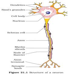
23.Distinguish between tendon & ligament.
Ans.
| TENDON | LIGAMENT |
| i) It is formed of white fibrous connective tissue | i) It is formed of yellow fibrous tissues |
| ii) Fibroblasts are arranged in rows between the bundles of white fibres | ii) Fibroblasts are scattered in matrix |
| iii) It is tough & non flexible | iii) It is elastic & flexible. |
| iv) It joins muscles to bones | iv) It joins bones together. |
24What is mucosa?
Ans. Mucosa is the mucous secreting epithelial tissue alongwith the supporting connective tissue beneath it. It lines some hollow organs or cavities of the body eg. alimentary canal, nose, trachea & lungs etc
3 Marks Questions
1.(a) Give the common name of Periplaneta Americana.
(b) How many spermathecae found in cockroach?
(c) What is the position of ovaries in cockroach?
(d) How many segments are present in the abdomen of cockroach?
(e) Where do you find malpighian tubules?
(f) What is mosaic vision?
Ans. (a) American Cockroach.
(b) One pair, present in 6th segment.
(c) Between 2nd and 6th abdominal terga.
(d) 10 segments.
(e) At the be of ileum in cockroach.
(f) Vision where several images of an object arc formed by compound eye. Helps to detect movement of objective very efficiently.
2. (i) Give three differences between frogs & toads?
(ii) What do you understand by open type of circulatory system?
Ans. (a)
| FROGS | TOADS |
| i) Scientific name of frog is Rana tigrina | i) Scientific name of toad is Bufo melanostictus |
| ii) Frogs are diurnal | ii) Toads are nocturnal |
| iii) Parotid glands absent | iii) Parotid glands present |
| iv) Skin moist & slipper | iv) Skin dry & rough. |
| V) amphibious animals | v) Terrestrial for egg laying. |
(b)In open circulatory system the blood vessels are poorly developed & open into spaces rather capillaries. All the vesceral organs are bathed in blood (haemolymph) e.g. cockroach.
3.What are the cellular components of blood?
Ans. The blood consists of two parts- plasma (liquid part) & corpuscles (solid parts). The blood corpuscles float in the plasma & are of three major types:-
(a)Red Blood cells or Erythrocytes:- They are circular disc–shaped biconcave cells without nucleus. They contains a pigment haemoglobin which has great affinity towards oxygen. In normal healthy individuals, the number of RBCs / mm3 ranges between 4.5-5 millions in females & 5.5-6.0 millions in males.
(b)White Blood cells or Leucocytes :- These are colourless nucleated corpuscles & can pass through the capillary walls into lymph & tissue fluid. Their count is 6000 to 10,000 per mm3 of blood. Their main function is protection against any foreign substance. Leucocytes are further of two types:-
(i)AGRANULOCYTES:- They have clear cytoplasm without granules & a bilobed nucleus they are further divided into two types lymphocytes & monocytes.
(ii) GRANULOCYTES:- They are of three main types
Basophils :- which are stained with basic dye (methylene blue) & have a bilobed nucleus.
Neutrophils:- which are stained with neutral dye & have multilobed nucleus.
Eosinophils :- which are stained with acidic dye (eosin) & have bilobed nucleus.
4.How do erytherocytes transport oxygen & carbon dioxide in the blood?
Ans. During development of erythrocytes there is formation of special substance called haemoglobin which is capable of transporting oxygen. It is a complex protein & is composed of two components a) a protein called globin & b) a Fe2+ porphyrin ring called heme. This haemoglobin when exposed to high partial pressure of oxygen combines with oxygem to form oxy-haemoglobin which carries 4 molecules of oxygen loosely bound to four Fe2+ ions. When this oxy haemoglobin reaches the tissues where there is low oxygen pressure oxyhaemoglobin dissociates into oxygen & deoxyhaemoglobin. In this way, erythrocytes transports oxygen from lungs to tissue. Similarly haemoglobin also transports carbon dioxide from tissues to lungs.
5.Describe the different types of connective tissues & give examples?
Ans. On the Basis of matrix, connective tissues are of two main types:-
(I)Connective tissue proper:- It connects & supports many tissues & organs. Its matrix is dense. Eg.
(i)Areolar tissue :- It consists of three types of cells & types of fibres, all distributed in the matrix. Fiborblasts are irregularly shaped flat cells with long protoplasmic processes, they secrete collagen & elastin proteins for the fibres.
(ii)Adipose tissue:- It consists of collagen fibres, elastin fibres, fibroblast, maerophages, & adipocytes which stores fat. It prevents heat loss by forming one insulating layer beneath skin.
(II)Supportive connective tissues :- It consists of following types of connective tissue:-
(i)Cartilage:- It is the endoskeletal material of the vertebrates, it is in the form of solid matrix formed of chondrin with few collagen fibres & chondrioblast cells.
(ii)Bone:- The matrix consists of bone cells, osteocytes, fibres & a ground substance impregnated by calcium phosphate, calcium carbonate, magnesium phosphate & calcium fluoride. Due to these salts, it becomes very hard & forms skeletal support of body.
6.Describe briefly the structure of voluntary muscles
Ans. A voluntary muscle is a bundle of numerous striated muscle fibres. Each fibres is long, unbranched measuring 40 mm in length & 20ym in thickness. Each fibre is enclosed in a membrane called sarcolemma & its cytoplasm is called sarcoplasm. The sarcoplasm contains many myofibrils that are long, thin, unbranched & cross striated. Each myofibril consists of alternating thick A & light I-band. A band is formed of protein myosin & I-band with actin protein. The thick filament bands lie parallel to one another. The thin filament extends between them upto a considerable distance in an orderly manner. At the center of the I-band is a fine, dense, dark, Z-line. Each segment of myofibril from one Z-band to the next functions as a contractile unit & is called sarcomere.
7.How does blood gets coagulated on coming out from an injured vessel. How coagulation is normally prevented uninjured vessels.
Ans. When a blood vessel is injured & blood comes out of it, the thrombocytes clump together, break & release the coagulation promoting substances called thromboplastin. Thromboplastin helps in the formation of enzyme thrombokinase. This enzyme thrombokinase hydrolyses prothrombin in the plasma into thrombin ca2+ ions are needed for both activation & functioning of thrombin. Thrombin catalyses the hydrolysis of soluble fibrinogen in the plasma into insoluble fibrin. The fibrin precipitates as a network of fibres & traps many blood cells to form a red solid mass called blood clot. The clot seals the wound in the blood vessel to stop bleeding.
However in uninjured tissues & blood vessels don’t release thromboplastin. That’s why coagulation is prevented in an uninjured vessel.
8.Name the various fibres of connective tissue & compare them.
Ans.
| Nature | Collagen fibres | Elastin fibres | Reticular fibres |
| i) colour | White | Yellow | White |
| ii) Protein | Formed of protein, tropho- collagen | Elastin protein | Reticulin protein |
| iii) Occurrence | Found in bundles | Singly | Singly |
| iv) Nature | unbranched | Branched & anastomosing | Branched but form a network |
| v) fibres | Thick, long wavy | Thin, long straight | Short |
| vi) elasticity | Tough, non elastic | Elastic | Delicate |
| vii) Location | Abundant in tendon | Abundant in ligament | Abundant in ambryo in lymphoid as well as blood forming tissues. |
9.Give an account of alimentary canal of frog.
Ans. ALIMENTARY CANAL OF FROG:-
It is a short tube starting from mouth to cloaca. Mouth opens into buccopharyngeal cavity. It has many maxillary teeth as the margin of upper jaw. Vomerine teeth lie at the floor of this cavity. The tongue is bilobed & muscular. It is used to capture the prey. Gullet opens into the oesophagus which is distended into stomach. Stomach follows small & large intestine. The rectum opens into the cloaca. Liver & pancreas are digestive canals.
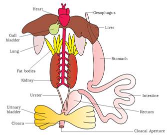
NCRT TEXTBOOK QUESTIONS SOLVED
1. Answer in one word or one line.
(i) Give the common name of Periplaneta americana.
(ii) How many spermathecae are found in earthworm?
(iii) What is the position of ovaries in cockroach?
(iv) How many segments are present in the abdomen of cockroach?
(v) Where do you find Malpighian tubules?
Solution: (i) Cockroach.
(ii) Four pairs.
(iii) In cockroach two large ovaries, lie laterally in the 2nd – 6th abdominal segments’.
(iv) Abdomen of cockroach consists of 10 segments.
(v) Malpighian tubules are present at the junction of midgut and hindgut in cockroach.
2. What are the following and where do you find them in animal body?
(a) Chondrocytes
(b) Axons.
(c) Ciliated epithelium
Solution: (a) Chondrocytes – Chondrocytes are the only cells found in cartilage. They are present in spaces called lacunae and they produce and maintain the matrix of cartilage. Bending ability of cartilage is due to chondrocytes. Cartilage is present at tip of nose, pinna of ear, epiglottis etc.
(b) Axon – Axon is one of the processes of neuron, which is the structural and functional unit of nervous system. The part of cyton – n’here axon arises is axon hillock and axon ends in group of branches called terminal arborizations. It conducts impulses away from the cyton. Neurons (nerve cells)
are present in brain and spinal cord.
(c) Ciliated epithelium – If the columnar or cuboidal cells bear cilia on their free surface they are called ciliated epithelium. Their function is to move particles or mucus in a specific direction over the epithelium. They are mainly present in the inner surface of hollow organs like bronchioles and Fallopian tube.
3. Draw a labelled diagram of the reproductive organs of an earthworm.
Solution: 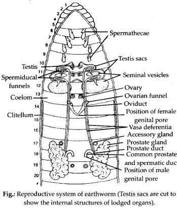
4. Answer the following.
(i) What is the function of nephridia?
(ii) How many types of nephridia are found in earthworm based on their location?
Solution: (i) Nephridia are excretory organs of earthworm, which perform the function of excretion and osmoregulation. Nephridia regulate the volume and composition of the body fluids. A nephridium is a coiled tubular and microscopic structure which starts out as a funnel that collects excess fluid from coelomic chamber. The funnel connects with a tubular wastes through a pore to the surface in the body wall or into the digestive tube.
(ii) In earthworm, nephridia are present in all segments except the first two. There are three types of nephridia on the basis of their location:
(a) Septal nephridia, present on both the sides of intersegmental septa from segment 15 to the last that open into intestine.
(b) Integumentary nephridia, attached to lining of the body wall of segment 3 to the last that open on the body surface and
(c) Pharyngeal nephridia, present as three paired tufts in the 4th, 5th and 6th segments.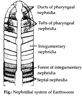
5. Draw a labelled diagram of alimentary canal of a cockroach.
Solution: 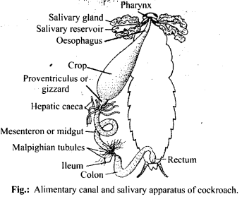
6. What are the cellular components of blood?
Solution: Blood is a fluid connective tissue. It is composed of plasma (fluid) and blood cells (corpuscles). Cellular components of blood (blood corpuscles) constitute about 45% of blood volume.
Three types of blood cells are:
(i) Erythrocytes or red blood cells: They are most abundant blood cells. Normal RBC count is 5-5.5 million/mm3 in males and 4.5-5 million/mm3 in females) RBCs help in transport of gases and maintain blood pH.
(ii) Leucocytes or white blood cells: The normal WBC count is 5000-6000/mm3 of blood. They are involved in immune response of body and act as soldiers and scavangers.
(iii) Thrombocytes or blood platelets: There are about 2,50,000 platelets/mm3 of blood. They are involved in blood clotting.
7. Distinguish between the following:
(a) Prostomium and peristomium
(b) Septal nephridium and pharyngeal
Solution: (a) Differences between prostomium and peristomium are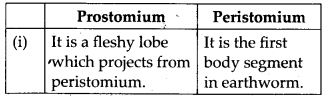
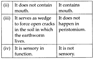
(b) Differences between septal and pharyngeal nephridia are: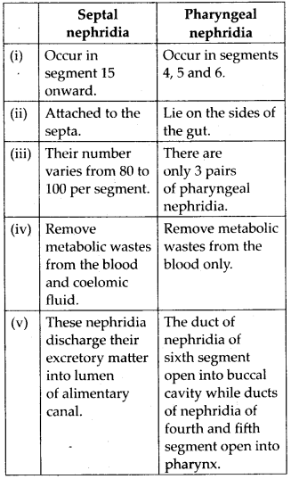
8. Mark the odd one in each series.
(a) Areolar tissue; blood; neuron; tendon
(b) RBC; WBC; platelets; cartilage
(c) Exocrine; endocrine; salivary gland; ligament
(d) Maxilla; mandible; labrum; antennae
(e) Protonema; mesothorax; metathorax; coxa.
Solution:
(a) Neuron: Areolar tissue, blood and tendon are connective tissues while neuron is a part a nervous tissue.
(b) Cartilage: RBC, WBC and platelets are parts of vascular connective tissue while cartilage is skeletal connective tissue.
(c) Ligament: Ligament is a connective tissue.
(d) Antennae: Maxilla, mandible and labrum are mouth parts of cockroach while antennae are sense organs.
(e) Protonema: Protonema is a filamentous juvenile stage in life cycle of Bryophytes, while mesothorax, metathorax and coxa are appendages of cockroach.
9. Match the terms in column I with those in column II.
Column I | Column II |
| (a) Compound epithelium (b) Compound eye (c) Septal nephridia (d) Open circulatory system (e) Typhlosole (f) Osteocytes (g) Genitalia | (i) Alimentry canal (ii) Cockroach (iii) Skin (iv) Mosaic vision (v) Earthworm (vi) Phallomere (vii) Bone |
Solution: (a) – (iii), (b) – (iv), (c) – (v), (d) – (ii), (e) – (i), (f) – (vii), (g) – (vi)
10. Mention briefly about the circulatory system of earthworm.
Solution: Earthworm possesses a closed type of blood vascular system, as the blood flows through closed blood vessels. Blood is red in colour due to respiratory pigment haemoglobin. Prominent blood vessels in earthworm includes dorsal, ventral, sub- neural, lateral oesophageal and supra- oesophageal blood vessels. There are four pairs of tubular hearts, provided with valves. The anterior two pairs of hearts, known as lateral hearts lie in the 7th and 9th segments and connect the dorsal blood vessel with the ventral blood vessel. They receive blood from the dorsal blood vessel and convey it to the ventral blood vessel. The posterior two pairs of hearts are called latero-oesophageal hearts and are situated in the 12th and 13th segments. The latero-oesophageal hearts apart from connecting the dorsal and ventral blood vessels are also joined with the supra oesophageal blood vessel. Latero-oesophageal hearts carry blood from the dorsal vessel and the supra oesophageal vessel to the ventral blood vessel.Contractions keep blood circulating in one direction. Blood glands are present in the 4th, 5th and 6lh segments which produce blood cells and haemoglobin which is dissolved in blood plasma. Blood cells are phagocytic in nature.
11. Describe various types of epithelial tissues with the help of labelled diagrams.
Solution: Epithelial tissue is a tissue made of one or more layers of compactly arranged cells that covers external surface and internal free surface of body organs and which is underlined by a basement membrane. The various types of epithelial tissue along with the diagram are given below:
(i) Simple epithelium : It is composed of single layer of cells which rest on basement membrane. Simple epithelium generally occurs over secretory and absorptive surfaces and forms lining of body cavities, ducts and tubes. Simple epithelium is of several types.
(a) Squamous epithelium: It consists of single layer of flat cells, tightly linked together and have centrally located oval or spherical nucleus. It is also called pavement epithelium. It is found in walls of blood vessels, air sacs of lungs, and lining of eye lens.
(b) Cuboidal epithelium: Cells of cuboidal epithelium are as tall as wide, with centrally placed nucleus. Its main functions are secretion and absorption. It lines sweat gland, thyroid follicles, salivary glands. Brush bordered cuboidal epithelium, i.e., cells having microvilli on their free surface lines proximal part of uriniferous tubule, pancreatic duct, testis and ovary.
(c) Columnar epithelium: Cells are with basally located nucleus. It helps in secretion and absorption. It occurs in lining of intestine, stomach, gall bladder.
(d) Ciliated epithelium: Free surface of columnar and cuboidal cells are covered with cilia. Cilia help in moving fluids, particles, mucus, etc. in a specific direction. It occurs in the inner surface of Fallopian tubules, nasal passage, bronchioles.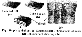
(e) Pseudostratified epithelium: It consists of single layer of cells but some cells are shorter than others. Due to difference in size of cells, the epithelium appears 2-3 layered. Pseudostratified columnar epithelium occurs in urethra and parotid salivary gland. Pseudostratified columnar ciliated epithelium (only larger cells ciliated) occurs in lining layer of nasal’ chambers, trachea and large bronchi. It helps in moving mucus and foreign particles.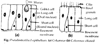
(ii) Compoundepithelium/stratifiedepithelium: It is multilayered epithelium where cells of only the lowermost or basal layer are in contact with basement membrane. It provides protection against mechanical and chemical stresses and has limited role in secretion and absorption. It covers dry surface of skin, moist surface of buccal cavity, pharynx, etc. Different types of compound epithelium are:
(a) Stratified squamous epithelium: The cells of outer layer are flattened and squamous while the inner layers are cuboidal cells. It is of two types: Non- keratinised lining oesophagus, pharynx, buccal cavity, cornea, vagina and anal canal and keratinised (comified): forming epidermis of skin, hair, horn and nail.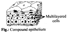
(b) Stratified cuboidal epithelium: The outer layer of cuboidal cells and basal layer of columnar cells. It lines ducts of sweat glands, large salivary and pancreatic ducts.
(c) Stratified columnar epithelium: Both upper and basal layers are made of columnar cells, e.g., epiglottis covering, part of urethra.
(d) Stratified ciliated columnar epithelium: Outer layer consists of ciliated columnar cells and basal layer of columnar cells, e.g., larynx.
(iii) Transitional Epithelium: This is stratified epithelium which contains cuboidal or columnar shaped cells, which are thin and stretchable. No basement membrane is present as it would impede stretchability. It lines the inner surface of renal calyces, urinary bladder, ureter. Because of its t distribution, it is also called urothelium.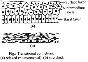
(iv) Glandular epithelium: It consists of specialised epithelial cells which synthesise intracellular macromolecules (protein in pancreas, lipids in adrenal glands, glycoprotein in salivary glands and all the three in mammary glands) and pour out the same in the form of a useful fluid secretion which is different from blood or any other extracellular fluid. Glands can be unicellular or multicellular on the basis of number of cells.
(a) Unicellular glands: Single-celled, e.g., goblet (mucous) cells of respiratory tract and alimentary canal.
(b) Multicellular glands: Consist of cluster of cells, e.g., Salivary glands.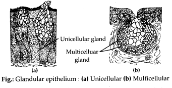
On the basis of presence or absence of duct glands can be:
(a) Exocrine glands : These glands pour their secretion through a duct. They secrete milk, saliva, mucus, earwax. e.g., goblet cells, salivary glands, tear glands, gastric glands, intestinal glands.
(b) Endocrine glands: They are ductless glands, which pour their secretions into blood or lymph for reaching the target region. Their secretion is called hormone e.g., pituitary gland, thyroid gland, parathyroid glands, adrenal glands.
(c) Heterocrine glands: Both exocrine and endocrine, e.g., pancreas.
On basis of mode of secretion glands can be:
(a) Merocrine: Secretion is discharged
through diffusion, e g., goblet cells, sweat glands.
(b) Apocrine glands: Glandular secretion accumulates in the terminal part of the cell which is pinched off, e.g., mammary glands.
(c) Holocrine glands : The cell filled with secretory product disintegrates during discharge of the product, e.g., sebaceous gland.
(v) Modified epithelium : It is of following types:
(a) Germinal epithelium (generally cuboidal, produces gametes), (b) Glandular epithelium (columnar or cuboidal secretes chemicals and mucus), (c) Sensory epithelium or neuroepithelium. Epithelial cells having sensory hair on free surface and connected with nerve fibres on the other surface (generally columnar, receives and conveys stimuli), e.g, nasal epithelium, taste buds, retina, sensory spots of internal ear. (d) Pigmented epithelium – The cells possess melanin granules, e.g, retinal layer in contact with choroid of eye.
12. Distinguish between
(a) Simple epithelium and compound epithelium.
(b) Cardiac muscle and striated muscle.
(c) Dense regular and dense irregular connective tissues.
(d) Adipose and blood tissue.
(e) Simple gland and compound gland.
Solution:
(a) Differences between simple and compound epithelium are as follows: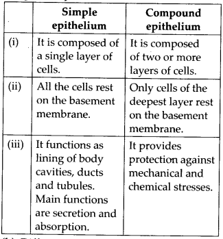
(b) Differences between cardiac and striated muscles are as follows: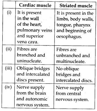
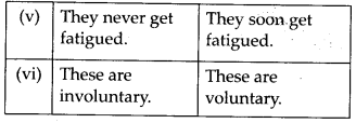
(c) Differences between dense regular and dense irregular connective tissues are as follows: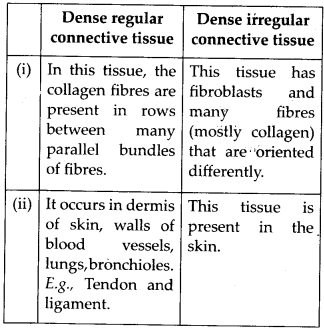
(d) Differences between adipose tissue and blood tissue are as follows: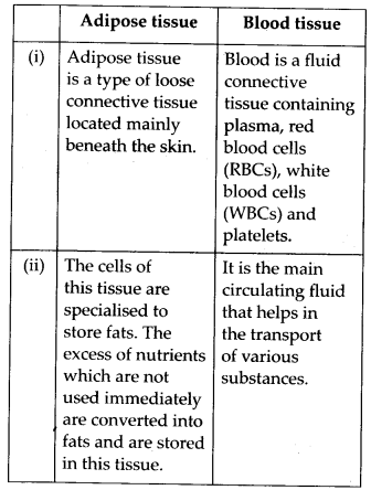
(e) Differences between simple gland and compound gland are as follows: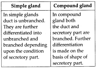
13. Draw a neat diagram of digestive system of frog.
Solution: 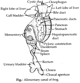
14. Mention the function of the following:
(a) Ureters in frog
(b) Malpighian tubules
(c) Body wall in earthworm.
Solution:
(a) Ureters in frog: Ureter is a transparent duct which arise from outer portion of kidney. In the “male frogs, ureter acts as urinogenital duct which runs backwards from kidneys and opens into the cloaca. It carries both urine and spermatozoa from kidney to the cloaca. In female, ureter conducts only urine from kidneys to the cloaca.
(b) Malpighian tubules: Malpighian tubules are excretory organs present in cockroach. These are present at junction of mid gut and hindgut. These are fine, long, unbranched, yellowish and blind tubules and are 100-150 in number. They help in the removal of excretory products from haemolymph.
(c) Body wall in earthworm: It consists of cuticle, epidermis, muscular layer and parietal peritoneum.
(i) It maintains the characteristic shape of’ the body.
(ii) It protects the internal organs.
(iii) The cuticle prevents excessive evaporation.
(iv) It serves as an ideal respiratory organ.
(v) The receptor cells play a vital sensory function.
(vi) The albumen helps in the formation of cocoon. It also serves as a food for the developing earthworm inside the cocoon.
(vii) Setae and muscles are responsible for locomotion.
(viii) Excretory matter is passed out through nephridiopores.