Cell The Unit of Life Class 11 Notes Biology Chapter 8
Topics and Subtopics in NCERT Solutions for Class 11 Biology Chapter 8 Cell The Unit of Life:
| Section Name | Topic Name |
| 8 | Cell The Unit of Life |
| 8.1 | What is a Cell? |
| 8.2 | Cell Theory |
| 8.3 | An Overview of Cell |
| 8.4 | Prokaryotic Cells |
| 8.5 | Eukaryotic Cells |
| 8.6 | Summary |
Topic 1 Cell :An Overview
An organism consist of one or more cells, accordingly there are two types of organisms, i.e., unicellular (composed of single cell) and multi cellular (composed of many cells).
Cell Theory
The cell theory was formulated by two German Scientists, Matthias Schneider and Theodore Schwinn independently. Schneider (1838) examined a large variety of plant tissues and observed that all plants are composed of different kinds of cells. At about the same time, Schwinn (1839), closely studied different types of animal cells and found that the animal cell had a very thin outer layer known as plasma membrane.
He also concluded, from his studies based on plant tissues that animal cells differ from plant cells in lacking cell wall.
Objections to Cell Theory
Cell theory failed to explain how and from where the new cells were formed. All these observations lead to a major expansion of cell theory that was expressed by Rudolf Virchow in 1855 modified the hypothesis of Schneider and Schwinn and explained in his statement that cells divide and new cells are formed from pre-existing cells, i.e., Omnis cellula-e-cellula.
Thus, the cell theory states that
Outer membrane, the boundary of the cell, which provides protection to the cell and controls the exchange of ions, molecules and other components in and out of the cell.
The outer membrane of a cell contains cell wall (only in plant cells) and plasma membrane.
Details of the above mentioned cell components and organelles is given later in the chapter in the second topic.
Microscopes allow us to study the structure of cells, two types are l commonly used, i.e., light microscopes and electron microscopes, Cells are divided into compartments that help segregate functions, leading to more efficient performance; human cells consist of two l major compartments, i.e., the cytoplasmic and nuclear.
Size of a Cell
The cells exhibit an endless variation in size, life span and cellular activities, e.g., Mycoplasma (smallest cell) or PPLOs (Pleuro-Pneumonia Like Organisms) is only 0.3 Jim in length and bacteria are approx. 3-5 Jim in size.
An ostrich egg, which is known to be the largest isolated single cell measures about 170 X 135 mm. Human Red Blood Cells (RBCs) are about 7 Jim in diameter and the nerve cell of human being is the longest cell having length of 90-100 cm.
Shape of a Cell
The cells also vary in their shapes. They may be polygonal, disc-like amoeboid, thread-like, cuboid or irregular. The cell shape is always related and vary with the function they perform.
(i) All living organisms are composed of cells and their products.
(ii) All cells arise from pre-existing cells.
(iii) Cells show similarity in chemical composition and metabolic activities.
(iv) Cells are the structural and functional unit of living organism.
Structural Outline of a Cell
The onion cell, which is a typical plant cell has a distinct cell wall as its outer boundary and just within it is the cell membrane. The human and animal cell has an outer membrane, inside which is a dense membrane bound structure called nucleus. Each cell consists of
(i) Nucleus, the central part of the cell, which is spherical in shape. Its number can be one or more per cell. It is denser than the surrounding cytoplasm.
The nucleus is composed of chromosomes (contains the genetic material, i.e., DNA), nuclear membrane and centrioles (non-membrane bound organelle present in only animal cells, which helps in cell division).
(ii) Cytoplasm, a semi-fluid matrix that occupies the volume of the cell. It is mainly composed of water with free floating molecules.
Inside the cytoplasm all cellular activities like gaseous exchange, elimination of wastes, hereditary mechanisms, etc occur.
Eukaryotic cells also contain other cell membrane bound distinct structures called cell organelles, like mitochondria, vacuoles, Endoplasmic Reticulum (ER), Golgi complex, etc.
The prokaryotic cells lack all these membrane bound organelles. It is to be noted that as ribosomes are not bounded by membrane and are found in all cells.
Ribosomes are also found in chloroplasts (in plants) and mitochondria and on rough ER other than cytoplasm.
Types of Cell
On the basis of the organisation, complexity and variety, all cells can be grouped into two types,i. e., prokaryotic cells and eukaryotic cells.
Prokaryotic Cell
Cell which do not have a nuclear membrane and other membrane bound organelles, is called prokaryotic cell.
Occurrence
Prokaryotic cells are placed in kingdom-Monera. These cells are represented by bacteria, cyanobacteria (blue-green algae), mycoplasma or PPLO. Bacteria are the simplest and common most type of organism amongst prokaryotes. They are generally smaller and multiply more rapidly than the eukaryotic cell.
The bacteria are found in almost every place like deep in the soil, human intestine, deep in seawater, etc.
Size
Bacteria tends to vary greatly in size. It normally ranges from 0.3-1.5p.m with some exceptions.
Shape
The four basic shapes of bacteria are bacillus (rod-like), coccus (spherical), vibrio (comma-shaped) and spirillum (spiral). All prokaryotic cells are similar in their organisation although they exhibit a wide variety of shapes and functions.
Components of a Prokaryotic (Bacterial) Cell
A bacterial cell is composed of various components as genetic material, cell envelope, cytoplasm, nucleoid, inclusion bodies, ribosomes, flagella, pili, fimbriae, etc.
Genetic Material
Nucleoid represents the genetic material incase of prokaryotes that is naked, not enveloped by a nuclear membrane. Many bacteria contain a small circular DNA known as plasmid other than the chromosomal or genomic DNA.
These plasmid confer certain unique characters to the bacteria like antibiotic resistance, sex factor, etc.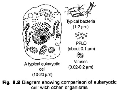
Cell Envelope and Its Modifications
Cell envelope is the outermost covering of protoplasm of the bacterial cell. It is known to protect the cell from mechanical shocks and injuries.
It is composed of following three layers, which perform specialisedfunction
i- Glycocalyx (Mucilage Sheath)
It is the outermost layer, made up of macromolecules that gives sticky character to the cell. Glycocalyx differs in composition and thickness among different bacteria. It could be in the form of loose mucilaginous sheath called slime layer or thick and tough covering called capsule.
Function help in resisting phagocytosis.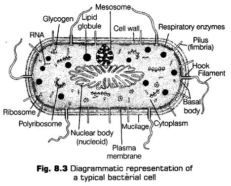
Cell Wall
It is present just below the glycocalyx made up of peptidoglycan or murein in all eubacteria and cyanobacteria. It is a rigid and solid covering that gives shape and strong structural support to the cell.
Cell wall performs the following functions
(a) It helps in preventing cell from bursting or collapsing.
(b) It allows the material to pass in and out of the cell.
(c) It wards off the attack of pathogens like viruses, bacteria, fungi, protozoans.
(d) Provides mechanical support to the cell against gravity.
Gram Positive and Gram Negative Bacteria
According to Christian Gram (1884) various types of reactions are shown by the cell walls of different bacteria. Thus, on the basis of the differences in the cell wall and the |
response to the staining procedure developed by Gram, bacteria are classified into following two types
(i) Gram positive (+ve) bacteria are those that take up the Gram stain and retain blue or purple colour, e.g., Bacillus subtilis, Clostridium, etc.
(ii) Gram negative (-ve) bacteria are those that do not take up Gram stain and loose the blue or purple icolour, e.g., Escherichia coli, (E.coli), Acetobacter, etc.
Plasma Membrane
It is the innermost layer of the cell envelope. It is semi-permeable in nature and is responsible for the interaction of the cell with the outside environment.
It performs a number of functions as follows
(a) It helps in the regulation of the exchange of specific materials between the cytoplasm and extracellular medium.
(b) Selectively permits particular molecules to pass and prevents others.
(c) Prevents loss of components from the cells through leakage.
Note:
* The plasma membrane is vital to cellular homeostasis and therefore, the health and welfare of all living organisms.
* Molecules move through membranes either passively, flowing down concentration gradients or actively, being pumped in or out of cells.
* Membrane in prokaryotes is . structurally similar to eukaryotes.
Membranous Structures
Prokaryotic cells lack the complex membrane bound organelles (such as chloroplast, mitochondria, etc). However, some other special membranous structures are found in them (i.e., mesosomes and chromatophores).
Mesosomes
These are formed by the extensions of the plasma membrane into the cell in the form of vesicles, tubules and lamellae.
Mesosomes are equal to mitochondria in eukaryotes, as these structures participate in aerobic cellular respiration in prokaryotes.
Mesosomes perform the following junctions in bacterium
(a) Helps in respiration, cellular secretion, etc.
(b) Helps in increasing the enzymatic content and surface area of the plasma membrane.
(c) Helps in the formation of a cell wall.
(d) Helps in the replication of DNA and distribution of genetic material to daughter cells during fission.
Chromatophores
They are another membranous structures present in some prokaryotes like cyanobacteria, etc.
They are internal membrane systems of photosynthetic forms, which possess photosynthetic pigments. These pigments are light reflecting.
Flagella
Bacteria can be motile or non-motile. Thus, motile bacteria possess one or more thread-like appendages extending from their cell wall called flagella (sing, flagellum). Bacteria are also classified according to the number and arrangement of flagellum in them.
Each flagellum is about 1-7 nm long covered by a protein coat.
The bacterial flagellum is differentiated into the following three parts
(i) Filament, the longest portion, extending from the cell surface to the-outside. It is made up of protein called flagellin.
(ii) Hook, a curved and tubular structure made up of protein subunits.
Pili Fimbriae
They occur only in Gram negative bacteria. They occur in both Gram positive and Gram negative bacteria.
They are longer and broder. They are shorter and narrower.
They are responsible for sex duction (conjugation) in bacteria. They are specialised for attachment of bacteria to its host (e.g., Bacteria, Salmonella typhimurium, Neisseria gonorrhoea, etc).
They are tubular structures. They are bristle-like solid structures.
(iii) Basal body, the most complex part of flagellum.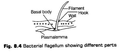
They help in locomotion i.e., movement from one place to another.
Pili and Fimbriae
These are also surface structures, but does not play any role in locomotion of bacteria. The pili are the elongated, tubular structues made of a special protein called pilin and fimbriae are the small bristle-like fibres coming out of the cell.
The pili helps in forming conjugation tube during transfer of genetic material between donor and recipient cell. While, the fimbaiae help the bacteria to attach to solid surfaces.
Differences between Pili and Fimbriae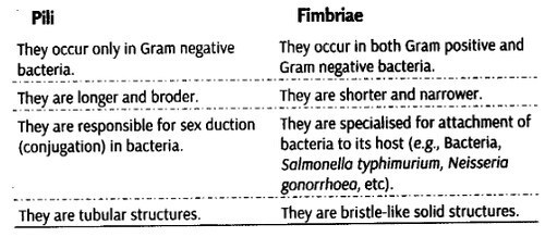
Ribosomes and Inclusion Bodies
Cytoplasm in prokaryotes appear granular, due to the presence of following structures:
i. Ribosomes
Like eukaryotes, ribosomes are also found in prokaryotes and serves a common function, i.e., acts as a site of protein synthesis. Ribosomes are small, but are complex both in structure and chemical composition. They are about 15-20 nm in size.
In prokaryotes, ribosomes are found in association with the plasma membrane of the cell (as it lack endoplasmic reticulum) in the cytoplasmic matrix. The prokaryotic ribosomes are of 70S type.
It has following two sub-units
(a) Smaller subunit (30S)
(b) Larger subunit (50S)
Ribosomes generally occur in helical groups called polysome or polyribosomes. In each polysome 4-8 ribosomes are attached to a single strand of wRNA. The ribosomes of a polysome helps in the translation (mechanisms to synthesise several copies of the same protein) of wzRNA into protein.
ii. Inclusion Bodies
They are non-living structures present in the cytoplasm and not bounded by any membrane system. They may either lie free in the cytoplasm (e.g., Cyanophycean granules, glycogen granules) or may be covered by 2-4 nm thick, non-protein membrane (e.g., Gas vacuoles, sulphur granules, etc).
Note:
* Gas vacuoles are gas storing vacuoles that do not have any covering of their own. They are found in cyanobacteria (blue-green algae), purple and green photosynthetic
bacteria.
* These are named so, because they are permeable to atmospheric gases but not to water.
Topic 2 Eukaryotic Cell
A cell which has a well organised nucleus with a nuclear envelope and several membrane bound organelles is called eukaryotic cell.
Internal organisation of eukaryotic cells is more advanced and elaborate, than the prokaryotic cells. All eukaryotic cells are not identical. Except monerans, eukaryotic organisation is seen in all the protists, plants, fungi and animals. Eukaryotic cell is larger than the prokaryotic cell (i.e., around 10-100 cm in size).
Generalised Structure
An extensive compartmentalisation of cytoplasm is seen through the presence of membrane bound organelles. Eukaryotic cells also possess a variety of locomotory and cytoskeletal structures.
All eukaryotic cell are not-identical, instead they differ from each other on the basis of structure and function. Cell wall is a special membrane, being present in plants, fungi and some protists. Plants cells also contains a large vacuole and plastids, which are absent in animal cells, while animal cells possess centrioles, which are absent in plant cells.
Differences between Plant and Animal Cell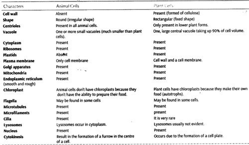
Components of a Eukaryotic Cell
An eukaryotic cell is composed of various cell components as cell membrane, cell wall (only in plants), mitochondria, chloroplast, Golgi bodies, ribosomes, centrioles (only in animals), etc.
All these are described here under in detail.
Cell Membrane
Every living cell is covered by a thin, elastic, transparent, semi-permeable and regenerative membrane called cell membrane also called plasma membrane or plasmalemma. The plasma membrane separates the internal environment of the cell from external environment. As this membrane helps in regulating the entrance and exit of molecules into and out of the cell.
In 1950s with the advancement of electron microscope the detailed structure of the membrane was studied. Most of the initial studies on cell membrane structure, i.e., especially on the human Red Blood cells (RBCs), which enabled the scientists to deduce the possible structure of plasma membrane.
Human RBCs are considered to be the best material for the : study of biochemical composition of the cell membrane because they lack nucleus as well as cytoplasmic organelles.
Structure
Studies on human RBCs concluded that the cell membrane is composed of lipid which forms a bilayer with protein molecules embedded in it at places. Later it was revealed that cell membranes also possess protein and carbohydrates.
Lipid
The lipid molecules are amphipathic in nature and are arranged within the membrane by the help of two types of ends. These are as follows
(i) Polar Hydrophilic End This region is in the form of (water loving) head, which faces towards the outer sides of the cell membrane to interact with the aqueous environments on both sides.
(ii) Non-polar Hydrophobic End This region is in the form of (water repelling) tail, both ends of which faces each other that occur towards the centre of the cell membrane.
The proportion of lipid molecules varies in plasma membrane of different cell types. These are formed of cholesterol (25-32%) and mainly of phospho- glycerides or phospholipids (55-75%).
Outside of cell Phosphatidylcholine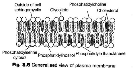
Proteins
Depending upon the ease of extraction, the ratio of protein and lipid varies considerably in different cell types. In human beings, the membrane of the erythrocytes (RBCs) has approximately 52% protein and 40% lipid.
The membrane proteins can be classified as
(i) Integral Proteins (intrinsic protein) They have stronger association and bound firmly to the membrane. These proteins are buried partially or totally in the phospholipid bilayer.
(ii) Peripheral Proteins (extrinsic protein) They have weaker association and are bound to lipids of membrane by electrostatic interactions.
Carbohydrates
These constitute about 1-5% of chemical composition of plasma membrane. These are associated with the phospholipids or with the peripheral proteins to form glycolipids and glycoproteins respectively.
To understand the structure of plasma membrane various models are given out of which the most accepted model is Fluid Mosaic Model.
Fluid Mosaic Model
This model was given by Singer and Nicholson (1972). According to this model, the lipid bilayer and integral proteins appear like a mosaic arrangement and the quasi-fluid nature of lipid enables the lateral movement of the proteins within the overall bilayer.
This ability of proteins to move within the membrane indicate the fluidity of the lipid part.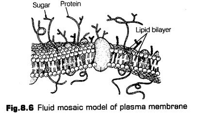
Fluidity of Membrane
The fluid nature of the membrane is important from the point of view of interactions of molecules within the membrane as well as other functions like formation of inter cellular junctions, cell growth, secretion,
endocytosis, cell division, etc.
Passage of substances across the membrane occurs mainly by two methods
i- Active Transport
Active transport is the movement of the molecules across the membrane against their concentration gradient, i.e., from lower to the Tighter concentration. It is an energy dependent process, in which ATP is utilised. It occurs in few ions and molecules,
e.g., Na+ / K+ pump.
Polar molecules requires a carrier protein of the membrane to facilitate their transport across the membrane because they cannot pass through the non-polar lipid bi-layer.
ii- Passive Transport
Passive transport is the mode of movement of molecules or substances across the membrane without any requirement of energy.
It can be further of following three types
(a) Osmosis It is the process by which water molecules pass through a membrane from a region of higher concentration to a lower concentration.
(b) Simple Diffusion In this process, neutral molecules move across the membrane along the concentration gradient (from higher to lower concentration), e.g., Gases and small molecules.
(c) Facilitated Diffusion In this process, the molecules are transported along concentration gradient by the help of ion channels and permeases. Energy is not required in this process.
Differences between Active and Passive Transport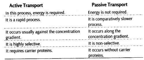
Functions
Cell membrane possess the folbwing functions
(i) It is a selectively permeable or semi-permeable membrane, allows only selected substances to pass inwardly.
(ii) It protects the cell from injury.
(iii) Membranes have carrier proteins for active transport.
(iv) Cell membrane contain enzymes which perform certain reaction on their surface, e.g., ATPase, phosphatase, etc.
Cell Wall
It was first discovered by Robert Hooke (1665). It is a rigid and a non-living structure which forms an outer covering of the plasma membrane in plants and fungi. It is absent in animal cells.
Cell wall is metabolically active in nature and is capable of growth. Its thickness varies from 0.1-10 pm.
Cell wall not only gives shape to cell and protects the cell from mechanical damage and infection, it also helps in cell to cell infraction and provides barrier to undesirable macromolecules.
Chemical Composition
The cell wall of algae is made up of cellulose, galactans, mannans and minerals like calcium carbonate, etc., while cell wall of plant is composed of cellulose, hemicellulose, pectins and proteins.
i.Structure of Cell Wall
On the basis of the structure, cell wall is differentiated into the following three parts
Middle Lamella
It is the layer mainly made up of calcium and magnesium pectates. It cements the cell walls of two adjoining cells together. It is absent on the outer side of surface cells middle lamella along with a cell wall transversed by plasmodesmata which connects cytoplasm of ^ neighbouring cells.
ii. Primary Cell Wall
It is produced inner to the middle lamella in a young and growing cell. It is capable of growth and extension. It tends to diminish gradually as the cell attain maturity.
iii. Secondary Cell Wall
The thick secondary wall is formed inner towards membrane to the primary wall. As the cell gets fully matured. Its composition is similar to the primary wall.
Functions
Cell wall possess the following Junctions
(i) It helps in providing a definite shape to the cell and also protects protoplasm against any mechanical injury, i.e., damage and infection.
(ii) It also helps in cell-to-cell interaction.
(iii) It provides barrier to undesirable macromolecules and attack of pathogens.
Endomembrane System
The endomembrane system consists of nuclear envelope, Endoplasmic Reticulum (ER), Golgi complex, lysosomes and vacuoles suspended in the cytoplasm.
These are considered together as an endomembrane system because their functions are coordinated with each other, inspite of this that each membranous organelles is distinct in terms of its structure and functioning.
Endoplasmic Reticulum (ER)
The endoplasmic reticulum is a complicated system of membranous channels and flattened vesicles. It is physically continuous with the outer membrane of the nuclear envelope. It is revealed from the electron microscopic studies of eukaryotic cells that there is a presence of a network or reticulum of tiny tubular structures that are being scattered in the cytoplasm.
ER is known to be absent in prokaryotes but is present in all eukaryotic cells except germinal cells and mature human RBCs.
Endoplasmic reticulum divides the intracellular space into two main compartments
(i) Luminal (inside ER) compartment
(ii) Extra-luminal (cytoplasm) compartment,
Types of Endoplasmic Reticulum
Endoplasmic reticulum are mainly of two types, depending upon the nature of its membranes
(i) Smooth Endoplasmic Reticulum (SER) These are smooth because they do not bear ribosomes in the form of granules on their surfaces. It is present in cells where they acts as a major site for the synthesis of lipid and also helps in synthesis of steroidal hormone in animal cells.
(ii) Rough Endoplasmic Reticulum (RER) They are found extensive and continuous with the outer membrane of nucleus. These have rough membrane because they bear ribosomes being attached to their surfaces.
They are actively being seen in the cells which have their involvement in the synthesis and secretion of proteins.
Functions
Endoplasmic reticulum possess the following functions
(i) It provides support to the colloidal cytoplasmic matrix.
(ii) Helps in the rapid intracellular transport of the material.
(iii) ER membranes contains a variety of enzymes for various metabolic processes, e.g., ATPase,
phosphatases, etc.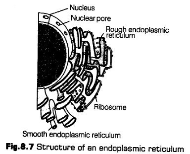
Differences between SER and RER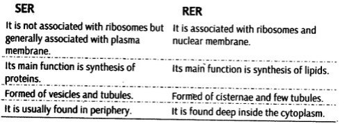
2. Golgi Apparatus
It was first discovered by Camillo Golgi (1898), when he was observing the densly stained reticular structures being present near the nucleus of the cell. These structures were named Golgi bodies after his discovery.
Golgi complex or Golgi apparatus is a major complex protoplasmic structure being made up of many flat, disc-shaped sacs or cisternae (0.5-1.0 nm) in diameter.
Occurrence
Golgi complex, occurs in all cells except prokaryotes (i.e., PPLO, bacteria, cyanobacteria) and some eukaryotes such as human RBCs, sieve tubes of plants, etc.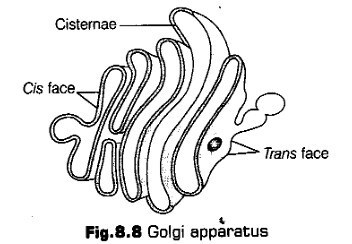
Cisternae of Golgi apparatus are found stacked parallel to each other. They vary in number in a Cell. They are often curved-like a shallow bowls to give Golgi complex a definite polarity.
They are concentrically arranged near the nucleus with two distinct faces
(i) Cis face {forming face) This is convex in shape that lies towards the cell membrane and is responsible for receiving secretory materials through the transitional vesicles, which are pinched off from the SER.
(ii) Trans face (maturing face) This is concave in shape that lies towards the nucleus and is responsible for releasing the material, which is being secreted by cis face and modified in the cisternae.
Note:
* Although, the cis and the trans faces of the organelle are entirely different in origin, but they inter connect each other.
* Proteins that are synthesised by ribosomes on ER are first modified in cisternae before they released from its trans face.
The Golgi apparatus acts as a site where the material to be released is being packaged in the form of vesicles delivered either to the intracellular targets or secreted outside the cell.
Functions
Golgi apparatus possess the following functions
(i) The Golgi apparatus is involved in the formation of lysosomes, vesicles that contain proteins and remains within the cell.
(ii) It performs the function of packaging material.
(iii) It acts as an important site for the formation of glycoproteins and glycolipids.
(iv) It helps in the production of complex carbohydrates other than glycogen and starch.
(d) It helps in the formation of cell wall.
3. Lysosomes
These are membrane bounded vesicles that are produced by the Golgi apparatus. They are rich in several hydrolytic digestive enzymes (hydrolases-lipases, proteases, carbohy- drases, etc). As these are optimally active at the acidic pH (less than 7). Therefore, are also called acid hydrolases and are capable of digesting macromolecules from various sources like carbohydrates, lipids and nucleic acids.
Functions
Lysosomes possess the following junctions
(i) They digest the food contents (intra cellular digestion).
(ii) They also perform extracellular digestion.
(iii) They also digest the old and useless organelles of the cells.
(iv) They also have functioning in cell division.
These are called suicidal bags due to the presence of hydrolytic enzymes.
De Duve observed the rounded bodies in liver cells and called them pericanalicular dense bodies (1949).
4. Vacuoles
Vacuole are a large membranous sac found in the cytoplasm. These store substances that are not essentially useful for the cell (like water, sap, excretory product and other materials). Plant vacuoles contain not only water, sugars and salts but also contain pigments and toxic molecules and also occupy up to 90% of the volume of the cell.
The vacuole is bounded by a single membrane structure known as tonoplast which in plant cells, facilitates the transport of materials and some ions against the concentration gradient inside the vacuole. Thus, the concentration of material is tend to be the higher in vacuole, than to be in the cytoplasm
Animal cells also have vacuole, but they are much more prominent in case of plant cells. Thus, plant cells have typically large central vacuole filled with a watery fluid that gives added support to the cell.
Following types of vacuoles are being found in different organisms
(i) Contractile Vacuole They play an important part in osmoregulation and excretion in Amoeba, etc. It occurs mosdy in protistan and algal cells that are found mainly in water.
(ii) Food Vacuole They occur in the cells of mainly protozoan protists. These are formed by engulfing the food particles, i.e., by the fusion of lysosome and phagosome. The digested material thus, passes out into the surrounding cytoplasm.
Air vacuoles and sap vacuoles are the another types of vacuoles being formed by the cells.
Mitochondria
Mitochondria are membrane bound cell organelles, essential for aerobic respiration of eukaryotic cells. These are also known as power house of the cell. Thus, they produces cellular energy in the form of ATP.
Occurrence
Mitochondria are present in all living cell except, prokaryotic cell and certain specialised eukaryotic cell such as anaerobic cells and mature RBCs.
It is revealed from the studies that mitochondria is not easily visible, unless it is specifically stained.
Shape and Size
Mitochondria vary considerably according to the shape and size. They have varying shape such as granular fibrillar, spherical, oval, discodial, etc. Average size of mitochondria is 2-6 |im in length and 0.5 pm in diameter (typical cylindrical or sausage-shaped mitochondria has diameter of 0.2-1.0 Jim).
Ultrastructure
A mitochondrion contains two membranes, i.e., outer and inner. Out of which outer membrane is smooth and forms the continuous boundary of the organelle. The inner membrane is semipermeable to some metabolities. It is infolded into the matrix as incomplete partitions called cristae.
The cristae are responsible for increasing the physiological active area or surface area. The density of cristae determines the intensity of respiration.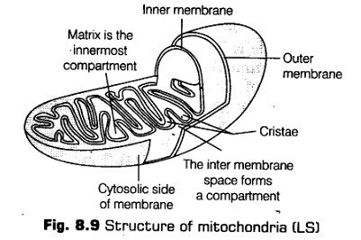
The outer and the inner membranes divides its lumen into two aqueous compartments separately, i.e., the outer and the inner compartment.
Inner compartment is also called matrix, which forms the inner core of the mitochondrion. The matrix also possesses single circular DNA molecule, a few RNA molecules, ribosomes (70S) and the components required for the synthesis of proteins. The mitochondria divide by fission.
The two membranes of mitochondria have their own specific enzymes associated with mitochondrial function.
Functions
Mitochondria possess the following functions
(i) Mitochondria provide important intermediates for the synthesis of several biochemicals like pyrimidines, alkaloids, etc.
(ii) The inner chamber matrix of the mitochondria has enzyme for the synthesises of fatty acids.
(iii) Helps in regulation of cellular metabolism.
(iv) Helps in apoptosis (programmed cell death).
(v) Each of membrane potential.
Mitochondrion is the second largest cell organelle and are more in animal cells than in plant cells.
Plastids
These are semi-autonomous organelles that have double membrane envelope. Plastids have their own genetic material (i.e., DNA). Due to their large size, they are easily seen under the microscope.
Occurrence
Plastids are found in all plant cells and euglenoides except in some protistans (e.g., Euglena, Dinophyceae, etc).
Types
Plastids are differentiated into three different types on the basis of the colour, i.e., type of pigments found in them.
Leucoplasts
These are the colourless plastids of varied shapes and sizes with stored nutrients in the form of carbohydrates lipids and proteins.
These are of following three types
(а) Amyloplasts are the carbohydrates (starch) containing leucoplast, e.g., Rice, wheat, potato, etc.
Amyloplasts are larger than the normal/original size of leucoplast.
(b) Elaioplasts are the leucoplast which stores oils and fats, e.g, Tuberose endosperm of castor seeds, etc.
(c) Aleuroplasts are the protein storing leucoplast.
e.g., Maize (aleurone cells).
ii. Chromoplasts
These are the leucoplast, whioh are yellow or reddish in appearance because of the presence of fat soluble carotenoid pigment carotene.
Xanthophyll and some other pigments are also present as the fat soluble carotenoid pigment other than carotene, e.g., Orange colour of carrot, etc.
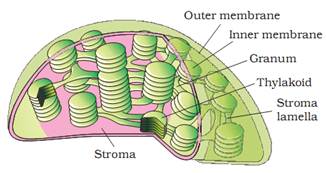
iii. Chloroplasts
These are the plastids which are greenish in colour containing photosynthetic pigments chlorophyll and carotenoids. These pigments are responsible for trapping the light energy, essential for the photosynthesis, i.e., the synthesis of organic food from an inorganic raw materials in the presence of sunlight.
Occurrence
Chloroplasts occur in major number in the photosynthetic mesophyll cells of leaves and green stem.
Shape and Size
• They may be lens-shaped, oval, spherical, discoid or even ribbon-like organelles. They also have variable length (5-10 mm) and width (2-4 mm).
Number
Their number also varies from one per cell of the Chlamydomonas (a green alga) to 2-40 per cell in mesophyll.
infrastructure
Chloroplasts are also bounded by double membrane envelope like mitochondria, the two membranes are smooth and are thick of about 90-100 A. The inner membrane of chloroplast is less permeable than the other one.
The inner membrane is grounded by a space known as stroma or matrix, a dense, colourless and a granular substance mainly formed of soluble proteins. It also contains enzymes which are essential for the synthesis of carbohydrates, lipids and proteins.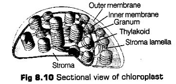
Thylakoids are number of membranous like flattened structures that run throughout the matrix or stroma. When several thylakoids are arranged or organised in the stack (like the piles of coins), called grana or the intergranal thylakoids. Many flat membranous tubules interconnect the thylakoids of different grana known as stroma lamellae.
Functions
Chloroplasts possess the following functions
(i) Helps in photosynthesis, i.e., formation of organic compounds.
(ii) In consumption of C02 and release of 02 in photosynthesis.
(iii) May also change into chromoplast in order to provide colour to many flowers and fruits.
(iv) Helps in storing fat and lipids.
(v) Functions in transduction of energy.
Note:
* The sum total of all plastids in a cell is called plastidome.
* The chloroplast with nitrogen fixing genes are called nitroplast.
* The space between the two membrane is called intermembrane space, which separates the two membrane. This space contains a narrow fluid. Stroma also contains small, double-stranded circular DNA, molecules and ribosomes.
* Ribosomes of chloroplasts are smaller (70S) than the ribosomes of cytoplasm (80S).Ribosomes
These are the small sub-spherical granular organelles, not bounded by any membrane. Ribosomes were first observed by George Palade (1953), as the dense particles under the electron microscope. Hence, are also called Palade particles.
Ribosomes are mainly composed of ribonucleoproteins (i. e., RNA-t- proteins) and are also known as protein factories,
as they are primarily involved in the synthesis of proteins or polypeptides.
As studied earlier, the prokaryotic ribosomes are 70S type, while the eukaryotic ribosomes are 80S type. Here, ‘S’ (Svedberg’s unit) stands for sedimentation coefficient (measure of density and size).
Both 70S and 80S ribosomes contains two sub-units, i.e., the smaller and the larger sub-unit .
Differences between 70S and 80S Ribosomes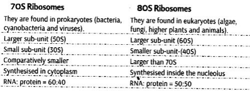
Cytoskeleton
The network of interconnected proteinaceous filaments and tubules, which extends from the nucleus to the plasma membrane in eukaryotic cells.
Functions
Cytoskeleton possess the following functions
(i) The cytoskeletal structures maintain the shape of the cell and its extensions.
(ii) It is also involved in many functions in a cell as mechanical support, motility, etc.
Cilia and Flagella
These are hair-like projections of cell membrane. Both cilia and flagella are almost identical in structure but differ somewhat in length. As cilia are small structures, working as oars (causing the movement of either the cell or the surrounding fluid), while flagella are comparatively longer in size than cilia and are responsible for the movement of cell.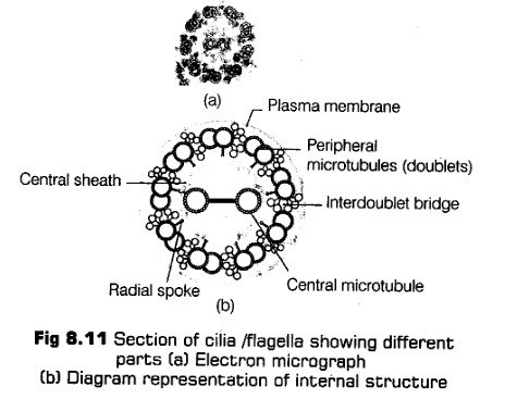
According to the electron microscopic studies it is predicted that the cilium or the flagellum are covered with plasma membrane. Their core called the axoneme, contains a number of microtubules, running parallel to the long axis.
Usually, the axoneme has nine pairs of doublets of peripheral microtubules that are radially arranged and a pair of centrally located microtubules. This arrangement of axonemal microtubules is referred to as the (9 + 2) array.
The central tubules are connected by bridges and is also enclosed by a central sheath, which is connected to one of the tubules of each peripheral doublets by a radial spoke.
Thus, it has been estimated that there are nine radial spokes. The peripheral doublets are also interconnected by linkers. Both the cilium and flagellum emerge from centriole like structure called the basal bodies.
Differences between Cilia and. jrgella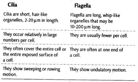
Flagella are also present in prokaryotic bacteria but these are ’ structurally different from that of eukaryotic flagella.
Centrosomes and Centrioles
Centrosome is an organelle that generally have two cylindrical structures known as centrioles. They are basically surrounded by an amorphous pericentriolar materials.
Both the centrioles in a centrosome lie perpendicular to each other in which each has an organisation like the cartwheel.
They are usually made up of nine evenly spaced peripheral fibrils (triplet in nature) of tubulin protein. With which adjacent triplets are also being linked.
The centre part of the proximal region of the centriole possess rod-shaped proteinaceous mass known as hub, which is connected with tubules of the peripheral triplets fibrils known as radial spokes (made up of protein).
From the basal body of cilia or flagella the centrioles and spindle fibres give rise to spindle apparatus during cell division in animal cells.
Functions
Centrosomes and centrioles possess the following functions
(i) These forms spindle fibres and move to the poles, at the time of cell divisidn, which thus, help in the movement of chromatids in daughter cells.
(ii) Help in the formation of cilia and flagella of the cells.
Nucleus
It is a specialised and principle cell organelle of the cell, which contains all the genetic information for controlling all essential processes related to metabolism and transmission.
Nucleus was first described by Robert Brown as early as 1831.
Later the name chromatin was given by Flemming when the material of the nucleus was stained by the basic dyes.
Nucleus is known to be the largest cell organelle also known as brain of the cell.
Occurrence
A nucleus is known to be present in all eukaryotic cells except a few cell types such as RBCs of humans, sieve cells of vascular plants, etc.
Prokaryotic cell lack a well organised nucleus, instead they have a nucleoid.
Ultrastructure
The interphase nucleus (nucleus of a cell when it is not dividing) has highly extended and elaborate nucleoprotein fibres called chromatin, nuclear matrix and one or more spherical bodies called nucleoli.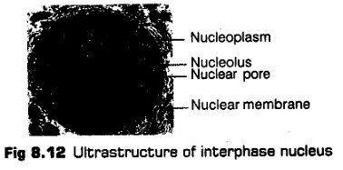
Microscopic Structure
It has been revealed from the studies of electron microscopy that the nuclear envelope, consists of two parallel membranes with a space between 10-50 nm called the perinuclear space, which forms a barrier between the materials present inside the nucleus and that of the cytoplasm.
The outer membrane usually bears ribosomes on it andremains continuous with the endoplasmic reticulum. The nuclear envelope is interrupted by minute nuclear pores, at a number nuclear of places, which are produced by the fusion of its two membranes. These nuclear pores are the passages through which movement of RNA and protein molecules takes place in I both directions between the nucleus and the cytoplasm.
Normally, there is only one nucleus per cell, but variations in the number of nuclei can also be seen in various organisms.
Nucleus is differentiated into following four parts
i. Nuclear Envelope
It is a double membrane bound envelope that surround the nucleus and separates the latter from the cytoplasm.
ii. Nucleoplasm
It is a clear, non-staining, fluid material present in the nucleus, which contains raw materials (nucleotides), enzymes (DNA/RNA polymerases) and metal ions for the synthesis of RNAs and DNA. The nuclear matrix or the nucleoplasm is composed of nucleolus and chromatin (spherical structures present in the nucleoplasm).
iii. Nucleolus
It is a naked, round and slightly irregular structure, which is attached to the chromatin at a specific region. The content of nucleolus is continuous with the rest of the nucleoplasm as it is not a membrane bound structure.
It is a site for active ribosomal RNA synthesis. Larger and more numerous nucleoli are present in cells actively carrying out protein synthesis.
Chromatin
It is named so, because it has the ability to get stained with certain basic dyes. It is known to be the hereditary DNA protein fibrillar complex. The chromatin fibres are distributed throughout the nucleoplasm.
It has two distinct regions
(a) Euchromatin (lightly stained)
(b) Heterochromatin (darkly stained)
Functions
Nucleus possess the following functions
(i) It stores information that control cellular functions.
(ii) It controls the synthesis of structural proteins.
(iii) It also stores the genetic information for development reproduction and behaviour.
(iv) It also induces genetic variations.
Chromosomes
It has been already studied in the chapter that the nucleus in the interphase has a loose and indistinct network of nucleoprotein fibres called chromatin. But during different stages of*cell division cells show structured chromosomes in place of the nucleus. The chromosomes are meant for the equal distribution of genetic material. Their number is fixed and is same in all individuals of a species.
Chromatin is composed DNA and some basic proteins called histones. Some non-histone proteins and RNA are also present in the chromatin.
A single human cell has approximately two metre long thread of DNA distributed among its 46 (23 pairs) chromosomes.
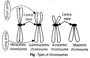
Each and every chromosome is composed of a primary constriction or the centromere. On the sides of which the disc-shaped structures are present known as kinetochores.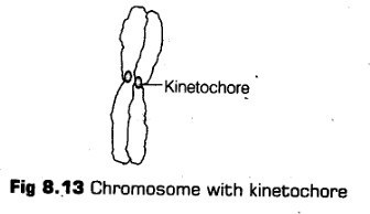
On the basis of the position of the centromere, the chromosomes can be classified into four following types
i. Metacentric Chromosome
It has chromosome with equal arms and centromere lies in the centre.
ii. Sub-metacentric Chromosome
It has one shorter arm and one longer arm with centromere slighdy away from the middle of the chromosome.
iii. Acrocentric
It forms one extremely short and one very long arm and centromere is located near the end of the chromosome.
iv. Telocentric
It has the terminal centromere, i.e., centromere is placed at an extreme end.
Telocentric chromosomes are not present in humans.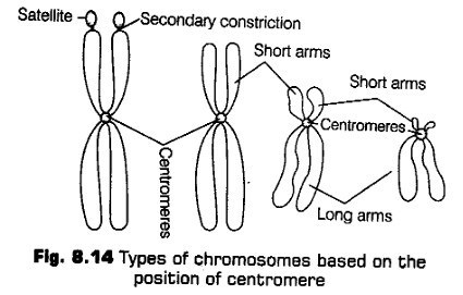
Few chromosomes have a non-staining secondary constrictions being present at a constant location at some or the other time which gives the appearance of a small fragment known a satellite.
Functions
Chromosomes possess the following functions
(i) Control cellular differentiation.
(ii) Contains all hereditary information located in the genes.
(iii) Forms a link between the offspring and the parents.
(iv) Introduce variations, through the process of crossing over.
(v) Control cell metabolism.
Microbodies
These are the membrane bound cytoplasmic elements that are composed of enzymes and other substances. These are minute vesicles found in both plant cells and animal cells, e.g., In the liver, kidney, Protozoa, yeast and many other types of cells. Their shape can be ovoid, spherical, granular, etc.
Peroxisomes and glyoxysomes are the types of microbodies being found in plant cell and animal cell respectively.
CBSE Class 11 Biology Chapter-8 Important Questions
1 Marks Questions
1.Define totipotency?
Ans. Each vegetative plant cell has capacity to develop into a full plant. This characteristic of plant is called totipotency.
2.Name two cell organelles which contain their own DNA?
Ans. Mitochondria & chloroplast.
3.Which cell organelle functions as “seggregation apparatus”?
Ans. Endoplasmic Reticulum (ER)
4.Which structure is called little nucleus?
Ans. Nucleolus.
5.What is the function of contractile vacuole?
Ans. Water balance or osmoregulation.
6.Name the enzymes present in peroxysomes?
Ans. Catalase & B- hydroxyoxidase.
7.Who gave the statement “Omnis cellular cellula”?
Ans. Rudolf Virchow.
8.Which organelle is called the engine of the cell?
Ans. Ribosomes where protein synthesis occurs
9.What is mycoplasma ?
Ans. Mycoplasma is aerobic prokaryote. Cell wall is absent in them & they have a nucleoid.
10.Why is karyotype done at metaphase?
Ans. Because metaphase chromosomes with two chromatids strands of each double chromosome held together at the centromere are clearly seen.
11.Expand PPLO.
Ans. Pleuropneum onia like organisms.
12.Name the parts of bacterial flagella.
Ans. Filament, hook, basal body.
13. What do elaioplasts and aleuroplasts store?
Ans. Elaioplasts: fats and oils.
Aleuroplasts: proteins.
14.Who first saw and described a live cell?
Ans. Anton Von Leeuwenhoek
15.Which is the largest single cell?
Ans. Egg of ostrich.
16.Who firs explained that Cell arose from pre-existing cells?
Ans. Rudolf Virchow.
17.What is the composition of plasma membrane of human erythrocyte.
Ans. 52% proteins, 40% lipids.
18.Eukaryotic ribosome are 80S. What does ‘S’ stand for.
Ans. Sedimentation coefficient.
19.What is the function of cytoskeleton in a cell?
Ans. Mechanical support, motility, maintenance of shape of cell.
2 Marks Questions
1.Give two examples of gram positive bacteria?
Ans. Mycobacterium & clostridium tetani.
2.What is the significance of plasma membrane?
Ans. Significance of plasma Membrane:-
(i)It forms the outer boundary of cell thus giving cell a definite shape
(ii) It protects inner contents of the cell.
(iii) It forms a molecular boundary between cell & its environment.
3.Differentiate between gram positive and gram negative bacteria?
Ans.
| GRAM-POSITIVE BACTERIA | GRAM-NEGATIVE BACTERIA |
| i) Their cell wall is only single layered & 100-200 A0 thick. | i) Their cell wall consists of two layers & is 70-120 A0 in thickness. |
| ii) They are stained by gram stain | ii) They are not stained by gram stain |
| iii) They do not have pilli. | iii) They have pilli |
| iv) Mesosomes present | iv.) Mesosomes absent |
4.Why lysosomes are called “suicidal bags”?
Ans. Lysosomes are sac-like structures bounded by a single membrane which contains several digestive enzymes. These enzymes when released from lysosomes bring about breaks down of various cytoplasmic structures. It helps in digestion of food particles, other foreign bodies, old worn out organelles of cell often resulting in death of cell hence are referred as suicidal bags of cell.
5.Explain the functions of centrosome?
Ans. Function of Centrosomes :-
(a) Centrioles form basal bodies.
(b)At the time of cell division, they organize spindle and form asters.
(c) They give rise to cilia and flagella.
(d) Out of the two centrioles, the distal centrioles of sperms forms the axial filament or axoneme of sperm tail.
6.What is meant by active transport across a cell membrane?
Ans. When molecules moves from a region of lower concentration to a region of higher concentration i.e. against concentration gradient, the process is known as active transport. The energy is required for the movement of molecules or ions in opposite direction. The enzyme responsible for the pumping of compounds into or out of cell believed to be a component of the membrane eg. Na+– K+ pump.
7.“Both lysosomes & vacuoles are endomembrane structures yet they differ in terms of their functions” comment.
Ans. Lysosomes & the vacuoles are endomembranous structures yet these differ in terms of their functions:-
(i)Lysosomes contains hydrolytic enzymes eg. lipase, protease which are able to digest lipids, proteins, nucleic acid & carbohydrate.
(ii)Vacoules are membrane bound spaces which facilitates transport of many ions & other materials against the concentration gradient.
8.Who proposed cell theory? Give its postulates?
Ans. M. J. Scheilden & Theodore Schwann gave the famous cell theory which states as follows:-
(i) All living things are made of cells & cell products.
(ii)The cell is the structural & functional unit of all living organisms.
(iii) All metabolic reactions in the living things take place with in the cell
The cell theory was later modified by Rudolf Virchow who stated that “all new cells arise from the pre- existing cells”.
9.Which cell organelle is known as powerhouse of cell & why?
Ans. The double membrane mitochondria are actively associated with aerobic respiration & the release of energy for cellular activity. The biological oxidation of the fats & carbohydrates release much amount of energy which is utilized by mitochondria for ATP synthesis. When required energy is released form ATP molecules for various cell processes in cells so they are termed as “Power house of the cell”
10.What are the main functions of cell wall?
Ans. FUNCTIONS OF CELL WALL:-
(i)It provides a definite shape to the cell.
(ii)It protects inner contents of cells
(iii)It protects delicate plasma membrane present below it.
(iv)It allows transport of various substances to & from the cell.
(v) It prevents cell contents from drying up.
11.State differences between SER & RER?
Ans.
| SER | RER |
| i) SER do not have ribosomes & is composed of vesicles & tubules | i) RER have ribosomes on its outer surface & is composed of cisternal |
| ii) It synthesizes steroids & lipids eg. fat cell lipid secretory cells of liver | ii) Its main function is protein synthesis due to the presence of ribosomes. |
| iii) Gives rise to sphaerosomes | iii) Gives rise to Golgi bodes, vacuoles as well as lysosomes. |
| iv) Free of ribosomes. | iv) Bears ribosomes. |
12.What are nuclear pores? State their functions?
Ans. Nuclear envelope contains two parallel membranes & the thickness is 10-50 nm. Outer membrane has small pores called the nuclear pores formed by fusion of two membranes. These pores are the passages through which movement of RNA & protein molecules occurs in both directions between nucleus & cytoplasm.
13.Give differences between cell wall & cell membrane?
Ans.
| CELL WALL | CELL MEMBRANE |
| i) present in plant cell exclusively | i) present predominantly in animal cells |
| ii) Made up of cellulose | ii) Made up of proteins fats & water |
| iii) Thick & tough in nature | iii) Extremely thin & elastic in nature |
| iv) Thickening of various kinds present | iv) No thickenings |
| v) it is not selectively permeable | v) selectively permeable membrane |
14.Which organelle is responsible for increasing the surface area of absorption in a cell? How?
Ans. The endoplasmic reticulum is responsible for increasing the surface area for absorption. It remains in the form of convulated tubule in the cytoplasm in the form of network. This provides more area for chemical reactions and increases the surface area of absorption.
15.What is mesosome in a prokaryotic cell? Mention the function that it performs?
Ans. Mesosome in a prokaryotic cell is formed by extensions of plasma membrane into the cell it may be in form of vesicle, tubule or lamella. They help in cell wall formation. They help in replication of DNA & distribution of it to daughter cells. They help in secretion respiration, & increase plasma membrane surface area.
16.“plasma membrane is described as” protein iceberg in sea of lipids”. why ?
Ans. The plasma membrane as described by singer & Nicolson is of fluid mosaic model type. The lipid & proteins are arranged in a mosaic fashion. The matrix is highly viscous fluid of two layers of phospholipids molecules having two types of globular proteins i) peripheral or extrinsic proteins & ii) integral or intrinsic proteins. The proteins present superficially or tightly with the membrane are enzymatic can move across the matrix & help in the active & passive transport of ions through the membrane.
17. What are nuclear pores ? State their function.
Ans. Minute pores present in the nuclear envelope; provide passage for movement of RNA and proteins between nucleus and cytoplasm.
18. Differentiate between the electron microscopic structure of cell/flagella and centriole.
Ans.
| Flagella/Cilia | Centriole |
i)Possess (9+2) pattern of axoneme microtubules enclosed by a membrance ii) Each tubule is a doublet | i) Possess (9+0) pattern, membrace less organelle ii) Each tubule is a triplet |
19. Give the specific terms for the following
(a)Cluster of ribosome’s found in cytoplasm
(b)Extensive in folding to the inner membrane of mitochondria.
(c) Stacks of closely packed thylakoids
(d)Stalked particles on the inner membrane of mitochondria.
Ans. (a) Polyribosome/polysome
(b) Cristae
(c) Grana
(d) Fe F particles
20. (a) What is the function of inclusion bodies in prokaryotic cells?
(b)Where are they present?
(c) Give two examples of inclusion bodies.
Ans. (a) Reserve materials att stored.
(b) They alt free in the cytoplasm
(c) e. g., Phosphate granules, cyanophycean granules, glycogen granules.
3 Marks Questions
1.Describe the ultrastructure of a cillium or flagellum?
Ans. Cilia & flagella have fundamentally the same structures. Each cilium or flagellum consists of eleven microtubules. These microtubules are arranged in two radii. Of these, nine are doublets. These are situated at the periphery & the remaining two are single microtubules situated in the centre. The microtubules are enclosed in a cytoplasmic matrix to form an axial filament. The outer tubules are 360 A0 in diameter & are composed of two sub- units. The smaller of these have two arms in A- tubule & the smaller is B- tubule. These are found around the cylinder. The central microtubules are enclosed in a common sheath. From the centre arise nine secondary filaments. These are connected with tubules of the outer doublets.
2.Distinguish between prokaryotic & eukaryotic cell?
Ans.
| PROKARYOTIC CELL | EUKARYOTIC CELL |
| i) It lacks well organized nucleus. The genetic material is present in the form of nucleoid. | i) Nucleus is well developed. |
| ii) DNA is in circular form & is not packed into chromosomes. | ii) Linear DNA packed into chromosomes |
| iii) Nuclear membrane is lacking | iii) Nuclear membrane is present. |
| iv) Mitochondria absent | iv) Mitochondria present. |
| v) Chloroplast absent | v) Chloroplast is present in plant cell only. |
| vi) Membrane bound organelles are absent | vi) Membrane bound organelle are present. |
| vii) The ribosomes are of 70stype | vii).The ribosomes are of 80s type |
| viii) Cell wall consist of mucoptides | viii) Cell wall is absent in animal cells in plant cell, cell wall is made up of cellulose, hemicelluloses, lignin etc. |
| ix) Flagella are simple | ix) Flagella are specialized. |
3.Explain the fluid mosaic model of plasma membrane.
Ans. The fluid mosaic model was proposed by G.Nicholson & s. singer. According to this each phospholipids layer is bimolecular & their hydrophilic ends are pointed towards top & bottom respectively.
In this, proteins are of two categories- peripheral (extrinsic) & integral (intrinsic). The integral proteins are tightly held in place by strong hydrophilic or hydrophobic interactions or both and are difficult to remove from the membranes. Two peripheral proteins are superficially arranged on either side membrane selectively permeable thus this model explains cell membrane is quasifluid & is made up of “protein icebergs in the sea of lipids”.
4.Describe the structure of a typical eukaryotic chloroplast.
Ans. Chloroplasts are bounded by two membranes, about 3000 A0 in total thicknesses. Each membrane is 40-60 A0 thick. The inner membrane is very intricately elaborated to form a system of lamellae. Internally the chloroplasts is divisible into two parts
(a) stroma- colourless, ground substance
(b)Membrane system- made of closed flattened sacs called thylakoids. These thylakoids are closely packed & appears as piles of coins. These structures are called Grana. The arrangement can be in the form of simple parallel sacs running lengthwise, or may be in a complex interconnecting network of the sacs. The chloroplasts invariably have some starch granules which often accumulate near a special region known as pyrenoid in algae
5.Mention three similarities & three differences between mitochondria & chloroplasts?
Ans. SIMILARITIES BETWEEN MITOCHONDRIA & CHLOROPLAST
(i)Mitochondria & chloroplasts are semi-automous organelle & they possess their own DNA, RNA as well as ribosomes.
(ii)They both develop & originate in the same way, formed by division of pre-existing organelle
(iii)Both of them contain circular DNA.
DIFFERENCES BETWEEN MITOCHONDRIA & CHLOROPLAST
(i)Mitochondria occurs in all eukaryotic cells while chloroplast are present only in plant cells.
(ii) Pigments are absent in mitochondria but always present in chloroplast.
(iii)The inner membrane of mitochondria are folded into cristae where as cristae are absent in chloroplast.
6.“multicellular organisms have better survival than their cellular counterpart” why?
Ans. In unicellular organisms, there is no division of labour. The single cell of the organism is capable of performing all the vital activities of life respiration, movement, digestion & reproduction etc. Respiration, nutrition & excretion generally occur through general body surface no special organs for these are present in them because they are too small to need them.
In multicellular organisms all the body cells do not perform all the vital activities of life rather these cells play more specialized role in life activities eg. some cells of the body perform the function of movement some perform the function of digestion or respiration or removal of wastes from the body some cells perform the function of transport. These cells would perform no other function except for which they are specialized. The group of similar cells performing similar function is termed as tissues.
7. Differentiate between a prokaryotic and eukaryotic cell.
Ans. The distinction betweenprokaryotesandeukaryotesis considered to be the most important distinction among groups of organisms. Eukaryotic cells contain membrane-bound organelles, such as the nucleus, while prokaryotic cells do not. Differences in cellular structure of prokaryotes and eukaryotes include the presence of mitochondria and chloroplasts, the cell wall, and the structure of chromosomal DNA .
Prokaryotes were the only form of life onEarthfor millions of years until more complicated eukaryotic cells came into being through the process of evolution.
NCRT TEXTBOOK QUESTIONS SOLVED
1.Which of the following is not correct?
(a) Robert Brown discovered the cell.
(b) Schleiden and Schwann formulated the cell theory.
(c) Virchow explained that cells are formed from pre-existing cells.
(d) A unicellular organism carries out its life activities within a single cell.
Soln.(a) Robert Hooke discovered the celland Robert Brown discovered nucleus in the cell.
2.New cells generate from
(a) bacterial fermentation
(b) regeneration of old cells
(c) pre-existing cells
(d) abiotic materials.
Soln.(c)
3.Match the following.
Column I Column II
(a) Cristae (i) Flat membranous sacs in stroma
(b) Cisternae (ii) Infoldings in mitochondria
(c) Thylakoids (iii) Disc-shaped sacs in Golgi apparatus
Soln.a – (ii); b – (iii); c – (i).
4.Which of the following is correct?
(a) Cells of all living organisms have a nucleus.
(b) Both animal and plant cells have a well defined cell wall.
(c) In prokaryotes, there are no membrane bound organelles.
(d) Cells are formed de novo from abiotic
Soln. (c) Mature mammalian erythrocytes and sieve tube cells of vascular plants lack nucleus. Animals lack cell wall and only cell membrane is present. Prokaryotes are unicellular organisms which lack nucleus and other membrane bound organelles. All cells arise from pre-existing cells.
5.What is a mesosome in a prokaryotic cell? Mention the functions that it performs.
Soln. Mesosome is a membranous structure in prokaryotic cell, which is formed by the extensions of the plasma membrane into the cell in form of vesicles, tubules and lamellae. Mesosomes are equal to mitochondria in eukaryotes, as they perform aerobic cellular respiration in prokaryotes. It helps in DNA replication and distribution of genetic material to daughter cells. Mesosomes also help in respiration, increase the surface area of the plasma membrane and enzymatic content and cell wall formation.
6.How do neutral solutes move across the plasma membrane? Can the polar molecules also move across it in the same way? If not, then how are these transported across the membrane?
Soln. Neutral solutes move across the membrane by the process of simple diffusion along the concentration gradient i.e., from higher concentration to the lower concentration. Polar molecules cannot pass through the nonpolar lipid bilayer, they require carrier proteip of the membrane to facilitate their transport across the membrane. In facilitated diffusion, molecules are transported along concentration gradient by help of ion channels and permeases and it does not involve energy expenditure (passive transport).
7. Name two cell-organelles that are double membrane bound. What are the characteristics of these two organelles? State their functions and draw labelled diagrams of both?
Solution: Mitochondria and chloroplast are double membrane bound organelles. Mitochondria: Mitochondria are cylindrical or sausage shaped cell organelles and contains two membranes, outer and inner. The inner compartment is called the matrix containing DNA, RNA, ribosomes, enzymes of Krebs cycle etc and outer membrane forms the continuous limiting boundary of the organelle. Inner membrane forms number of infoldings called the cristae which increases the surface area. Oxysomes are present on inner mitochondrial membrane. Mitochondria are semiautonomous organelles, i.e., have their own DNA and ribosomes.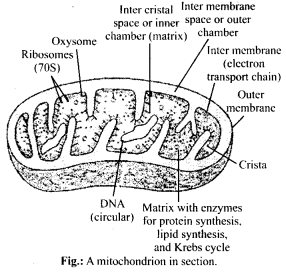
Functions of mitochondria:
- Mitochondria are essential for aerobic respiration.
- Mitochondria provide intermediates for synthesis of important biomolecules such as chlorophyll, cytochrome, steroids etc.
- Mitochondria regulate the calcium ion concentration in the cell.
- Mitochondrial matrix contains enzymes for the synthesis of fatty acids.
- Synthesis of many amino acids takes place here
Chloroplast: They are green coloured plastids which are disc shaped. The space limited by inner membrane of chloroplast is called as stroma. Stroma has organised flattened membranous sacs called the thylakoids. Thylakoids are arranged in stacks called grana. Matrix of a chloroplast contains DNA, RNA, ribosomes and enzymes. Chloroplast is also a semiautonomous organelle.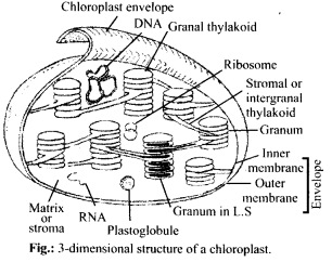
Functions of chloroplast:
- Photosynthesis is performed by chloroplast.
- Chloroplast stores starch grains.
- Maintains balance of C02 concentration in the air.
- Keeps oxygen balance constant in atmosphere by liberating 02 into the atmosphere, used during respiration and combustion.
8.What are the characteristics of prokaryotic cells?
Solution: Characteristics of prokaryotic cells are as follows:
- The prokaryotic cell is essentially a single – envelope system.
- Prokaryotes lack membrane bound cell organelles.
- Prokaryotes have 70S ribosomes.
- DNA is naked and lies coiled in cytoplasm. It is not covered by nuclear membrane and is termed as nucleoid.
- Nuclear components, like, nuclear envelope, nucleolus, nucleoplasm are absent.
- Cell wall is present in bacteria and cyanobacteria, but absent in mycoplasma.
- Multiplies by asexual reproduction.
- Transcription and translation takes place in cytoplasm.
9.Multicellular organisms have division of labour. Explain.
Soln. Division of labour is differentiation of certain components or parts to perform different functions for increased efficiency and higher survival. Multicellular organisms often possess millions of cells. Various cells are grouped together to form specific tissue, organ or organ system, with each specialised to perform particular function. Every cell of a multicellular organism cannot obtain food from outside. The organism requires a system for obtaining food, its digestion and distribution. Therefore, a digestive system and system of transport are also required. Certain cells of the body take over the function of reproduction. Others take part in repair and replacement of worn out or injured portions. For optimum functioning of cells, a multicellular organism also requires an internal favourable environment. Therefore, multicellular organisms come to have division of labour.
10.Cell is the basic unit of life. Discuss in brief.
Soln. Cell is fundamental, structural and functional unit of life, as no living organism can have life without being cellular. All life begins as a single cell. An organism is either made of single cell (unicellular) or many cells (multicellular). In unicellular organism, single cell is capable of independent existence and perform all essential functions of life, while in multicellular organism, each group of cells is specialised for specific function. Life passes from one generation to the next in form of cells, and new cell always arise from division of pre-existing cells. Cells are totipotent, i.e., single cell has ability to form whole organism. The activities of an organism are sum total of activities of its cells, therefore, cell is the basic unit of life.
11.What are nuclear pores? State their function.
Soln. Nuclear envelope bounds the nucleus from outside and separates it from cytoplasm. It consists of two membranes, with outer membrane continuous with endoplasmic reticulum. The nuclear envelope is interrupted by minute nuclear pores, at a number of places, which are produced by the fusion of its two membranes. These
nuclear pores are the passages through which movement of RNA and protein molecules takes place in both directions between the nucleus and the cytoplasm.
12.Both lysosomes and vacuoles are endomem-brane ‘structures, yet they differ in terms of their functions. Comment.
Soln. Organelles of endomembrane system such as lysosome and vacuoles function in close coordination with one another but are specialised to perform different functions. Lysosomes breakdown the ageing and dead cells, they help in digestion of food as they contain hydrolytic digestive enzymes. They are involved in cell division also. Vacuoles on other hand, help in excretion and osmoregulation in Amoeba (contractile vacuole) or provides buoyancy, mechanical strength in prokaryotes (air vacuoles).
13.Describe the structure of the following withthe help of labelled diagrams.
(i) Nucleus (ii) Centrosome
Soln.(i) Nucleus: Nucleus is double membrane bound principle cell organelle which contains all genetic information for controlling cellular metabolism and transmission of genetic information.
Nucleus is differentiated into following four parts:
(a) Nuclear envelope: It is a double membrane bound envelope that surround the nucleus and separates the latter from the cytoplasm.
(b) Nucleoplasm: Itis clear, non-staining, fluid material present in the nucleus, which contains raw materials (nucleotides), enzymes (DNA/RNA polymerases) and metal ions for the synthesis of RNAs and DNA. The nuclear matrix or the nucleoplasm is composed of nucleolus and chromatin.
(c) Nucleolus: It is a naked, round and slightly irregular structure, which is attached to the chromatin at a specific region. It is a site for active ribosomal RNA synthesis.
(d) Chromatin : It has the ability to get stained with certain basic dyes. It is known to be the hereditary DNA protein fibrillar complex. The chromatin fibres are distributed throughout the nucleoplasm.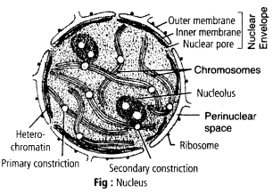
(ii) Centrosome: Centrosome is an organelle usually containing two cylindrical structures called centrioles. They are surrounded by amorphous pericentriolar materials. Both the centrioles in a centrosome lie perpendicular to each other. They are made up of nine evenly spaced peripheral fibrils of tubulin protein. Each of the peripheral fibril is a triplet. The adjacent triplets are also linked. The hub of centriole is connected with tubules of the peripheral triplets by radial spokes made of protein.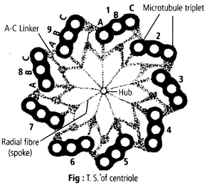
14.What is a centromere? How does the position of centromere form the basis of classification of chromosomes. Support your answer with a diagram showing the position of centromere on different types of chromosomes.
Soln. A chromosome consists of two identical halves, the chromatids held together at one point called the centromere. The centromere is also called as primary constriction. On its side a disc shaped structure called kinetochore is present. Chromosomes are classified into four types according to position of centromere on the chromosome.
(i) Metacentric chromosome: In this chromosome, centromere is in the middle and the two arms are almost equal in length.
(ii)Submetacentric chromosome: The centromere is slightly away from middle point so one arm is slightly shorter than the other.
(iii)Acrocentric chromosome: The centromere is near the end and one arm is extremely short and other arm is extremely long.
(iv)Telocentric chromosome: Centromere is at the tip of chromosome. These chromosomes are not present in humans.