IB DP Biology Topic 11: Animal physiology : 11.2 Movement HL Paper 2

Question

This light micrograph shows skeletal muscle
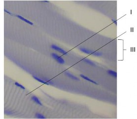
image

    1. Identify

      1. the dark structure indicated by I [1]

      2. the protein producing the thick filament in the dark band indicated by II. [1]

      3. the structure indicated by III [1]

    2. Discuss whether the tissue shown in the micrograph consists of cells or not [2]

    3. Explain how calcium is involved in muscle contraction [3]

▶️Answer/Explanation

Ans:

a i nucleus 

a ii myosin   

a iii muscle fibre/muscle cell  Reject myofibril because it would be much narrower – diameter 1 to 2 μm.

b

a «muscle fibres are» multinucleate/contain many nuclei «whereas cells are expected to have only one/so muscle fibers are an exception to the cell theory»

b one cell membrane/sarcolemma enclosing a whole muscle fibre «as expected for cells» 

c very large/much larger/longer/than most cells 

d muscle fibres formed by fusion of cells/are syncytia 

c

a action potential/nerve impulse causes release of calcium 

b from sarcoplasmic reticulum/specialized endoplasmic reticulum 

c binds to troponin 

d causes tropomyosin to move/be removed «from binding sites» 

e exposes myosin-binding sites on actin/allows myosin «heads» to bind to actin

Question

Defence occurs on the micro and macro levels.

Describe the functioning of immunoglobulins.

[3]
a.

Outline how antibiotics offer protection from certain forms of infectious disease.

[4]
b.

Coughing to clear the airways is accomplished by muscle contractions. Explain muscle contraction.

[8]
c.
▶️Answer/Explanation

Markscheme

a. «immumoglobulins are/function as» antibodies 

b. variety of binding sites / variable regions for binding 

c. specific to antigens on bacteria/viruses/pathogens 

d. constant region aids destruction of the bacteria/virus/pathogen 

e. attracts phagocytes/macrophages to engulf pathogen 

f. bursting pathogen cells/agglutination/neutralizing toxins/other example of the action of antibodies

Award marks for an annotated diagram.

a.

a. protect against/kill/inhibit growth of microorganisms/bacteria/prokaryotes 

b. bacteria/prokaryote processes blocked but not processes in eukaryotes/other organisms 

c. block metabolic pathways/DNA replication/DNA transcription/translation/ribosome functioning/cell wall formation 

d. do not protect against viruses as they have no metabolism/are non-living 

e. antibiotics fail to protect if bacteria have resistance 

f. can be used in humans/animals because antibiotics do not affect eukaryotic cells/bacterial metabolism is different

b.

a. myofibrils «in muscle fibers/cells» 

b. sarcomeres «are the repeating units in muscle/myofibrils» 

c. sarcomeres arranged end to end / sarcomeres shorten during muscle contraction 

d. actin and myosin/overlapping protein filaments/diagram to show sarcomere with actin and myosin overlapping 

e. dark and light bands «in sarcomeres»/diagram to show this/light bands narrower when muscle is contracted 

f. thick filament is myosin and thin filament is actin/diagram to show this 

g. nerve impulses stimulate contraction/cause depolarization of sarcolemma/T-tubules/trigger release of calcium from sarcoplasmic reticulum 

h. calcium ions released from sarcoplasmic reticulum/bind to troponin 

i. troponin causes tropomyosin to move/exposes binding sites on actin 

j. myosin «heads» form cross bridges with/bind to actin 

k. myosin heads move/change angle/swivel/cock / myosin heads cause the power stroke 

l. myosin filaments pull actin towards center of sarcomere/more overlap between actin and myosin/Z-lines move closer 

m. ATP is used «to provide energy»/cause cross-bridges to break/cause movement of myosin heads/cause filaments to slide/cause muscle contraction 

n. intercostal/abdominal/diaphragm muscles contract «to cough»

Marks can be awarded for any point made clearly on an annotated diagram.

c.

Question

Explain how skeletal muscle contracts.

[8]
b.

Active skeletal muscle requires a good supply of oxygen. Outline the mechanism of ventilation in the lungs.

[6]
c.
▶️Answer/Explanation

Markscheme

Remember, up to TWO “quality of construction” marks per essay.

a. sliding filament model / filaments/actin and myosin slide past each other;
b. action potential/depolarisation/nerve impulse arrives at end of motor neurone;
c. neurotransmitter/acetylcholine released causing action potential (in muscle fibre);
d. sarcoplasmic reticulum releases calcium ions;
e. calcium ions cause binding sites on actin/for myosin to be exposed;
f. myosin heads bind to sites on actin/form cross-bridges;
g. myosin (head) moves actin filament using energy from ATP;
h. actin moved towards the centre of sarcomere/M line/M band;
i. sarcomeres shortened;
j. (binding of) ATP causes release of myosin head from actin;
k. conversion of ATP to ADP and Pi causes myosin heads to change angle;
l. cycle (of events) repeated (during muscle contraction);
Accept the above points in annotated diagrams.

b.

Remember, up to TWO “quality of construction” marks per essay.

during inhalation:
a. external intercostal muscles contract moving rib cage up and out;
b. diaphragm contracts becoming lower/flatter;
c. increase in volume and decrease in pressure (of thorax);
d. air flows into lungs as atmospheric pressure is higher;

during exhalation:
e. internal intercostal muscles contract so ribs move in and down;
f. diaphragm relaxes and returns to domed shape;
g. decrease in volume and (therefore) increase in pressure (of thorax);
h. air moves out until pressure in lungs falls/is equal to atmospheric pressure;
i. abdominal muscles can be used to make a stronger/forced exhalation;

c.

Question

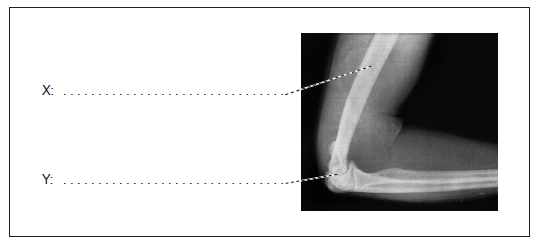
Label the structures indicated on the X-ray of a human elbow.

[2]
a(i).

Explain the role of calcium in muscle contraction.

[3]
b.

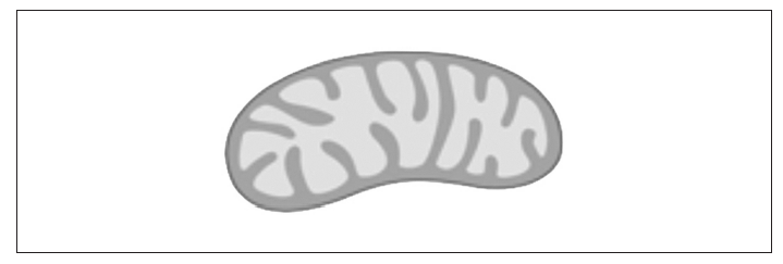
One of the stages of aerobic respiration is called the link reaction.

Label the diagram to indicate where the link reaction occurs.

[1]
c(i).

Outline the role of coenzyme A in aerobic respiration.

[2]
c(ii).
▶️Answer/Explanation

Markscheme

X: humerus;

Y: synovial fluid / cartilage / joint capsule / elbow joint;

a(i).

action potential/nerve impulse/motor neuron causes release of calcium;

calcium released from sarcoplasmic reticulum;

calcium causes binding sites on actin to be exposed;

myosin heads bind to binding sites/to actin and push actin (inwards);

b.

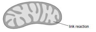
Accept a line or arrow pointing to any part of the matrix, or a circle in it. It is not necessary to state link reaction unless more than one area is indicated.

c(i).

accept/bind acetyl group/acetate / acetyl coenzyme A/acetyl CoA formed;

passes acetyl group/acetate to Krebs cycle;

c(ii).
Scroll to Top