The diagram shows the ventilation system in humans.
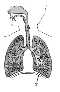
What is the function of the structure labelled X?
A. Protect the lungs
B. Contract to cause inhalation
C. Become flatter to move the ribcage up
D. Relax in order to increase the thoracic capacity
▶️Answer/Explanation
Markscheme
B
The diaphragm is a large, dome-shaped muscle that separates the chest cavity from the abdominal cavity. During inhalation, the diaphragm contracts and flattens out, increasing the volume of the thoracic cavity and decreasing the pressure inside the lungs. This decrease in pressure causes air to rush into the lungs, filling the expanded space. When the diaphragm relaxes, it returns to its dome shape, decreasing the volume of the thoracic cavity and increasing the pressure inside the lungs. This increase in pressure causes air to be expelled from the lungs during exhalation.
Question
The diagram shows a section through an alveolus.
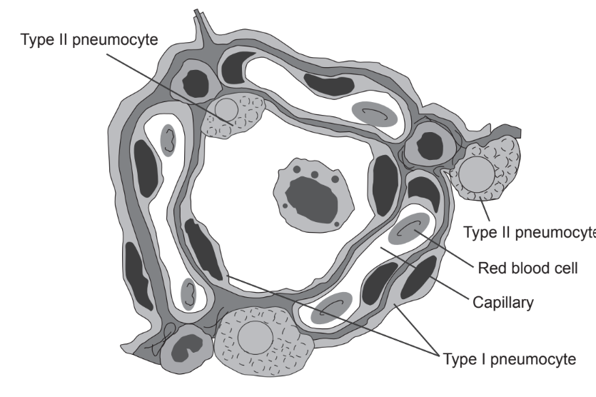
What are the functions of the following labelled structures?
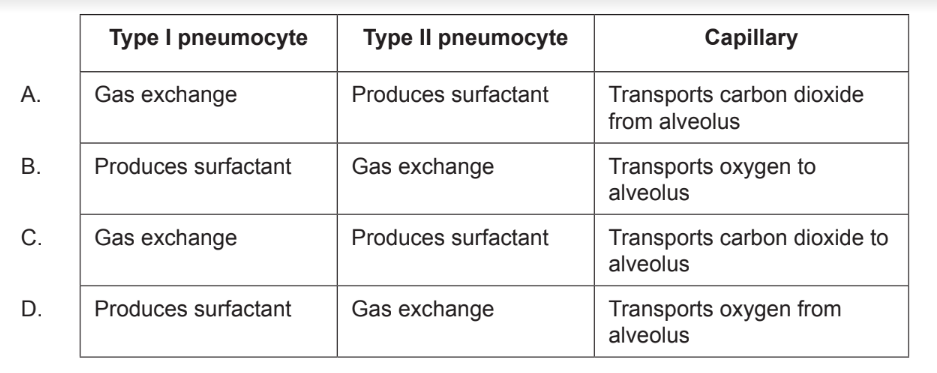
▶️Answer/Explanation
Ans:C
Type 1 pneumocytes are thin flattened cells that are responsible for the gas exchange between alveoli and capillaries. Type 2 pneumocytes are smaller cells that are cuboidal in shape. They are responsible for the secretion of pulmonary surfactants in order to reduce the surface tension in the alveoli. There are many capillaries around every alveoli. This good blood flow maintains a steep concentration gradient between the oxygen and carbon dioxide in the alveoli and the blood so that the rate of diffusion is faster.