Body Fluids and Circulation
Table of Content
- Blood
- Blood Groups
- Coagulation of Blood
- Lymph
- Circulatory Pathways
- Human Circulatory System
- Cardiac Cycle
- Heart Sound
Blood
It is a fluid connective tissue consists of matrix, plasma and formed elements.
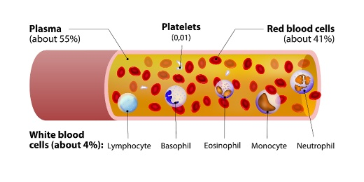
Fig. 1. Elements of the blood
Plasma
It is a straw colored, viscous fluid which constitutes about 55% of the blood plasma. The major proteins found in plasma are fibrinogen, globulins, and albumins. Fibrinogens functions during blood clotting. Albumins maintain osmotic balance in the body. Globulins are defensive in nature. Minerals such as Sodium Ions, Calcium Ions, Magnesium Ions, Bicarbonate Ions. Apart from this amino acid, glucose is also present in plasma.
Formed Elements
Formed Elements include erythrocytes, leucocytes, and blood platelets.
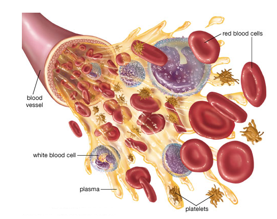
Fig. 2. Formed elements in blood
Erythrocytes also known as Red Blood Cells. They are the most abundant cells in the blood. They are formed in red bone marrow of adults. They are biconcave in shape and enucleated (without the nucleus). They carry iron containing protein known as Hemoglobin. Hemoglobin helps in the transport of oxygen and carbon-dioxide in blood. The average life span of RBCs is 120 days.
Leucocytes also known as White Blood Cells. They are nucleated cells which do not contain hemoglobin, so they appear colorless. They are of two types- granulocytes and agranulocytes.
Neutrophilsb, Basophils and Eosinophils are granulocytes. Lymphocytes and Monocytes are agranulocytes.
Neutrophils are known as Polymorphonuclear Leucocytes. Out of all the three granulocytes, neutrophils are most abundant. They are phagocytic cells.
Basophils are least in number in comparison to other granulocytes. They secrete serotonin, histamine, and basophils. So, basophils are involved in inflammatory reactions.
Eosinophils are involved in allergic reactions.
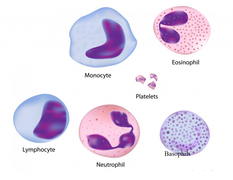
Fig. 3. White blood cells and platelets
Blood platelets are also known as Thrombocytes. They are involved in blood clotting. Decrease in the number of blood platelets can lead to loss of blood in the body.
Blood Groups
Blood grouping is divided into- ABO blood group and Rh group.
ABO blood grouping
ABO blood grouping is done based the presence or absence of certain antigens on the surface of RBCs. The antigen present on the surface of RBCs can be A or B. There are 4 types of blood group are A, B, AB and O group.

Fig. 4. Blood groups and antigen on red blood cells
The above table depicts blood groups and donor compatibility. As O blood group do not have any antigen on their surface, they are said to be universal donor whereas AB are considered universal recipients as they contain both the antigen of their surface. Blood transfusion is done only based on blood group of the donor and recipients.
Rh grouping
Rh is also an antigen like rhesus monkeys. Individuals having Rh antigen on RBCs are considered as Rh positive. Those which are without Rh antigen are considered Rh negative. If Rh -ve person receives Rh +ve blood, the Rh -ve individual will start producing antibodies against it. So, Rh group should also be tested at the time of blood transfusion.
An important case of Rh mismatching has been observed between the Rh -ve blood of a pregnant mother with Rh +ve blood of the fetus. Rh antigens of the fetus do not get exposed to the Rh-ve blood of the mother in the first pregnancy, due to placenta. But during the delivery of the first child, there is a possibility of mixing of the blood of mother with child.
In such cases, mother starts to produce antibodies against the Rh antigen. So, in the next pregnancy, the Rh antibodies from the Rh -ve mother can leak into the blood of the Rh +ve fetus and can destroy the fetal RBCs. This leads to agglutination of red blood cells. This condition is known as Erythroblastosis Foetalis. Fetus will anemic and suffers from jaundice. This can be avoided by injecting anti-Rh antibodies to the mother instantly after the delivery of the first child.
Coagulation of Blood
Blood coagulation is also known as Blood Clotting. Blood responds by clotting or coagulation after any injury or trauma. This helps in preventing excess loss of blood. When we get injured, after some time reddish brown scum is formed at the site of injury. This is known as clot which is made up of network of threads known as Fibrils. This network contains dead and damaged formed elements of blood. Fibrils are formed from the conversion of inactive fibrinogen in presence of enzyme thrombin. Thrombin is also formed from inactive form known as Prothrombin. Thrombokinase is required for the conversion of prothrombin into thrombin.
Platelets release certain factors to begin blood clotting. Calcium ions play an important role duriBlood coagulationclotting.
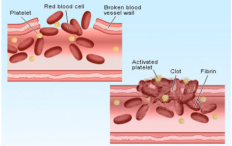
Fig. 5. Blood coagulation
Lymph
Blood circulates in blood capillaries in tissues. Some water along with some water-soluble substances leak out and enter the interstitial space. This is known as Tissue Fluid or Interstitial Fluid. Exchange of gases, nutrients between the blood and the cells occurs through this fluid.
A network of vessels that collects this fluid and drain it to major veins is known as Lymphatic System. Lymphatic system contains a fluid known as Lymph. Lymphocytes which are an important cell of the immune system are present in lymph.
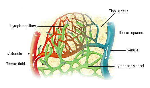
Fig. 6. Lymph
Circulatory Pathways
There are two types of circulatory system- Open Circulatory System and Closed Circulatory System.
When blood flow in open spaces known as Lacunae and Sinuses, it is known as Open Circulatory System it is present in molluscs, arthropods, etc.
When blood flow in closed vessels it is known as Closed Circulatory System. For Example, humans. Closed circulatory possess an advantage over open circulatory system, as blood can flow in a regulated manner in case of closed circulatory system.
Vertebrates have muscular, pumping organ known as Heart. Fishes have 2 chambered heart. Amphibians have 3 chambered heart except crocodiles (4 chambered heart). Birds, reptiles, and humans have 4 chambered heart.
Human Circulatory System
It includes heart, vessels, and blood. Heart is mesodermal in nature. It is located in the thoracic cavity, in between the two lungs.
The double membrane that surrounds the heart is known as Pericardium. Pericardium encloses the pericardial fluid. Heart is 4 chambers with 2 atria and 2 ventricles. A thin wall separates the left and the right atria is known as Intra-atrial Septum whereas left and right ventricle are separated by thick intra-ventricular septum.
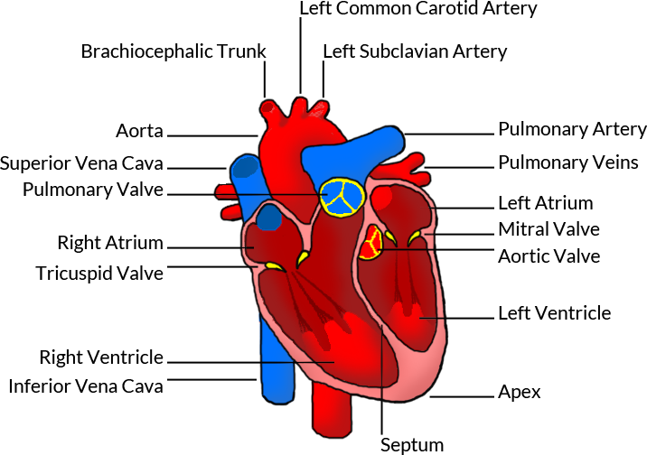
Fig. 7. Human heart
The opening between the right atrium and the right ventricle is guarded by a valve known as Tricuspid Valve, whereas a bicuspid or mitral valve guards the opening between the left atrium and the left ventricle. The openings of the right and the left ventricles into the pulmonary artery and the aorta respectively are provided with the semilunar valves. Valves in the heart allows the blood to flow in one direction and thus preventing backflow of blood.
Heart is a muscular organ and the muscles of the heart are known as cardiac muscles. A specialized cardiac musculature called the nodal tissue is also dispersed in the heart. One which is present in the upper right corner of the right atrium is known as Sinoatrial Node or SA Node. Another tissue is present in the upper left corner of the right atrium is known as Atrio-Ventricular Node or AV Node.
A bundle of nodal fibres, Atrioventricular Bundle (AV Bundle) continues from the AVN which passes through the atrio-ventricular septa and divides into a right and left bundle. These branches give rise to minute fibers known as Purkinje Fibers. SA node has the ability to get excited and generates the action potential. So, SA node is known as the Pacemaker of the Heart.
Cardiac Cycle
Sequence of electrical and mechanical events during every heart beat is known as Cardiac Cycle. It is divided into two phases – Diastole and Systole. During diastole heart ventricles relaxes due to which it is filled with blood. During systole, ventricles contract to pump the blood into the arteries.
Phases of Cardiac Cycle are as follows:
Atrial Systole includes contraction of the left and the right atria followed by electrical stimulation. This increases blood pressure in the both left and right atria. This allows blood to pump into the ventricles. During this AV valves are open and semilunar valves are closed. It takes about 0.1 seconds.
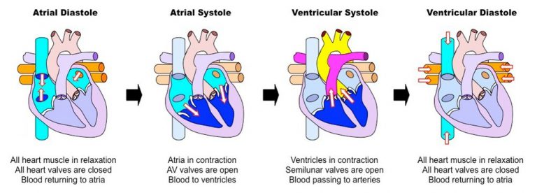
Fig. 8. Cardiac cycle
Ventricular Systole includes contraction of the left and right ventricles which is followed by electrical stimulation. During this AV valves are closed and semilunar valves are open. It takes about 0.3 seconds.
Cardiac diastole is a period when heart relaxes to fill the blood. When atria and ventricles are relaxing together, they form complete cardiac diastole. During ventricular diastole, pressure in the ventricles drops below the left atrial pressure, mitral valve opens and left ventricle gets filled with the blood. Similarly, when the pressure in the right ventricle drops below that in the right atrium, the tricuspid valve opens, and the right ventricle gets filled with blood. During diastole, the pressure within the left ventricle is lower than that in aorta, which allows the blood to circulate in the heart itself with the help of the coronary arteries.
Heart Sound
The heart sound is known as “lubb-dubb” sound. The first heart sound lubb is produced when mitral and tricuspid valves gets closed at the beginning of the ventricular systole. The second sound dubb is produced when aortic and pulmonary valves gets closed at the end of ventricular systole.
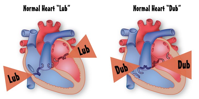
Fig. 9. Heart sound