| Cell Cycle and Cell Division Refresher Course |
| Cell Cycle and Cell Division Concepts Files |
| Cell Cycle and Cell Division Master Files |
| Cell Cycle and Cell Division Notes 1 |
Cell Cycle and Cell Division
Table of Contents
Define Cell Cycle
Cell Cycle is defined as the series of changes that occur in a cell which leads to division of cells into two daughter cells. It takes about 1 hour to complete the cycle.
Cell Cycle is divided into two phases – Interphase and Mitosis
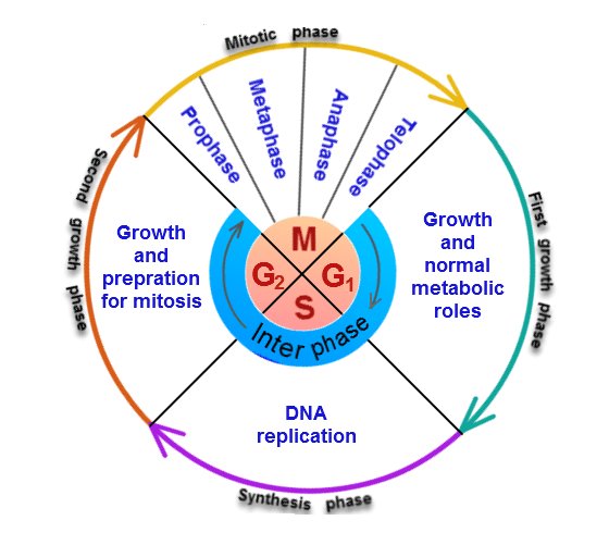
Fig. 1. Cell cycle
Interphase
It is a phase in which a cell prepare itself for cell division. It is further divided into following phases:
G1 phase is also known as First Gap Phase. During this phase, biosynthetic activities occur at a very fast rate. Cell synthesizes more proteins, increase the number of mitochondria and ribosomes.
S phase is the phase where DNA is replicated. At the end of DNA replication, each chromosome is with two sister chromatids. Thus, the amount of DNA gets doubled during this phase, but the ploidy remains same.
G2 phase is the phase where the cell prepare itself for mitosis.
Mitosis
Also, known as Equational Division because, the number of chromosomes will remain same in the daughter cell as present in parent cell. It is a phase where nuclear division occurs. This is known as Karyokinesis. It is divided into following phases:
Prophase
During this phase chromatin condenses to form chromosomes.There are two identical copies of each chromosome which are attached to centromere. During the end of the prophase, nucleolus dissolves. Nuclear membrane also disintegrates at the end. The centrosome moves to the opposite poles. Spindle fibers starts to appear.
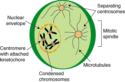
Fig. 2. Prophase of mitosis
Metaphase
During this phase, chromosomes aligned themselves on the equatorial plate. The analysis of the metaphase chromosome is used in cytogenetics and cancer studies.

Fig. 3. Metaphase of mitosis
Anaphase
It is the shortest stage of the cell cycle. During this phase, the replicated chromosomes move apart and daughter chromatids moves to the opposite poles. Chromosomes are maximally condensed during late anaphase. The chromosomes appear Y- shaped as they move to the opposite poles.
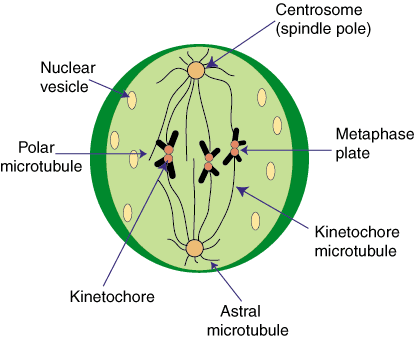
Fig. 4. Anaphase of mitosis
Telophase
This is where two daughter nuclei are formed. Nucleolus and nuclear membrane re-appear. The late telophase is marked by cytokinesis. This is last phase of mitosis.
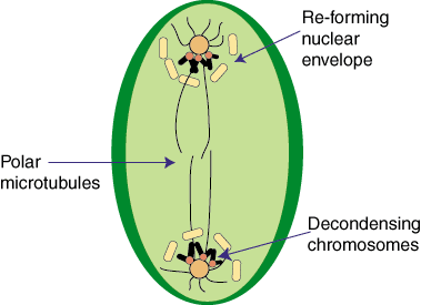
Fig. 5. Telophase of mitosis
Cytokinesis
The division of cytoplasm, organelles, and cell membrane to form two cells is known as Cytokinesis. It divides the cell into two daughter cells which are identical to the parent cell.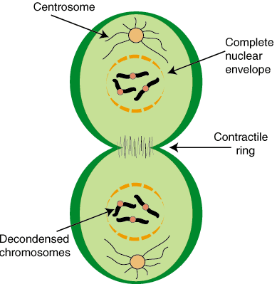
Fig. 6. Cytokinesis
Cell Cycle Exit
Some cells divide at a fast rate whereas some divides slowly and some even do not divide once they are formed. Those cells which do not divide once formed, enter into a phase known as GO phase. For Example, neuronal cells once formed will not divide.
Meiosis
Also, known as Reductional Division because it reduces the number of chromosomes to half the parent cell. It is of two types meiosis I and meiosis II. Gametes such as sperm or egg under meiosis.
Meiosis I
It is divided into following phases:
Prophase I
It is the longest phase of meiosis I. During this phase, homologous chromosomes pairs and DNA segments are exchanged (known as recombination). It is further divided into following-
Leptotene
- It is the first stage of meiosis.
- Each individual chromosome exists with two sister chromatids.
- Elements of synaptonemal complex assemble.
- Condensation and coiling of chromosomes occur.
Zygotene
- Chromosomes align as homologous pair of chromosomes.
- Synapsis of homologous chromosomes occur.
- The paired chromosomes are known as Bivalent or Tetrad.
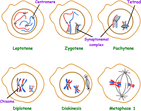
Fig. 7. Different stages of prophase I of meiosis I
Pachytene
- During this stage, crossing over and homologous recombination occurs.
- Chiamata is formed where homologous chromosomes exchange there segments.
Diplotene
- Homologous chromosomes start to separate.
- Synaptonemal complex disassemble.
- Chromosomes remain bound at the chiasmata.
Diakinesis
- Chromosomes condenses further, so that four parts of the tetrads are visible.
- The nucleoli disappear, nuclear membrane disintegrates.
- Mitotic spindle starts to form.
Metaphase I
Homologous chromosomes are aligned on the metaphasic plate. The replicated chromosomes are joined together via protein known as Cohesin.

Fig. 8. Metaphase I
Anaphase I
Kinetochore microtubules shorten, pulling homologous chromosomes to opposite poles. The cohesin (protein complex) from the chromosome arms is degraded while the cohesin surrounding the centromere remains safe. This allows the sister chromatids to attach together while homologs are segregated.
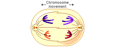
Fig. 9. Metaphase I
Telophase I
Each daughter cell now has half the number of chromosomes as compared to parent cell. The microtubules that forms the spindle starts to disappear. The chromosomes again forms chromatin. Sister chromatids remain attached during telophase I.
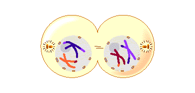
Fig. 10. Telophase I
Meiosis II
Meiosis II is the second meiotic division. It is similar to mitosis, but the genetic results are different. It results in the formation of four haploid cells from the two haploid cells produced in meiosis I.
The four main steps of Meiosis II are as follows: Prophase II, Metaphase II, Anaphase II, and Telophase II.
In prophase II nucleoli and nuclear membrane disappears. Shortening and thickening of the chromatids also occur during this phase. Centrosomes move to the poles and spindle fibers are formed for the second meiotic division.
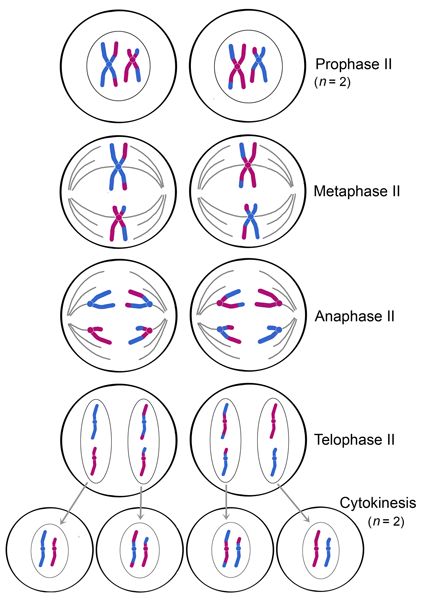
Fig. 11. Different stages of meiosis II
In metaphase II, the centromeres are present with two kinetochores that are attached to spindle fibers from the centrosomes at opposite poles. The metaphasic plate is rotated by 90 degrees when compared to meiosis I, perpendicular to the previous plate.
Anaphase II is accompanied by sister chromatid segregation. The remain of the cohesin is degraded to allow sister chromatid segregation.
Telophase II, which is like telophase I, de-condensation of chromosomes occurs. Nuclear envelopes reorganize and cleavage or cell plate formation starts to produce four daughter cells, each with a haploid set of chromosomes.