Cell: The Unit of Life
Table of Content
- What is cell?
- Cell theory
- Prokaryotes and Eukaryotes
- Plasma Membrane
- Ribosomes
- Endoplasmic reticulum
- Golgi apparatus
- Lysosomes
- Vacuoles
- Mitochondria
- Plastids
- Nucleus
- Peroxisomes
What is Cell in biology or what is a cell in human body or why is the cell the basic unit of life?
The basic structural and functional unit of life is the cell. It is capable of independent existence and performing essential functions of life. All organisms including plants, animals are made up of cell. So, cells are considered as basic unit of life.
Robert Hooke first discovered cells in a piece of cork.
Different types of cell/ types of cells.
There are different types of cells found in human body. This includes hepatocytes in liver, nephrons in kidney, neurons in brain, etc. Different cells are grouped together to form tissues which perform specific function.
Cell theory or Discovery of a cell
In 1839, Schleiden, German botanist, and Schwann, a British zoologist, led to the development of the cell theory or cell doctrine. The modern theory includes the following components-
- All living organisms are made up of cells.
- Cell is the structural and functional unit of life.
- All cells arise from the pre-existing cells. This was given by Rudolf Virchow.
- Energy flow occurs within the cells.
- Cells contains the heredity information which is passed from cell to cell.
- All cells have same chemical composition.
Functions of the cells
- Different cells have different function. For Example, Nerve cells are involved in neurotransmission.
- All metabolic reactions occur inside the cell, For Example, Glycolysis
- Genetic material is present inside the nucleus of the cell which is responsible for passing hereditary information.
- Cells participate in reproduction.
Prokaryotes and Eukaryotes
Prokaryotes | Eukaryotes |
| Cell wall is present which is made up of peptidoglycan. | Cell wall is present in plant cells which is made up of cellulose |
| DNA is generally circular | DNA is generally linear |
| No well-defined nucleus. Instead of nucleus nucleoid is present. For example, Bacteria | Well defined nucleus For example, Humans |
| Mitochondria are usually absent | Mitochondria are present. |
| Ribosomes are 70S | Ribosomes are 80S |
Structure of eukaryotic cells/ Parts of cell/ what is the cell made up of?
Plasma Membrane
It is a dynamic, fluid structure that forms the external boundary of the cell. It is a selectively permeable membrane. It allows only certain solutes to pass through it. In 1972, Jonathan Singer and Garth Nicolsan proposed fluid mosaic model. According to this model, membrane is viewed as quasi-fluid structure in which proteins are embedded throughout the lipid bilayer. The bilayer is composed of two leaflets of amphipathic molecules with polar head and non-polar tails.
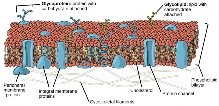
Fig. 1. Structure of the plasma membrane
The primary forces for organizing lipid bilayer are hydrophobic interactions. Three classes of lipids are present in plasma membrane- phospholipids, glycolipids and sterol. Membrane contain two types of protein- peripheral proteins and integral proteins. Proteins which are held with the bilayer loosely and can be removed easily is known as peripheral proteins. Proteins that are held in the lipid bilayer very tightly and cannot be released easily is known as integral proteins.
Ribosomes
Ribosomes are composed of RNAs and proteins. It translates genetic information stored in messenger RNA into proteins. Functional ribosomes consist of two subunits of unequal size, known as large and small subunits. Most eukaryotes contain two types of ribosomes: cytosolic and organellar. The ribosome found in prokaryotes if 70S and in eukaryotes it is 80S. S stands for sedimentation coefficient. It is the ratio of a velocity to the centrifugal acceleration.
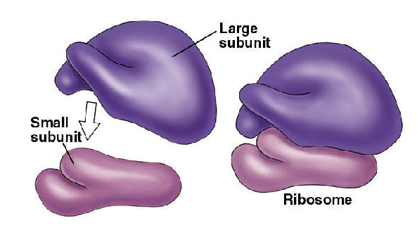
Fig. 2. Structure of the ribosomes
Endoplasmic reticulum
It is the largest single membrane bound intracellular compartment. It is an extensive network of closed and flattened membrane bound structure. The enclosed compartment is called Lumen. ER membrane can be rough or smooth based on the presence or absence of ribosomes. When ribosomes are attached to ER, it is known as Rough ER. When ER membrane do not contain ribosomes, it is known as Smooth ER.
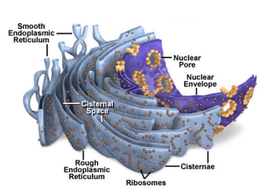
Fig. 3. Structure of endoplasmic reticulum
Proteins synthesized by ribosomes associated with the membrane of RER enter into the lumen and membrane of RER by the process of co-translational translocation. In the lumen of the RER, five principal modifications of proteins occur before they reach their final destination-addition and processing of carbohydrates, formation of disulfide bonds, proper folding, specific proteolytic cleavages and assembly into multimeric proteins. The SER acts as site for the lipid biosynthesis, detoxification and calcium regulation.
Golgi complex/Golgi apparatus
It is a single membrane bound organelle and forms a part of endomembrane system. It consists of flattened membrane sacs known as Cisternae. Each stack is known as golgi stack. Each golgi stack has two faces- the cis face or the entry face and the trans face or exit face. The golgi apparatus is especially prominent in cells that are specialized for secretion.
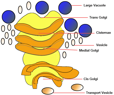
Fig. 4. Structure of golgi apparatus
Lysosomes
It is a single membrane enclosed organelle which contains hydrolytic enzymes. Hydrolytic enzymes include proteases, nucleases, glycosidases, lipases, phospholipases, phosphatase and sulphatases. The environment is acidic inside the lysosomes with a pH of about 5.0. There is a proton pump inside the lysosomal membrane which pumps proton inside the membrane using ATP as a source of energy. Lysosomes are responsible for the digestion of both intracellular as well as extracellular materials.
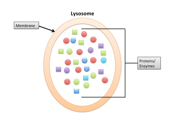
Fig. 5. Structure of lysosomes
Vacuoles
Fluid filled vesicles are known as Vacuoles. They are surrounded by a membrane known as Tonoplast. The pH of the lumen is acidic similar to that of lysosomes. Plant vacuole contains water, dissolved inorganic ions, sugars, enzymes etc. The vacuole is different from contractile vacuole. Contractile vacuole is an organelle involved in osmoregulation. It pumps excess of water out of the cell. For Example, it is found in Amoeba.
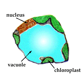
Fig. 6. Structure of vacuoles
Mitochondria
It is found in all eukaryotic cell. It is a site for aerobic respiration. They are known as the power house of the cell as it synthesizes ATP, energy currency of the cell. They are the double membrane bound cell organelle. It contains circular DNA molecule and ribosomes. The space between the outer and the inner membrane is known as Intermembrane Space. The inner membrane is convoluted to form cristae. The inner membrane is impermeable to solutes and they are rich in phospholipid known as Cardiolipin. Inner membrane contains enzyme complex known as ATP Synthase or F0-F1 ATPase.
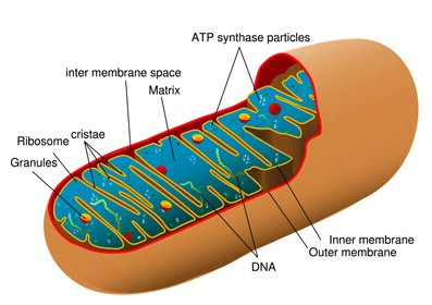
Fig. 7. Structure of mitochondria
Plastids
They are double membrane cell organelle found in plant cells. They also contain double stranded DNA and ribosomes. They are differentiated into – Leucoplast, Chromoplast and Chloroplast.
Chloroplast encloses the fluid filled structure known as Stroma. Stroma contains a stack of sacs. Each stack is known as Granum and each of the flattened sacs which makes up the granum is called a Thylakoid. Each granum is connected to each other by stromal lamellae.
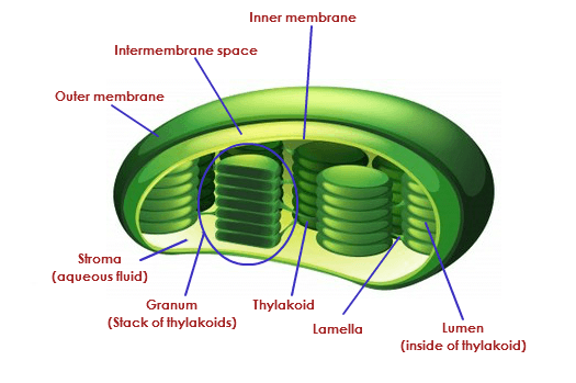
Fig. 8. Structure of chloroplast
Chromoplasts are plastids responsible for pigment synthesis and storage. They give yellow, orange, red colors to fruits and flowers. Leucoplast are colorless plastids and acts as storage organelles.
Leucoplast are colorless plastids that are divided into – Amyloplast that store starch, Elaioplast stores lipids in fats, proteinoplast stores proteins.
Nucleus
Nucleus is a double membrane structure found in eukaryotes. Eukaryotes have single nucleus except the red blood cells which do not contain nucleus. Nucleus contain genetic material known as DNA packed in the form of chromosomes with proteins known as Histones.
There are two types of chromosomes found – Euchromatin and Heterochromatin. Euchromatin is a less compact structure that can be transcribed (formation of messenger RNA from DNA). Heterochromatin is a compact structure that cannot be transcribed.
The fragment of DNA that codes for a protein is known as Gene. Nuclear membrane is impermeable to large molecules, so nuclear pores are present that regulates the movement of solutes in and out of the nucleus. Nucleolus is the largest structure found inside the nucleus of eukaryotes. It is involved in the assembly of the ribosomes.
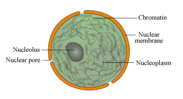
Fig. 9. Structure of nucleus
Peroxisomes
Peroxisomes are found in all prokaryotic cell. The major function of peroxisomes is to breakdown of fatty acids. Peroxisomes are derived from the endoplasmic reticulum. Proteins found in peroxisomes are known as Peroxins.
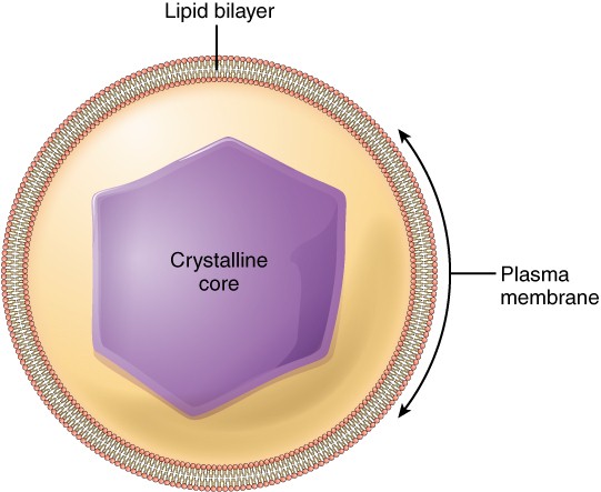
Fig. 10. Structure of peroxisomes