| Digestion and Absorption Refresher Course |
| Digestion and Absorption Concepts Files |
| Digestion and Absorption Master Files |
| Digestion and Absorption Handwritten Notes |
| Digestion and Absorption Note |
Digestion and Absorption
Table of Content
- What is Digestion?
- Types of Digestion
- Digestive system
- Digestion of food
- Digestion of Carbohydrates
- Digestion of fats
- Digestion of proteins
- Absorption of digested products
What is Digestion?
The process of breakdown of complex, water insoluble food molecules into simple water soluble molecules is known as Digestion. It is a complex process involving various enzymes, secretion etc.
Types of Digestion
Mechanical digestion is a process of physically breaking the food into smaller pieces. It includes the food that is chewed in the mouth.
Chemical digestion is complex process of digestion of food involving enzymes which breakdown complex food molecules into simple food molecules.
Digestive system
The human digestive system comprises of alimentary canal and digestive glands.
Alimentary canal
The anterior most part of the alimentary canal is known as mouth and the posterior most part is known as Anus. The mouth leads into the oral cavity or buccal cavity. Buccal cavity contains teeth and tongue. Each tooth is fixed in socket of jaw bond. This type of arrangement of teeth is known as Thecodont. For Example, Mammals.
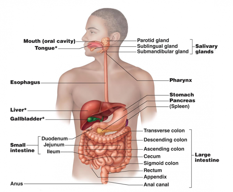
Fig. 1. Alimentary canal
In mammals, temporary teeth are replaced by permanent teeth in adults. This is known as Diphyodont. A human adult consists of 4 types of teeth which are 32 in number. The 4 types of teeth are- Incisors, Canines, Molars, and Premolars.
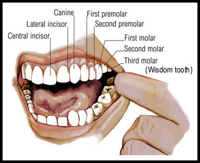
Fig. 2. Types of teeth
Surface of the teeth is covered by the hardest substance known as Enamel. The tongue is freely movable, muscular organ that helps in chewing of food inside the oral cavity. The oral cavity leads into the pharynx, which is common passage for food as well as air. Esophagus also known as Food Pipe helps in passage of the food to the stomach.
Stomach is a bag-like structure located in the upper portion of the abdominal cavity. It is divided into three parts – Cardiac, Fundic and Pyloric.
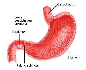
Fig. 3. Parts of stomach
Pyloric is the region which opens into the first part of the small intestine. Small intestine is divided into duodenum, jejunum and ileum. Pyloric sphincter guards the opening of stomach into the duodenum. Ileum opens into the large intestine.
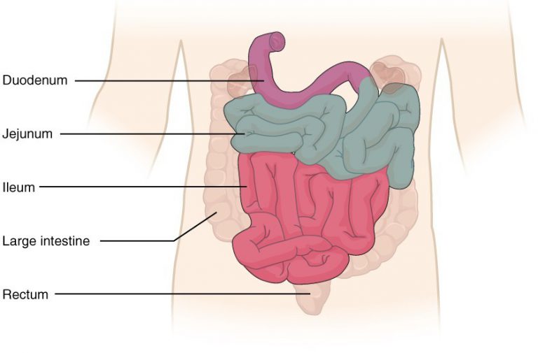
Fig. 4. Parts of small intestine
Large intestine is divided into caecum, colon, and rectum. Rectum finally opens into the anus. Symbiotic organisms reside in caecum. A vestigial organ known as Vermiform appendix arises from the caecum. Colon is divided into ascending, transverse, and descending part.
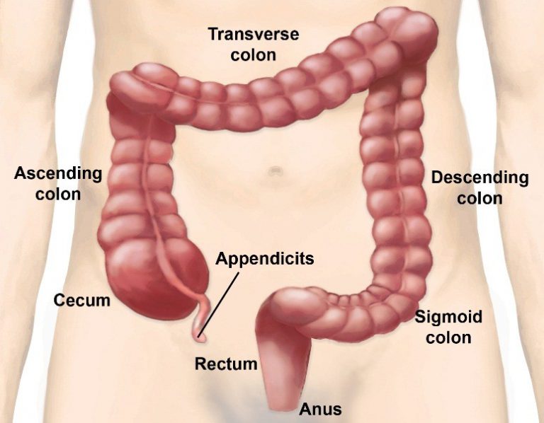
Fig. 5. Parts of large intestine
The wall of the alimentary canal starting from the esophagus to the rectum is lined by four layers- outermost layer serosa, inner to it lies the muscularis, next submucosa and innermost mucosa. Muscularis is made up of two types of muscles – Circular Muscles and Longitudinal Muscles. Mucosa for folds in the stomach and small finger-like projections in intestine known as Villi. Villi increases the surface area for efficient absorption of food.
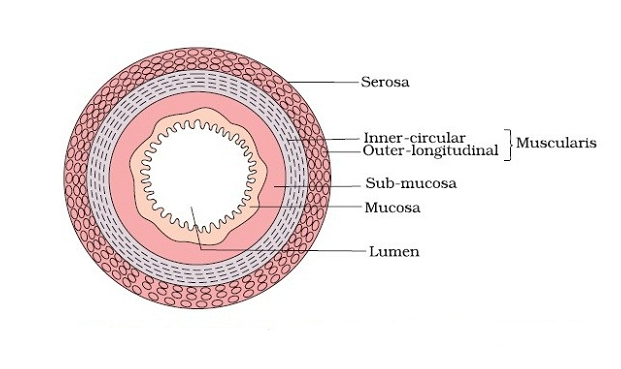
Fig. 6. Wall of the alimentary canal
Mucosa forms gastric glands in stomach, crypts in the intestine
Digestive glands
The three main digestive glands are – Salivary Glands, Liver and Pancreas.
There are three pairs of salivary glands present – the parotids glands located in cheek, the sub-maxillary/sub-mandibular located in lower jaw and the sublinguals, found below the tongue. These glands secrete salivary juice. Salivary juice contains an enzyme salivary amylase or ptyalin, that participates in digestion of carbohydrates which begin in the mouth.
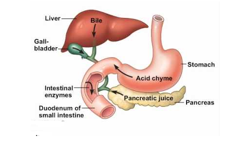
Fig. 7. Digestive glands
Liver is the largest gland found in the human body. The structural and the functional unit of liver is hepatic lobules. Each lobule is connected by a tissue known as Glisson’s Capsule. Liver secretes bile juice which is stored in muscular sac-like structure known as Gall Bladder. The duct of the gall bladder is known as Cystic Duct that combines with the duct of the liver to form common bile duct.
The bile duct and the duct of the pancreas open into duodenum.
Pancreas is an endocrine as well as exocrine gland. The endocrine function of the pancreas involves the secretion of insulin and glucagon which regulates the blood sugar level. Whereas endocrine function of pancreas includes secretion of pancreatic juice that participates in digestion of food.
Digestion of food
Digestion of food begins in the buccal cavity where two important functions are performed- mastication of food and swallowing. Teeth and tongue promote mastication of food. Saliva mixes the food thoroughly and forms bolus. The saliva in the mouth contains electrolytes such as K+, Na+ etc. and two important enzymes known as lysozyme and salivary amylase. Salivary amylase breakdown starch into maltose, a disaccharide.
The bolus is then passed to pharynx and then to esophagus. The movement of food inside the esophagus is known as Peristalsis. The gastro-esophageal sphincter guards the food into the stomach.
The mucosa in the stomach forms the gastric glands. Gastric glands consist of-
- Neck cells that secrete the mucus. Mucus lubricates the lining of the stomach against excoriation by hydrochloric acid.
- Peptic cells that secrete pepsinogen.
- Parietal or oxyntic cells that secrete hydrochloric acid, which creates acidic environment inside the stomach.
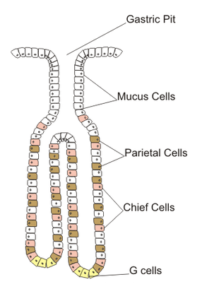
Fig. 8. Gastric gland
When food mixes with the stomach, it forms chyme. In presence of hydrochloric acid, pepsinogen is converted into pepsin. Pepsin is an enzyme that converts proteins into peptones and proteases.
Rennin is also secreted by gastric glands in infants which helps in digestion of milk proteins.
Small intestine receives secretions such as bile, pancreatic juice and intestinal juice. The pancreatic juice contains enzymes which are present in inactive form or pro-enzymes – Trypsinogen, Chymotrypsinogen, Procarboxypeptidases, Amylases, Lipases and Nucleases.
The intestinal mucosa has goblet cells that secretes mucus. The secretions of the brush border cells of the mucosa along with the secretions of the goblet cells forms the intestinal juice or succus entericus. It contains a variety of enzymes like disaccharidases (Example: Maltase), Dipeptidases, Lipases, Nucleosidases, etc.
Enterokinase, an enzyme secreted by intestinal mucosa activates trypsinogen to form trypsin.
Digestion of Carbohydrates
Digestion of Carbohydrates begins in the mouth. Salivary amylase helps in digestion of starch into maltose. Further digestion of carbohydrates occurs due to pancreatic amylase present in pancreatic juice in small intestine. Intestinal juice also contains enzymes such as maltase which digest maltose into glucose, sucrase which digest sucrose into glucose and fructose, lactase which digest lactose into glucose and galactose.
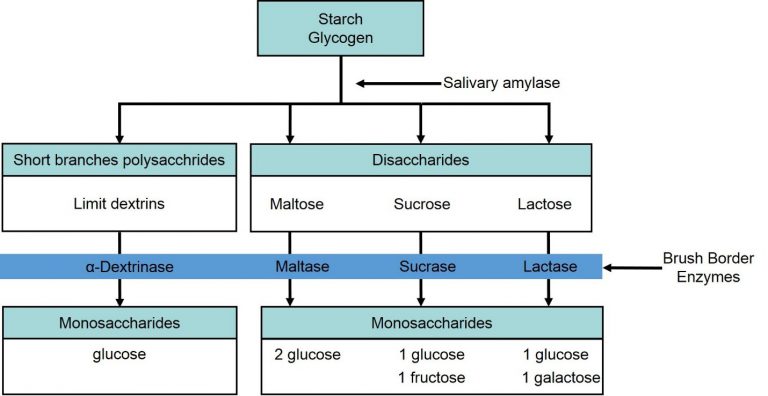
Fig. 9. Digestion of carbohydrates
Digestion of fats
Small amount of lipase is secreted by gastric glands where begins the process of fat digestion. Then the small intestine contains bile secreted by liver, which helps in emulsification of fats, that is, breakdown of fats into small micelles. Intestinal juice also contains lipases that aids in digestion.
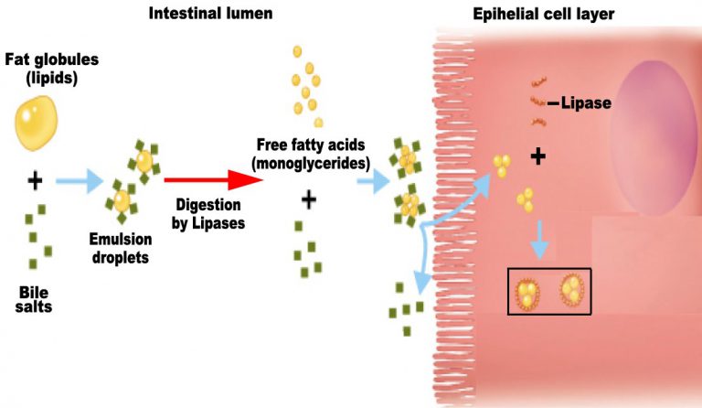
Fig. 10. Emulsification of fats
Digestion of proteins
This process begins in stomach, where, pepsin breaks proteins into peptones and proteases. Trypsin, chymotrypsin, carboxypeptidase breakdown peptones, proteases into dipeptides.
The undigested and unabsorbed substances are passed to large intestine, from where they can be removed from the body. No important digestion process occurs in large intestine.
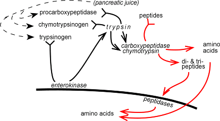
Fig. 11. Digestion of proteins
Absorption of digested products
The end products of digestion pass from intestine to the blood or lymph for absorption. Glucose, amino acids, electrolytes, are absorbed by simple diffusion. Some amino acids and glucose are transported with the help of carrier molecules, this type of transport is known as Facilitated Transport.
Transport of ions such as sodium ions occurs via active transport. Fatty acids and glycerol which are end products of fats are insoluble in water, so first gets converted into micelles which move into intestinal mucosa.
Maximum absorption occurs in small intestine.