Excretory Products and their Elimination
Table of contents
- Excretory organs in variety of organisms
- Human excretory system
- Urine formation
- Function of the tubules
- Regulation of kidney function
- Micturition
Plants and animals produce harmful substances formed due to metabolic reactions which must be removed from the body. It includes, urea, uric acid, ammonia, carbon-dioxide, water, ions etc. the 3 most important nitrogenous waste that should be removed from the body are urea, uric acid and ammonia. Out of these, ammonia is most toxic. Animals excreting ammonia as nitrogenous waste are known as Ammonotelic, such as most bony fishes, most aquatic amphibians etc. The excretion of ammonia occurs via diffusion. The organisms which excrete urea as nitrogenous waste, they are known as ureotelic. For Example, Mammals, Amphibians etc. are ureotelic. Those organisms which excrete uric acid as nitrogenous waste, they are known as Uricotelic. For Example, Birds, Reptiles, Snails etc.
Excretory organs in variety of organisms
- Amoeba and Paramecium have contractile vacuole for excretion.
- Sponges have canal system for excretion.
- Hydra have coelenteron.
- Platyhelminthes have flame cells for excretion.
- Annelids have nephridia for excretion.
- Prawns have green glands for excretion.
- Insects have malpighian tubules for excretion.
- Scorpions have Coxal glands for excretion.
Human excretory system
Human excretory system consists of a pair of kidneys, a pair of ureter, urinary bladder, and urethra. Kidneys are bean shaped structure located in the abdominal cavity. Towards the center of the inner concave surface of the kidney is a notch called hilum. Ureter, blood vessels and nerves enter through hilum. Inner to the hilum is a funnel-shaped space known as Renal Pelvis with projections known as Calyces. Kidney is divided into two zones known as – Outer Cortex and Inner Medulla. The medulla is divided into conical masses known as Medullary Pyramids. Extension of renal cortex which separates the pyramids is known as column of Bertini.
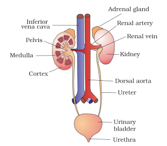
Fig. 1. Human excretory system
The structural and functional unit of kidney are nephrons. Each nephron is made up of two parts- glomerulus and renal tubule.
Glomerulus is the cluster of capillaries. The blood enters the glomerulus via afferent arteriole and leaves the glomerulus via efferent arteriole. Glomerulus is enclosed in a cup- shaped structure known as Bowman’s Capsule. Bowman’s capsule together with glomerulus is known as Malpighian body or renal corpuscles.
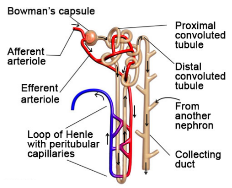
Fig. 2. Structure of the nephrons
The tubule continues to form a highly coiled structure known as Proximal Convoluted Tubule (PCT). The next part of the tubule is Henle’s loop which consists of ascending and descending limb. The ascending limb continues to form Distal Convoluted Tubule (DCT). The DCT opens into collecting duct.
There are two types of nephrons- cortical nephrons and medullary nephrons. When loop of Henle is short and extends only little to medulla are known as Cortical Nephrons. When loop of Henle is long and extends into the medulla are known as Medullary Nephrons.
Urine formation
It involves three main steps- glomerular filtration, tubular reabsorption, and secretion.
Glomerular Filtration– During this process blood enter the glomerulus via afferent arteriole. Water and nitrogenous waste move into the glomerulus and blood cells, proteins exit the glomerulus through efferent arteriole. On an average, kidney filters about 1100 ml to 1200 ml of blood per minute. The glomerular capillary blood pressure causes filtration of blood through 3 layers- endothelium that surround the glomerular blood vessels, the epithelium of Bowman’s capsule and a basement membrane in between these two layers. The epithelial cells present in the Bowman’s capsule are known as Podocytes which are arranged in an intricate manner to leave some minute spaces called Filtration Slits or Slit Pores.
The amount of filtrate produced by the kidneys per minute is known as Glomerular Filtration Rate.
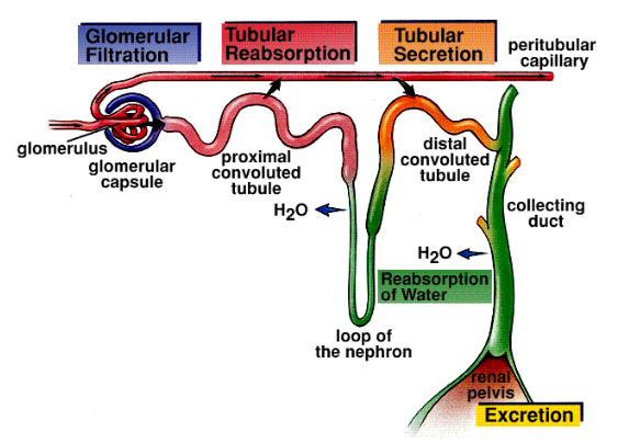
Fig. 3. Steps of urine formation
Tubular reabsorption– it involves the absorption of required molecules or ions such as sodium ions, glucose, amino acids etc. Some of the substances are absorbed actively and some are absorbed passively. Glucose, amino acids are absorbed actively whereas water is absorbed passively.
The last step of urine formation is secretion. Potassium ions, hydrogen ions, ammonia are secreted out to maintain the ionic and acid balance of the body fluids.
Function of the tubules
Proximal Convoluted Tubules (PCT) is lined by cuboidal brush border epithelium. 70-80% of electrolytes and water are reabsorbed in PCT. Secretion of hydrogen ions, potassium ions, ammonium ions also occurs into the filtrate.
Henle’s loop, very less reabsorption occurs in ascending limb. It is impermeable to water, but permeable to electrolyte. Descending limb absorbs most of the water, making the filtrate concentrated. Descending limb is almost impermeable to electrolyte.
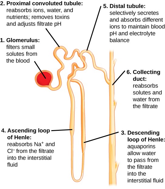
Fig. 4. Function of the tubules
Distal convoluted tubule, reabsorption of water, bicarbonate ions and sodium ions occurs. Secretion of potassium ions and hydrogen ions to maintain the electrolyte balance of the fluid.
Collecting duct, reabsorbs large amount of water to make the urine concentrated. Secretion of hydrogen ions and potassium ions also takes place in collecting duct. It maintains the ionic balance and pH of blood.
Regulation of kidney function
Kidney function is regulated by hypothalamus, Juxtaglomerular Apparatus (JGA) and heart. Changes in the blood volume, ionic balance activates the osmoreceptors in the body. This stimulates the hypothalamus to release Antidiuretic Hormone (ADH), also known as Vasopressin. This helps in the reabsorption of water from the tubules and prevents diuresis. This increase the blood volume and switch off the osmoreceptors for negative feedback mechanism.
The JGA is activated during fall in glomerular blood pressure. Juxtaglomerular cells release the renin which converts angiotensinogen in blood to angiotensin I which gets further converted to angiotensin II. Angiotensin II is a vasoconstrictor, so it increases the glomerular blood pressure. Angiotensin II also acts on adrenal cortex to release aldosterone to increase sodium ion and water reabsorption from the distal tubules. This mechanism is known as Renin-Angiotensin Mechanism.
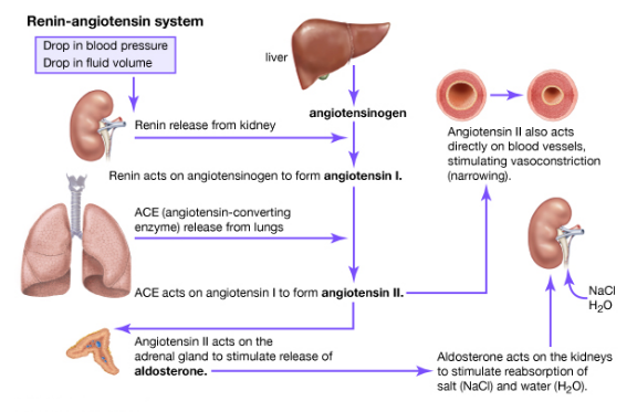
Fig. 5. Regulation of kidney function by renin-angiotensin-aldosterone system
Heart release Atrial Natriuretic Factor (ANF) which causes vasodilation to decrease the blood pressure. It acts as a negative feedback mechanism for renin-angiotensin mechanism.
Micturition
Urine formed in the nephrons is carried to the urinary bladder where it is stored until voluntary signal is given by the central nervous system. When urinary bladder is stretched, it gets filled with the urine. Stretch receptors present on the walls of the urinary bladder sends signal to the central nervous system. CNS initiates the contraction of the smooth muscles of the bladder allowing relaxation of urethral sphincter to release urine. This process of release of urine is known as Micturition. An adult human excretes, on an average, 1 to 1.5 liters of urine per day. The urine formed is a light yellow colored watery fluid which is slightly acidic (pH-6.0) and has a characteristic odor.