Human Reproduction
Table of Content
- Male Reproductive System
- Female Reproductive System
- Gametogenesis
- Spermatogenesis
- Oogenesis
- Menstrual Cycle
- Fertilization and Implantation
Humans are sexually reproducing animals. Human reproduction includes following processes:
- Formation of male and female gametes. This process is known as gametogenesis.
- Transfer of male gamete to female genital tract.
- Fusion of male and female gamete to form zygote. This process is known as fertilization.
- Attachment of zygote in the uterine wall. This is known as implantation.
- Embryo development.
- Parturition (delivery of baby outside the female body).
Male Reproductive System
It is in the pelvis region of the male body. Male reproductive system comprises of- a pair of testis, glands, accessary ducts, and male genitalia. The testis is located outside the abdominal cavity in a pouch-like structure known as scrotum. This location of testis is essential for proper development of sperms (male gamete) inside the testis. It helps to maintain a lower temperature than the body temperature needed for gametogenesis or spermatogenesis. Testis is divided into compartments known as testicular lobules.
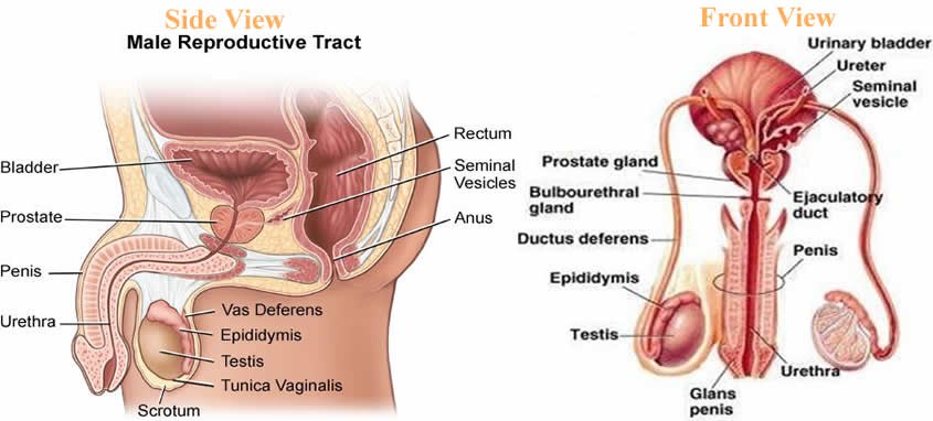
Fig.1. Parts of Male Reproductive System
Each testicular lobule consists of highly coiled structure known as seminiferous tubules. These are the site for sperm formation. Seminiferous tubules are lined by two types of cells known as- spermatogonia and sertoli cells. Spermatagonia also known as germ cells, give rise to sperms, the male gamete. Spermatogonia are diploid cells and they undergo meiosis to form sperms. Sertoli cells are elongated cells which nourishes the growing sperms. Outside the seminiferous tubule, the interstitial space contains cells known as leydig cells. Ledig cells are involved in synthesis of testicular hormone known as testosterone. Testosterone hormone is involved in development of secondary sexual characteristics such as moustaches and beard.

Fig.2. Section view of Seminiferous Tubule
Apart from these, male reproductive system consists of certain accessory ducts such as rete testis, vasa efferentia, epididymis and vas deferens. Rete testis are the network of tubules which are located in the hilum of the testis. It is a site where sperms are concentrated. Loss of this process leads to infertility.
Vasa efferentia are the small ducts that carry semen from the testis to the epididymis. It passes sperms to vas deferens. Vas deferens opens into urethra as the ejaculatory duct. The male external genitalia are known as penis. It facilitates insemination, that is, transfer of sperms to the female genitalia.
Male accessory glands include- prostate glands, seminal vesicle, and bulbourethral glands.
Seminal vesicles are paired glands that opens into the vas deferens near the junction with the urethra. Its secretion forms the part of semen. Semen is the fluid containing spermatozoa or sperm. The fluid of seminal vesicle is alkaline in nature. This alkaline nature helps in neutralization of alkalinity of the female vagina during insemination.
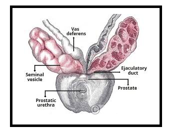
Fig.3. Accessory Glands
Prostate glands are in the neck of the urinary bladder. They secrete the fluid that makes up the semen and functions in nourishment as well as protection of the sperms.
Bulbourethral glands also known as Cowper’s glands. They are located inferior to the prostate gland. Their secretion is rich in glycoproteins. They also form a small component of semen.
Components of semen
Semen also known as seminal fluid contain sperms in it. It is made up of secretions from the prostate glands, bulbourethral gland, and seminal vesicle. It contains fructose, amino acids, citrate, proteins, proteolytic enzymes, calcium etc.
Female Reproductive System
The female reproductive system comprises of a pair of ovaries, uterus, cervix, vagina, and external genitalia. The primary female sex organ is a pair of ovaries. Ovaries produce female gamete known as ovum. It produces female hormones known as ovarian hormones such as estrogen, progesterone etc. Ovaries are located in the lower abdomen and they are 2 to 4 cm in length. The outermost layer is made up of germinal cells known as germinal epithelium. Each ovary is covered by a thin epithelium which encloses the ovarian stroma. The stroma is divided into outer cortex and inner medulla. The female accessory ducts consist of fallopian tubes, uterus and vagina.
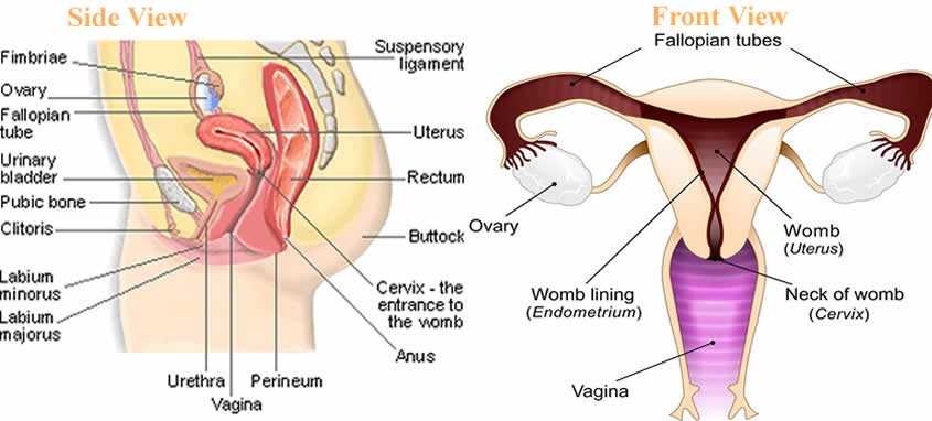
Fig.4. Female Reproductive System-side view and front view
Fallopian tubes also known as uterine tubes are fine tubes which are lined by ciliated epithelium. They lead from ovaries into the uterus. It allows the passage of egg from ovaries into the uterus. These tubes are also a site for fertilization.
Vagina is the tubular and muscular organ. It extends from the vulva to the cervix. It is covered by a membrane known as hymen. The vagina helps in sexual intercourse as well as child birth. The funnel shaped part closer to the ovary is known as infundibulum. Infundibulum contains finger-like projections known as fimbriae. They help in collection of ova after ovulation. Infundibulum opens into ampulla. The fallopian tube or oviduct ends at isthmus. Isthmus has a narrow lumen and it joins the uterus. The uterus appear pear shaped.
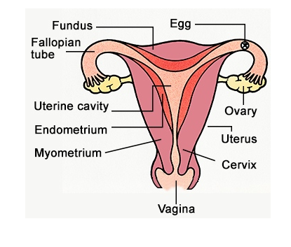
Fig.5. Diagrammatic section of Female Reproductive System
Uterus also known as womb is the site were fetus develop at the time of gestation. There are 3 layers of uterus- the external thin layer is known as perimetrium, middle layer made up of smooth layer is known as myometrium and the outer most layer which is glandular in nature is known as endometrium. It is the endometrium that lines the uterus. Myometrium brings about the contraction at the time of delivery of the baby.
The external female genitalia consist of – mons pubis, labia minora, hymen and clitoris. The fatty tissue that covers the pubic hair and skin is known as mons pubis. The folds that surround the vaginal opening is known as labia majora. The membrane that covers the opening of the vagina is known as hymen. The tiny finger-like projection that lies above the urethral opening is known as clitoris.
The characteristic feature of female mammal is the presence of mammary glands.
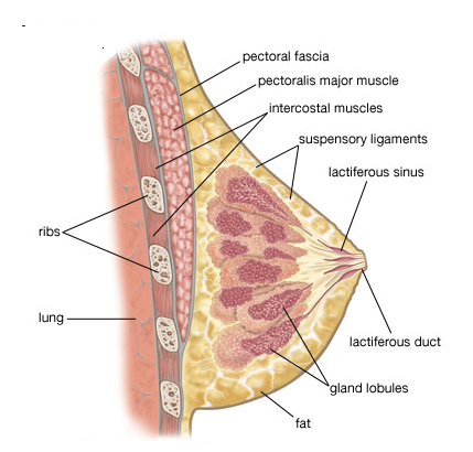
Fig.6. Mammary Glands
Mammary glands are paired structures that is made up of glandular tissue and fat. Glandular tissue comprises of mammary lobes that contain cells known as alveoli. The alveoli open into mammary tubules. These tubules then combines to form ducts.
Gametogenesis
The formation of male and female gametes, that is, sperms and ovum respectively is known as gametogenesis. Testis produce sperms in male whereas ovaries produces ovum or egg in females. The process of formation of sperms is known as spermatogenesis whereas formation of ova is known as testis.
Spermatogenesis
The process of spermatogenesis begins from diploid Spermatogonia in seminiferous tubules. Spermatogonia undergo mitotic divisions to increase their number. Some spermatogonia matures to form primary spermatocytes. Primary spermatocytes undergo meiosis to form secondary spermatocytes which are haploid in nature. One meiotic division of primary spermatocytes form two secondary spermatocytes. Now the second meiotic division begins, where secondary spermatocytes are converted into spermatids. The process of transformation of spermatids into sperm or spermatozoa is known as spermiogenesis. Then the sperms are finally released from seminiferous tubules by a process known as spermiation.
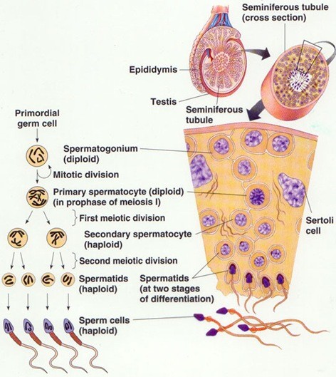
Fig. 7. Spermatogenesis
Sperm is uniflagellar structure which is motile in nature. They have a very short life span. Sperm is divided into head, middle piece, and the tail. Head contains the nucleus and it is surrounded by a structure known as acrosome. Acrosome contains the enzymes that degrades the wall of female ovum for fertilization. The middle piece contains the mitochondria that synthesizes ATP for the movement of the sperm into the female genital tract. The tail helps in movement of the sperm. It contains the flagella in it.
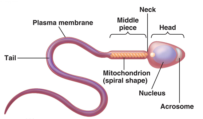
Fig. 8. Structure of the Sperm
Spermatogenesis is controlled by a gonadotropic hormone released from the hypothalamus. The two gonadotropins that are involved in spermatogenesis is luteinizing hormone and follicle stimulating hormone. Luteinizing hormone targets the leydig cells to secrete the male sex hormone known as testosterone. Follicle stimulating hormone targets the sertoli cells to nourish the sperms during spermatogenesis.
Oogenesis
The process of formation of female gamete or ovum is known as oogenesis. Oogenesis begins at the time of embryonic development of a female child. No oogonia or ovum is formed after the birth of a female child. These oogonia are arrested at prophase I of meiosis until a female child reaches the puberty. The oogonia which are arrested at prophase I of meiosis are known as primary oocytes.
Then the primary oocyte is surrounded by a layer of cells known as primary follicle.
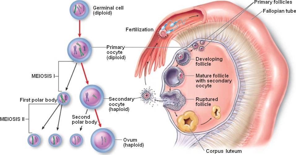
Fig. 9. Oogenesis
Primary follicle which are then later surrounded by a layer of cells is known as secondary follicles. The secondary follicle is transformed into tertiary follicle which is filled by a fluid known as antrum. At this stage the first meiotic division is complete. The first meiotic division forms a secondary oocyte and one small polar body. The secondary oocyte matures into graffian follicle. The graffian follicle is surrounded by a membrane known as zona pellucida. The graffian follicle then ruptures to release an oocyte from the ovary. The process of release of an oocyte from the ovary is known as ovulation.
Menstrual Cycle
The reproductive cycle of female primates such as apes, monkeys, females etc. is known as menstrual cycle. The onset of menstruation at puberty is known as menarche. Single ovum is released in a single cycle of menstruation. Menstrual cycle is of 28 days. It is divided into following phases-
- Menstrual phase (from day 1 to 5) – The body discharge the blood vessels from the vagina in the form of menstrual fluid. About 10 to 80 ml blood is lost in case of normal menstrual cycle.
- Follicular phase (from day 1 to 13) – In this phase, an ovum matures or prepares itself for ovulation. The lining of the uterus known as endometrium gets thickened due to lining of blood vessels. The secretion of luteinizing hormone and follicular stimulation hormone increases in this phase.
- Ovulation phase (day 14)- It is the phase where ovum is released from the ovary.
- Luteal phase (day 15 to 28) – The released ovum remains in the fallopian tube for about 24 hours. If the sperm does not enter the body, the ovum degenerates. The ovum or matured graffian follicle converts into corpus luteum. If fertilization does not corpus luteum degenerates. If fertilization occurs, corpus luteum will secrete a pregnancy hormone known as progesterone, required to maintain pregnancy.
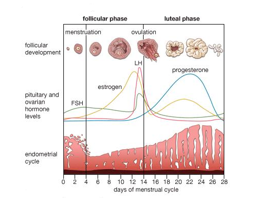
Fig. 10. Menstrual Cycle
Around 50 years the menstrual cycle ceases, this is known as menarche.
Fertilization and Implantation
The sperm that enters the female body via insemination, reaches to the female fallopian tube. The ovum released from the ovary also reaches the fallopian tube. Fallopian tube is a site where fertilization occurs. The process of fusion of male gamete, sperm with female gamete ovum is known as fertilization.
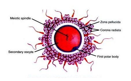
Fig. 11. Ovum surrounded by sperms during Fertilization
The sperm first meets zona pellucida layer of the ovum and induces membrane block to prevent entry of additional sperms. The secretions from the acrosome such as hyaluronidase enzyme help the sperm to enter the ovum. Now the second meiotic division of secondary oocyte is complete. This second meiotic division give rise to second polar body and an ovum.
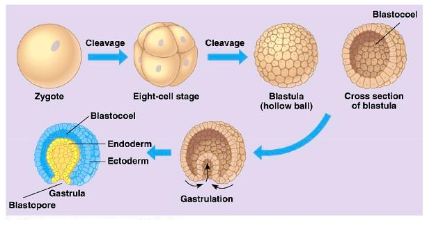
Fig.12. Embryo Development
Fertilization forms a diploid zygote. Zygote undergo various mitotic divisions to form cells known as blastomeres. These cells form a compact mass of cells known as morula (8-17 cells). Morula undergo further mitotic divisions to form blastocyst which moves further towards the uterus from the fallopian tube. The blastomeres of the blastocyst are arranged into an outer layer called trophoblast and an inner group of cells attached to trophoblast called the inner cell mass. The trophoblast layer will gets attached to the endometrium and the inner cell mass will differentiate as the embryo. The attachment of the blastocyst in the uterus is known as implantation. This marks the beginning of the pregnancy. Blastocyst then later transform into gastrula. It is the stage where 3 germ layers are formed- outermost ectoderm, middle mesoderm, and innermost ectoderm. These germ layers form different tissues and organs during embryonic development.
Pregnancy and Embryonic Development
After implantation, the uterine lining forms a structure known as placenta, for communication between the fetus and the maternal body. The placenta helps in the exchange of the nutrients and gases between the mother and the fetus. It also involved in removal of waste produced by the growing fetus. The placenta is connected to the fetus via umbilical cord. Placenta also produces different hormones needed during or after pregnancy. The hormones such as human chorionic gonadotropin (hCG), human placental lactogen (hPL), estrogens, progestogens, etc. In the later stages of pregnancy, a hormone called relaxin is also secreted by the ovary. This hormone helps in uterine contraction at the time of parturition.

Fig.13. The human fetus within the uterus
As shown from the above figure, amnion is the membrane that covers the embryo and the fluid filled in it is known as amniotic fluid. Amnion is protective in nature. It protects the growing embryo from mechanical shock or injury.
Parturition and Lactation
The process of delivery of the baby or better known as childbirth is known as parturition. There are various hormones that acts at the time of child birth such as – oxytocin and relaxin. Both promotes uterine contraction for the expulsion of the baby from the female external genital tract. During pregnancy, the mammary glands starts to produce milk. This process is known as lactation. Just after childbirth, the initial milk ejected from the maternal mammary glands is known as colostrum. The steps for the parturition are explained below in the form of the figure-
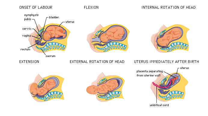
Fig.14. Parturition