| Locomotion and Movement Refresher Course |
| Locomotion and Movement Concepts Files |
| Locomotion and Movement Master Files |
| Locomotion and Movement Revision Note |
| Locomotion and Movement Note 1 |
| Locomotion and Movement Reference Book |
Locomotion and Movement
Table of Content
- Types of Movement
- Muscle
- Structure of Muscle
- Structure of contractile proteins
- Mechanism of muscle contraction
- Lactic Acid formation in muscles
- Skeletal system
What is Locomotion Movement? What is a Locomotion in biology?
Movement is one of the important feature of living organisms. When movement result in change in position or location, is known as Locomotion. For Example, Walking, Climbing, Running etc.
Types of Movement
The three main types of movement are – Ciliary, Amoeboid and Muscular.
What is the movement of cilia?
Ciliary movement occurs in internal tubular organs lined by ciliated epithelium. It is used to remove dust particles that enter during the inhalation, it also helps in movement of ova in the female reproductive tract.

Fig. 1. Ciliary movement in Paramecium
Amoeboid movement is observed in some immune cells such as macrophages, leucocytes etc. It is also observed in Amoeba, which moves through pseudopodia.
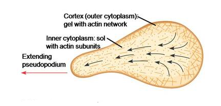
Fig. 2. Amoeboid movement in Amoeba
Muscular movement is observed in tongue, jaws, limbs etc. For Locomotion, Muscular, skeletal and neural system are involved.
Muscle
Muscle is a tissue of mesodermal origin. It is a tissue involve in movement of the body. There are three types of muscle – Skeletal Muscles, Visceral Muscles and Cardiac Muscles.
Skeletal Muscles are voluntary muscles which are under the control of somatic nervous system. It is also known as Striated Muscles because of the characteristic cross-striations present in the muscle tissue. Muscles are attached to bones by tendons. They are involved in different body movements and body posture.
Visceral muscles are also known as Smooth Muscles. They are non-striated in appearance. They control involuntary movements in the body. They line the internal organs such as alimentary canal, reproductive tract, etc.
Cardiac muscles are muscles of the heart that help in the rhythmic contraction and relaxation of the heart. They are involuntary muscles. They have cross-striations with branching pattern.
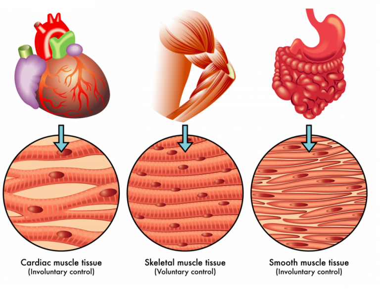
Fig. 3. Types of muscles
Structure of the muscle
Skeletal Muscle is made up of number of muscle bundles also known as Fascicles. Each muscle bundle is made up of number of muscle fibers. Sarcolemma is a type of plasma membrane which lined the muscle fiber. Sarcolemma encloses the sarcoplasm. Muscle fiber is made up of multiple nuclei, so it is known as Syncytium. The endoplasmic reticulum of the muscle fiber is known as Sarcoplasmic Reticulum. Sarcoplasmic Reticulum stores calcium ion that participates in muscle contraction. Muscle fiber contain parallelly arranged filaments known as Myofibrils or Myofilaments. The fibrous tissue that surround the skeletal muscle is known as Epimysium.
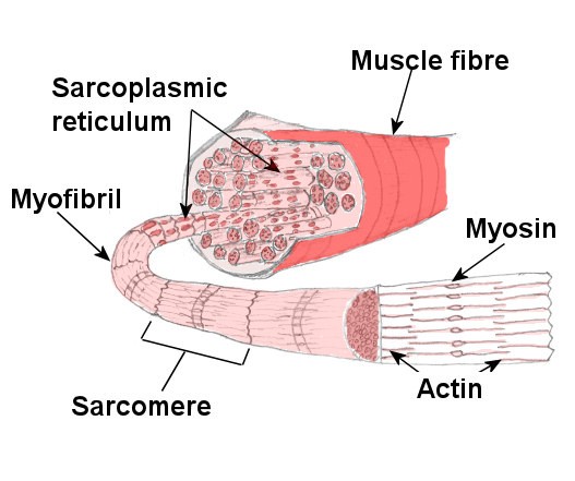
Fig. 4. Structure of the muscles
Myofibrils have characteristic cross-striations due to presence of two proteins – Actin and Myosin. The light bands also known as Isotropic, contain actin protein, whereas dark band known as Anisotropic which contains myosin. Actin filaments are thin filaments whereas myosin are thick filaments.
In the center of each actin band there is a stretch of the elastic fiber known as Z-line. The portion of the myofibril between the two successive Z lines is known as Sarcomere. Sarcomere is considered as the functional unit for muscle contraction.
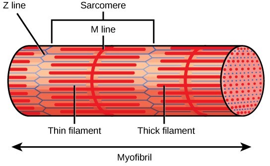
Fig. 5. Structure of sarcomere
Structure of contractile proteins
The monomeric unit of actin is known as G actin or globular actin. Polymers of G actin form F actin or F filaments. Two F filaments wound around each other to form actin. Another protein tropomyosin, run around the F actin. Another protein, troponin is distributed at regular intervals on tropomyosin.
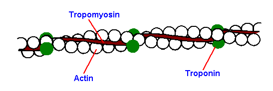
Fig. 6. Structure of the actin
Myosin protein is also made up of monomeric known as Meromyosins. Each meromyosin has two regions: globular head and a long tail.
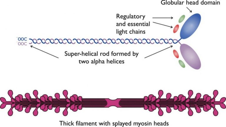
Fig. 7. Structure of the myosin
The globular head has ATPase activity and binding site for actin.
Mechanism of muscle contraction
Sliding filament theory explains the mechanism of muscle contraction. Sliding of the thin filaments over thick filaments drive the muscle contraction. Muscle contraction starts when a signal is sent from a central nervous system via motor neuron. The junction between the motor neuron and the muscle fiber is known as Neuromuscular Junction. Neurotransmitter such as acetylcholine is released at neuromuscular junction which generates action potential in the sarcolemma.
Action potential causes the release of calcium ions from the sarcoplasmic reticulum into the sarcoplasm. Increase calcium level cause calcium ions to bind to troponin on actin filaments and unveil the active sites for myosin binding. The ATPase activity of myosin exposes sites to allow cross bridge formation between actin and myosin. This causes shortening of sarcomere to bring the muscle contraction.
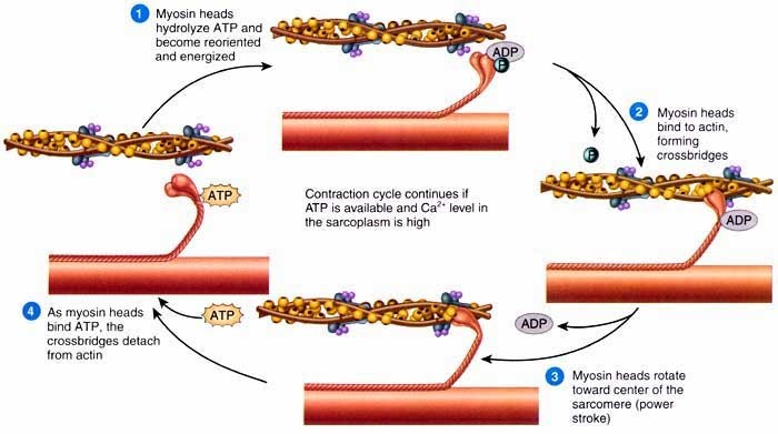
Fig. 8. Sliding filament theory
The process stops or the muscle relaxes when calcium ions are pumped back into the sarcoplasmic reticulum. This step masks the actin filaments bringing muscles to its original position.
Lactic Acid formation in muscles
Repeated muscle activation, For Example, during physical work such as running causes lactic acid accumulation in muscles. This occurs due to anaerobic breakdown of glycogen in muscles. This causes muscle fatigue. This is also due to insufficient oxygen supply to the muscles.
Skeletal system
It consists of bones and cartilages. It helps in the movement of the body. Bone is hard due to presence of calcium salts in it whereas cartilage is flexible due to presence of chondroitin sulphate. Humans contain 206 bones and few cartilages. The two divisions of skeletal system are – Axial Skeletal System and Appendicular Skeletal System.
Axial skeletal consists of 80 bones. It consists of skull, sternum, vertebral column, and ribs.
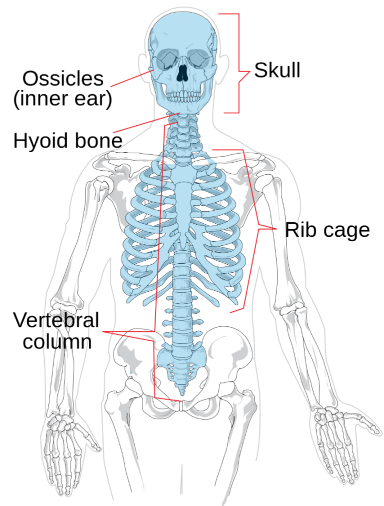
Fig. 9. Axial skeletal system
Skull consist of cranial and facial bones which are 22 in number. Cranial bones are 8 in number which protects the brain. The facial region consists of 14 skeletal elements which form the front part of the skull. Hyoid bone which is U-shaped in structure is present at the base of the buccal cavity. Each middle ear consists of three small bones – Malleus, Incus, and Stapes. These 3 bones together are known as Ear Ossicles.
Vertebral column is formed of 26 serially arranged units known as Vertebrae. It extends from
the base of the skull and forms the main framework of the trunk. Each vertebra has a central hollow part known as Neural Canal through which the spinal cord passes.
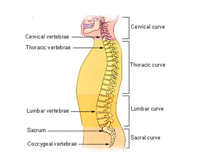
Fig. 10. Vertebral column
First vertebra is the atlas and it articulates with the occipital condyles. The vertebral column is differentiated into 7 cervical, 12 thoracic, 5 lumbar, 1 sacral and 1 coccygeal regions starting from the skull. The number of cervical vertebrae is conserved among the mammals.
Sternum is a flat bone on the ventral midline of thorax. There are 12 pairs of ribs. Ribs are curved bones that form the ribcage. Ribs are attached to sternum.
First seven pairs of ribs are known as True Ribs as they are directly attached to the sternum. The 8th, 9th and 10th pairs of ribs join the seventh rib instead of joined directly to sternum. These are known as False Ribs. Last 2 pairs (11th and 12th) of ribs are not connected directly to the sternum, they are known as Floating Ribs. Thoracic vertebrae, ribs and sternum combines to form rib cage.
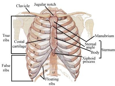
Fig. 11. Sternum
Appendicular Skeleton consists of bones of limbs and girdles. Each limb is made of 30 bones. The bones of the forelimbs or hands are humerus, radius and ulna, carpals (wrist bones which are 8 in number), metacarpals (palm bones which are 5 in number) and phalanges (digits which are 14 in number).
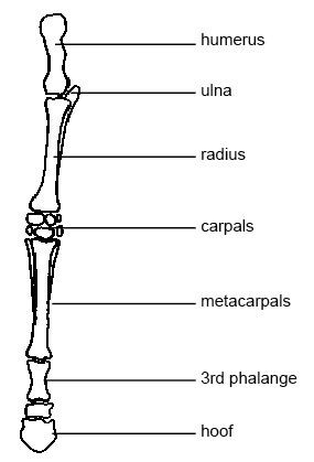
Fig. 12. Bones of forelimbs
The bones of the hind limbs or legs are- Femur is the thigh bone (longest bone), tibia and fibula, tarsals are ankle bones which are 7 in number, metatarsals are 5 in number and phalanges are 14 in number. Knee cap contains a cup shaped bone known as Patella.
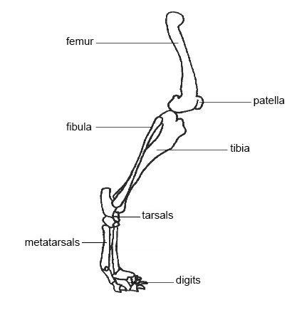
Fig. 13. Bones of hindlimbs
Pectoral Girdle consists of two bones clavicle and scapula. Scapula contains the cavity known as Glenoid Cavity which forms ball and socket joint with the humerus. It articulates with the forelimb bones.
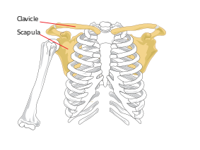
Fig. 14. Bones of pectoral girdle
Pelvic girdle contains a cup shaped cavity known as Acetabulum that forms ball and socket joint with the femur. It articulates with the hind limb bones. The muscles of the hip are attached to pelvic girdle.
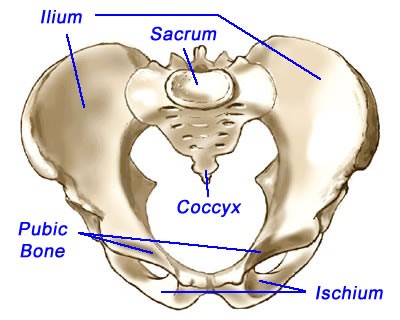
Fig. 15. Bones of pelvic girdle