Insect leg dissection
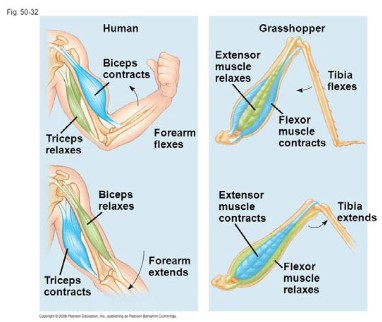
- Extensor muscle relaxes and flexor muscle contracts \(\rightarrow \) tibia contracts
- Extensor muscle contracts and flexor muscle relaxes \(\rightarrow \) tibia extends
- Step one and step two gives insect jump action.
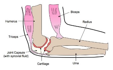
Elbow Joint
- The type of joint determines the amount of movement that is possible
- For ball and socket joints, such as the hip or the shoulder, movement through all three planes are possible. At the hip joint, the head of the femur is the ball the fits into the socket of the pelvis. The movements possible at the joint are flexion, extension, rotation, abduction and adduction.
- For hinge joints, such as the knee, flexions (bending) and extensions (straightening) are the possible movements (movement in one plane); however, slight side to side movements are possible
- Elbow joint is an example of a hinge joint
- Cartilage: reduces friction in the joint, provides high tensile strength and support, and absorbs compression
- Synovial fluid: reduces friction by providing lubrication between the cartilage and other tissues in joints during movement
- Joint capsule: seals the joint space and provides stability to the joint by limiting movements
- Tricep: contracts and causes extension (arm straightening)
- Bicep: contracts and causes flexion (arm bending)
- Radius: Lever attached to the biceps. When the biceps contract, the radius provides a solid structure for lifting
- Ulna: Lever connected to the triceps. When the triceps contract, the ulna provides support as a lever as the arm straightens out
- Humerus: arm bone
- Ligament: connects bones together
- Tendon: connects bones with muscles
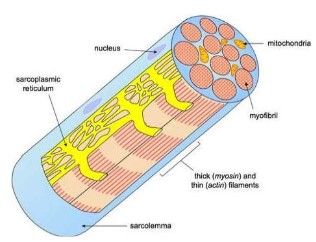
Muscle cells
- Skeletal muscles are composed of bundles of muscle fibers and have a striped appearance because of areas of thick and thin filaments (myosin and actin)
- Muscle cells have many nuclei(mutinucleated) and are long because the embryonic muscle cells fuse together.
- Muscle fibers are composed of many parallel elongated fibers called myofibrils.
- A modified endoplasmic reticulum, called the sarcoplasmic reticulum (fluid-filled membranous sacs), extends throughout the muscle fibre, wrapping around each myofibril, sending a signal to the all parts of the muscle fibre to contract at the same time
- Myofibrils – rod-shaped parallel bodies consisting of actin and myosin filaments
- Sarcolemma – plasma membrane of the muscle cell.
- Mitochondria – large numbers; found dispersed around individual myofibrils
Sacromere
- is the repeating unit of a striated muscle cells.
- Lies between two Z lines which are dense protein discs.
- Contains the thick filament (myosin) and thin filament (actin).
- Myosin contains a head which binds to the binding site on the actin; interaction between myosin and actin (cross-bridge) is responsible for muscle contraction.
- Myosin is seen as dark bands while actin is seen as light bands.
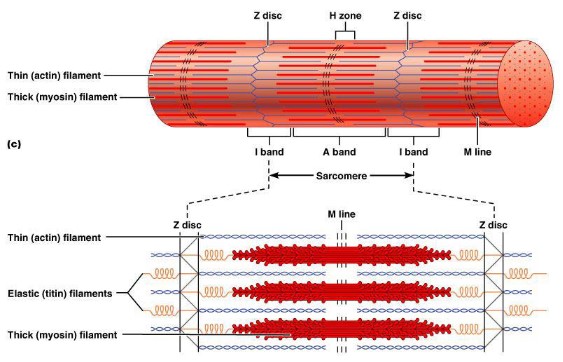
* no need to know titin; M line; H zone; I band, A band, M line. Only needs to remember Z line; myosin and actin filaments and their construction pattern
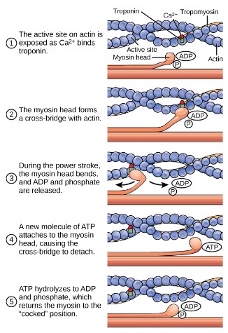
Muscle contraction mechanism
- During a muscle contraction, myosin filaments pull actin filaments towards the centre of the sarcomere
- This shortens the sarcomere and the overall length of the muscle fibre
- When this occurs, the myosin heads bind to sites on the actin filaments, creating cross-bridges, pulling (sliding) the actin filaments along the myosin filaments with energy from ATP
1) Action potential causes sarcoplasmic reticulum to release \(Ca^{2+}\)
2) Influx of \(Ca^{2+}\) into sarcoplasm and binds to troponin
3) Binding of troponin releases tropomyosin, which exposes myosin binding sites on actin
4) Myosin heads are bound to actin binding sites making a cross-bridge
5) ATP releases the myosin head
6) Hydrolysis of ATP to ADP causes a change in shape (power stroke)
7) The myosin heads bind to actin filaments forming cross-bridges at a site one position further from the centre of the sarcomere
8) Myosin head forms a new cross-bridge with a different actin binding site pulling the myosin towards the centre of the sarcomere, continuing the contraction.
9) The step 5,6,7,8 repeats, pulling the Z lines together and contracting the entire
muscle