Question
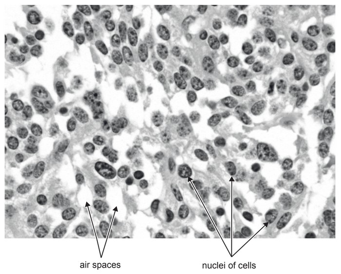
The light micrograph shows tumour tissue from a patient’s lung.
(a) State one cause of lung cancer.
(b) Suggest one difference between tissue taken from a lung cancer tumour and normal lung tissue that might be seen in micrographs.
(c) The lung tumour in the light micrograph was slow-growing. Predict with a reason what would have been visible in the micrograph if the tumour was growing rapidly.
Answer/Explanation
Answer:
(a)
a. smoking/tobacco;
b. passive smoking;
c. Radon/other radiation;
d. exposure to arsenic/asbestos/smoke from coal burning/fires/silica/rock
dust/vehicle exhaust fumes/nitrogen oxides;
(b)
a. fewer/smaller/lack of alveoli/air spaces;
b. many cells/nuclei per area / much denser tissue;
c. more cells undergoing mitosis (in the tumour);
(c)
See
a. more mitosis
OR
cells in prophase/metaphase/anaphase/telophase;
Why
b. more dividing cells/tumour cell divide uncontrollably
OR
a higher mitotic index;
Question
Draw the ultrastructure of a prokaryotic cell based on electron micrographs. [3]
▶️Answer/Explanation
Ans
a.cell wall;
b.plasma membrane; Clearly shown as a separate line under the cell wall or theinner line
c.cytoplasm AND 70S ribosomes; Do not allow (small) circles
d.nucleoid/naked DNA;
e.plasmid
OR
pili
OR
flagella/flagellum;

Question
a. Outline the cell theory[2]
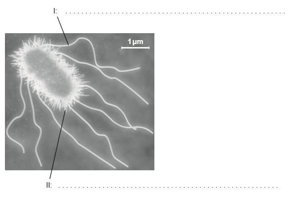
b (i) Annotate the electron micrograph of the Escherichia coli cell with the function of the structures labelled I and II.[2]
b (ii) falculate the magnification of the electron micrograph.[4]
▶️Answer/Explanation
Markscheme
a. living things are composed of cells;
b. cells are the basic/smallest unit of life;
c. cells come from pre-existing cells;
Do not accept cells are the “smallest organisms”.
Do not accept “cells are the building blocks” of life on its own.
a. I: locomotion;
b. II: attachment to surfaces / holds bacteria together / conjugation;
Do not accept “exchange material” on its own.
If more than one function is given, mark the first answer only.
×15 000 (accept answers in the range of × 14 000 to × 16 000)
Examiners report
Most gained both marks for their knowledge of cell theory in part a.
Students who read the rubric correctly in b scored well. However approximately half of the students read “label” instead of “function”.
A disappointing number had no idea how to calculate the magnification (15000X) and some answers showed no concept of scale, with answers such as 0.15 X or 5×10⁻⁷ X.
Question
The diagram shows how vesicles are used to transport materials in a cell.
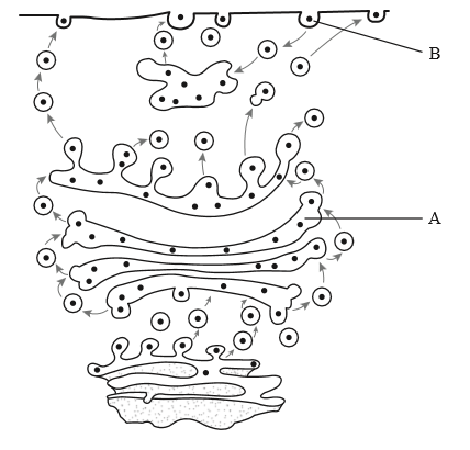
State the name of organelle A.
State the process occurring at B.
Describe how the structure of the membrane allows the formation of vesicles.
Explain active transport across membranes.
▶️Answer/Explanation
Markscheme
Golgi apparatus/complex/body
Reject Golgi vesicle and Golgi unqualified.
endocytosis/phagocytosis/pinocytosis
Reject exocytosis.
a. fluidity of membrane allows change of shape/invagination/formation of vesicles;
b. phospholipids can move / phospholipid bilayer makes membrane fluid/flexible;
c. weak bonding between phospholipid tails;
d. bends/kinks in the phospholipid tails prevent close packing;
e. cholesterol affects membrane fluidity;
a. moves substances up/against a concentration gradient / from lower to higher concentration;
b. protein/pump (in membrane) that moves material; (reject channels)
c. ATP is used; (reject energy alone)
d. example/labeled diagram showing mechanism;
Question
State the functions of the following organelles of a eukaryotic animal cell: lysosome, Golgi apparatus, free ribosomes, plasma membrane, rough endoplasmic reticulum.
Distinguish between anaerobic and aerobic cell respiration in eukaryotes.
Explain the mechanism of ventilation in the lungs in order to promote gas exchange for cell respiration.
▶️Answer/Explanation
Markscheme
lysosome:
a. (from Golgi apparatus) with digestive enzymes / break down food/organelles/ cell;
Golgi apparatus:
b. site that processes/modifies/packages and releases proteins;
free ribosomes:
c. site of synthesis of proteins (released to cytoplasm);
plasma membrane:
d. controls entry and exit of materials/substances in cell;
rough endoplasmic reticulum:
e. synthesis and transport of proteins; (both needed)
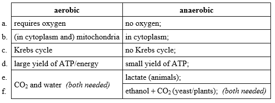
Award [1] for each contrasting characteristic.
Table format is not necessary for the marks.
a. inspiration/inhalation brings air into lungs;
b. external intercostal muscles contract;
c. and move rib cage upwards and outwards;
d. diaphragm flattens/contracts;
e. increasing thoracic volume;
f. pressure decreases from atmospheric pressure so air rushes into lungs;
g. expiration/exhalation forces air out;
h. internal intercostal muscles contract / external intercostal muscles and diaphragm relax;
i. abdominal/abdomen wall muscles contract and push diaphragm upwards;
j. decreasing thoracic volume;
k. increasing pressure in lungs so air is forced out;
l. a concentration gradient between air sacs and blood needs to be maintained;
Question
The diagram shows some of the structures in an animal cell.
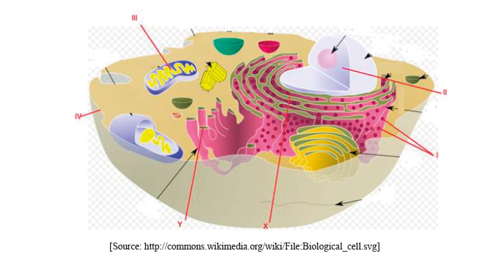
(i) Label structures I, II, III and IV.
I.
II.
III.
IV.
(ii) State one function of structure III.
Explain how materials are transported within a cell between structures X and Y.
▶️Answer/Explanation
Markscheme
(i) Award [1] for any two of the following correctly labeled.
I. ribosomes
II. nucleus (do not accept nuclear membrane)
III. mitochondrion
IV. plasma/cell membrane
(ii) ATP production/site of aerobic respiration (do not accept energy production)
(protein) material transported by vesicles;
from rER to Golgi apparatus/complex/body/membrane;
vesicles bud off from rER/fuse with Golgi membrane (due to membrane fluidity);
Do not accept vacuole(s).