Question
Which statement applies to cholesterol?
It is hydrophobic and found on the outside of the phospholipid bilayer.
It is hydrophilic and found inside the phospholipid bilayer.
It impacts membrane fluidity.
It is transported in association with glucose in the blood.
▶️Answer/Explanation
Ans: C
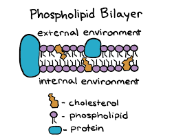
Cholesterol is a type of lipid that is found in the cell membrane. It has a hydroxyl group that can attach to the phosphate head of the phospholipids, and a hydrocarbon tail that can interact with the fatty acid tails of the phospholipids. Cholesterol affects the membrane fluidity by changing the space and interaction between the phospholipids.
At high temperatures, cholesterol decreases fluidity by making the phospholipids pack more tightly and reducing their movement. This also makes the membrane less permeable to water-soluble substances.
At low temperatures, cholesterol increases fluidity by preventing the phospholipids from packing too closely and becoming rigid. This helps the membrane to maintain its flexibility and function.
Question
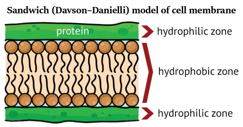
The Davson–Danielli model of membrane structure proposed that membranes were composed of a phospholipid bilayer that lies between two layers of globular proteins, as shown in this diagram.
What evidence supported this model?
An electron micrograph that showed two dark lines with a lighter band in between
Freeze-fracture electron microscopy
Evidence that all membranes are identical
The hydrophobic regions of protein would be in contact with water.
▶️Answer/Explanation
Ans: A

The Davson-Danielli model was a model of the plasma membrane of a cell, proposed in 1935 by Hugh Davson and James Danielli. The model describes a phospholipid bilayer that lies between two layers of globular proteins. The model was supported by electron microscopy, which showed three distinct layers within a cell membrane, with an inner white core and two flanking dark layers. The model also explained the selective permeability of the membrane and its thinness.
However, the Davson-Danielli model was later falsified by several lines of evidence, such as:
– Membrane proteins were discovered to be insoluble in water and varied in size, which contradicted the idea of a uniform and continuous protein layer around the membrane.
– Membrane freeze-fracturing revealed irregular rough surfaces in the membrane, representing trans-membrane integral proteins that spanned the lipid bilayer.
– Fluorescent antibody tagging of membrane proteins demonstrated their fluidity within the membrane, which was inconsistent with the rigid protein layers in the Davson-Danielli model.Therefore, the Davson-Danielli model is not supported by current evidence and has been replaced by the fluid mosaic model, which includes transmembrane proteins and accounts for the dynamic nature of the membrane.
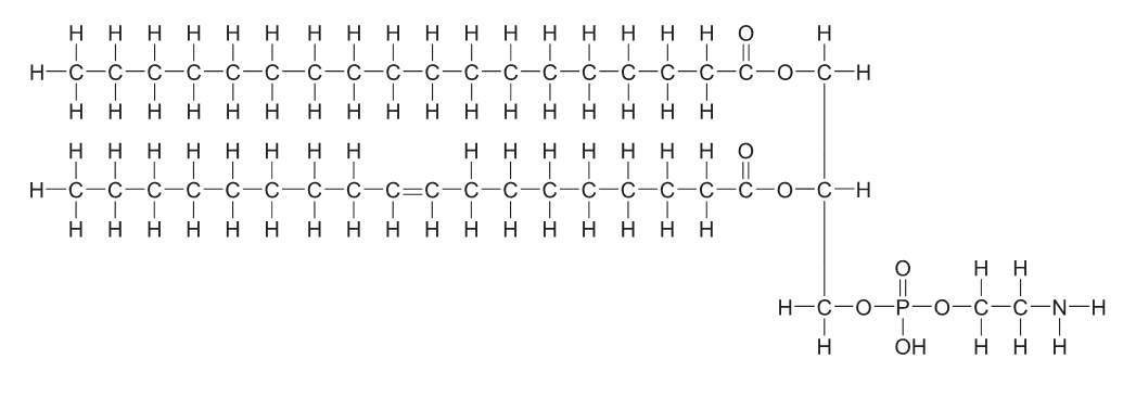
A diagram of a membrane

[Source: © International Baccalaureate Organization 2017]
In the diagram, which part of the membrane structure does the molecule below form?
▶️Answer/Explanation
Markscheme
A
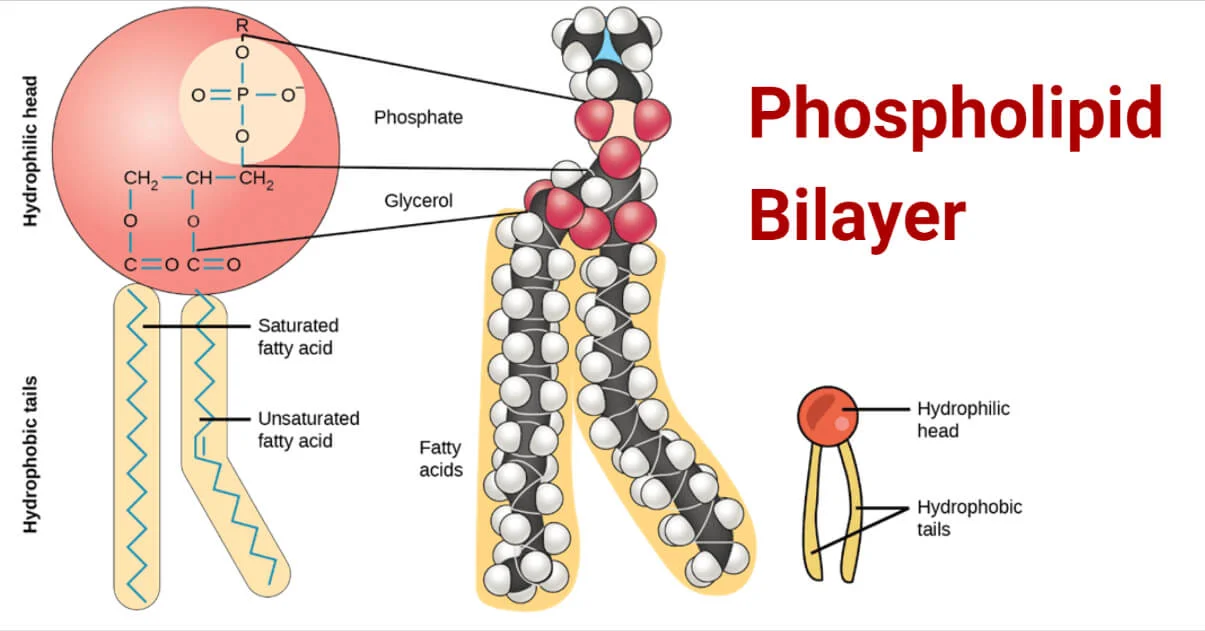
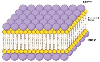
The polar head of a lipid bilayer is the hydrophilic region of a phospholipid molecule that forms the outer surface of the membrane. It contains a phosphate group that is electrically charged and can interact with water molecules. The polar head is attached to a glycerol backbone that links it to the nonpolar tail of the phospholipid. The tail is composed of two fatty acid chains that are hydrophobic and face inward in the bilayer. The polar head and the nonpolar tail give the phospholipid a amphiphilic property, meaning it has both water-loving and water-hating parts. This allows the phospholipids to form a stable barrier between the cell and its environment.
The cell membrane model proposed by Davson–Danielli was a phospholipid bilayer sandwiched between two layers of globular protein. Which evidence led to the acceptance of the Singer–Nicolson model?
A. The orientation of the hydrophilic phospholipid heads towards the proteins
B. The formation of a hydrophobic region on the surface of the membrane
C. The placement of integral and peripheral proteins in the membrane
D. The interactions due to amphipathic properties of phospholipids
▶️Answer/Explanation
C
The Singer-Nicolson model of lipid bilayer was proposed in 1972 by S.J. Singer and G.L. Nicolson to explain the structure and function of cell membranes. They suggested that membranes are dynamic structures composed of proteins and phospholipids, in which the phospholipid bilayer is a fluid matrix for proteins.
Some of the evidence that supported their model were.
Labeling experiments, x-ray diffraction, and calorimetry showed that integral membrane proteins diffuse at rates affected by the viscosity of the lipid bilayer, and that the molecules within the cell membrane are dynamic rather than static.
– Microscopy and thermodynamic data did not support previous models that had proteins present as sheets neighboring a lipid layer, or as repeating, regular units of protein and lipid.
– The Frye-Edidin experiment showed that when two cells are fused, the proteins of both diffuse around the membrane and mingle rather than being locked to their area of the membrane.
– Fluorescence microscopy and structural biology have validated the fluid mosaic nature of cell membranes and their involvement in various cellular processes.
Question
The Davson–Danielli model of membrane structure proposed that membranes were composed of a phospholipid bilayer that lies between two layers of globular proteins, as shown in this diagram.
What evidence supported this model?
An electron micrograph that showed two dark lines with a lighter band in between
Freeze-fracture electron microscopy
Evidence that all membranes are identical
The hydrophobic regions of protein would be in contact with water.
▶️Answer/Explanation
Ans: A
The Davson-Danielli model was a model of the plasma membrane of a cell, proposed in 1935 by Hugh Davson and James Danielli. The model describes a phospholipid bilayer that lies between two layers of globular proteins. The model was supported by electron microscopy, which showed three distinct layers within a cell membrane, with an inner white core and two flanking dark layers. The model also explained the selective permeability of the membrane and its thinness.
However, the Davson-Danielli model was later falsified by several lines of evidence, such as:
– Membrane proteins were discovered to be insoluble in water and varied in size, which contradicted the idea of a uniform and continuous protein layer around the membrane.
– Membrane freeze-fracturing revealed irregular rough surfaces in the membrane, representing trans-membrane integral proteins that spanned the lipid bilayer.
– Fluorescent antibody tagging of membrane proteins demonstrated their fluidity within the membrane, which was inconsistent with the rigid protein layers in the Davson-Danielli model.Therefore, the Davson-Danielli model is not supported by current evidence and has been replaced by the fluid mosaic model, which includes transmembrane proteins and accounts for the dynamic nature of the membrane.

A number of different proteins are involved in nerve function. Which of the following does not require a membrane protein?
A. Active transport of sodium
B. Diffusion of K+ into the cell
C. Diffusion of the neurotransmitter across the synapse
D. Binding of the neurotransmitter to the post-synaptic membrane
▶️Answer/Explanation
Markscheme
C
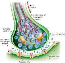
Diffusion of the neurotransmitter across the synapse is a process that involves the release of chemical messengers from the presynaptic neuron and their binding to receptors on the postsynaptic neuron. The neurotransmitter molecules then diffuse across the synaptic cleft, a fluid-filled space that separates the two neurons. This diffusion does not require the membrane protein because it is a passive process that depends on the concentration gradient of the neurotransmitter. The higher the concentration of the neurotransmitter in the presynaptic neuron, the faster it will diffuse across the synaptic cleft. The diffusion stops when the neurotransmitter binds to the receptor or is removed from the synaptic cleft by reuptake or degradation.
Question
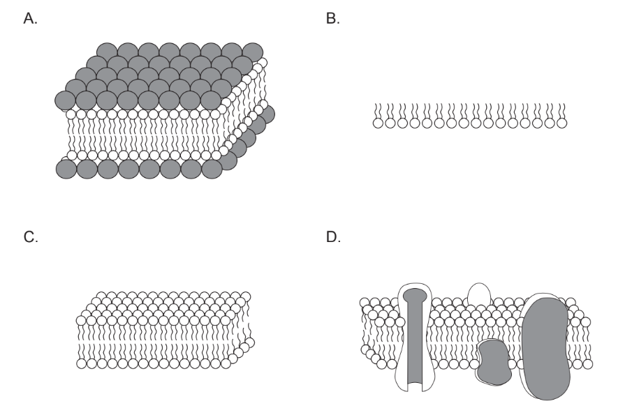
In 1925, Gorter and Grendel carried out an experiment to study the structure of cell membranes in different mammals. The total surface area of red blood cells was measured in a sample and compared to the surface area formed by a single layer of lipid extracted from cell membranes and floated on water.
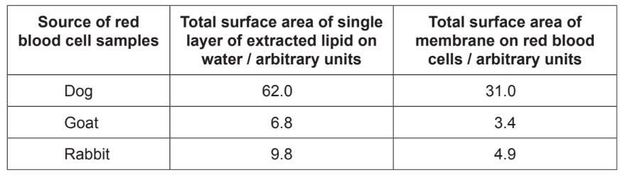
Which diagram best illustrates Gorter and Grendel’s conclusion drawn from this experiment?
▶️Answer/Explanation
Ans:B