Topic 1.6: cell division
In the Cell Cycle unit you will learn about the function and process of mitosis. Emphasis is placed on key checkpoints of the cell cycle, at which cancer cells behave differently that noncancerous cells.
The unit is planned to take 3 school days.
- Cell division is essential but must be controlled.
- Serendipity and scientific discoveries—the discovery of cyclins was accidental. (1.4)
- Outline the discovery of cyclins including the role of serendipity.
1.6.U.1 Mitosis is division of the nucleus into two genetically identical daughter nuclei . [The sequence of events in the four phases of mitosis should be known. To avoid confusion in terminology, teachers are encouraged to refer to the two parts of a chromosome as sister chromatids, while they are attached to each other by a centromere in the early stages of mitosis. From anaphase onwards, when sister chromatids have separated to form individual structures, they should be referred to as chromosomes.]
- State the function of mitosis.
- List four processes which involve mitosis.
- State the names of the four phases of mitosis.
- Draw typical eukaryotic cells as they would appear during the interphase and the four phases of mitosis.
- Outline four events that occur during prophase.
- Outline the process of metaphase, inclusive of the role of microtubules and the kinetochore.
- Outline the process of anaphase.
- Outline four events that occur during telophase.
What is (and is not) mitosis?
Mitosis functions as part of the process by which cells are cloned to make genetically identical daughter cells
There are four key reasons why a cell may be required to divide mitotically:
- Tissue repair / replacement – damaged or aged cells replaced with identical healthy ones
- Organismal growth – multicellular organisms derive new cells via mitotis
- Asexual reproduction – vegetative propagation in plants occurs via mitotic division
- Development (of embryos) – zygotes undergo mitosis and differentiate to become embryos
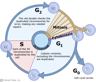
Mitosis is nuclear division plus cytokinesis, and produces two identical daughter cells during prophase, prometaphase, metaphase, anaphase, and telophase. Interphase is often included in discussions of mitosis, but interphase is technically not part of mitosis, but rather encompasses stages G1, S, and G2 of the cell cycle.
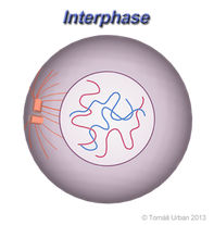
The cell is engaged in metabolic activity and performing its prepare for mitosis (the next four phases that lead up to and include nuclear division). Chromosomes are not clearly discerned in the nucleus, although a dark spot called the nucleolus may be visible. The cell may contain a pair of centrioles (or microtubule organizing centers in plants) both of which are organizational sites for microtubules.
This phase of the cell cycle is a continuum of three distinct stages:
- G1 – First intermediate gap stage in which the cell grows and prepares for DNA replication
- S – Synthesis stage in which DNA is replicated
- G2 – Second intermediate gap stage in which the cell finishes growing and prepares for cell division
Cell cycle checkpoints are mechanisms within Interphase that ensure the fidelity and continued viability of mitotic division in cells
- G1 Checkpoint: Determines appropriate growth conditions (nutrients, cell size, presence of growth factors, etc.). Assess the level of DNA damage from ionising radiation or UV.
- G2 Checkpoint: Determines the state of pre-mitotic cell. Suitable size required for successful cell division.
- Identify replication faults which may have occurred to changes in the DNA sequence distorting genetic fidelity of the daughter cells.
- Metaphase Checkpoint: Ensures proper spindle assembly and correct attachment to centromeres preventing non-disjunction events.
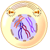
- Chromatin in the nucleus begins to condense and becomes visible in the light microscope as chromosomes. Chromosomes are comprised of genetically identical sister chromatids (joined at a centromere)
- The nucleolus disappears.
- Paired centrosomes move to the opposite poles of the cell and form microtubule spindle fibres
- Centrioles begin moving to opposite ends of the cell and fibers extend from the centromeres. Some fibers cross the cell to form the mitotic spindle.
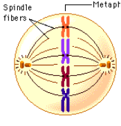
- Microtubule spindle fibres from both centrosomes connect to the centromere of each chromosome
- Microtubule depolymerisation causes spindle fibres to shorten in length and contract
- This causes chromosomes to align along the centre of the cell (equatorial plane or metaphase plate)
- This organization helps to ensure that in the next phase, when the chromosomes are separated, each new nucleus will receive one copy of each chromosome.
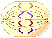
- The paired chromosomes separate at the kinetochores and move to opposite sides of the cell
- Continued contraction of the spindle fibres causes genetically identical sister chromatids to separate
- Motion results from a combination of kinetochore movement along the spindle microtubules and through the physical interaction of polar microtubules.
- Once the chromatids separate, they are each considered an individual chromosome in their own right
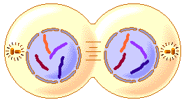
- Chromatids arrive at opposite poles of cell,
- Chromosomes decondense (no longer visible under light microscope)
- Nuclear membranes reform around each chromosome set
- Cytokinesis occurs concurrently, splitting the cell into two
- The chromosomes disperse and are no longer visible under the light microscope.
- The spindle fibers disperse, and cytokinesis or the partitioning of the cell may also begin during this stage
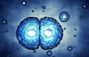
- Cytokinesis is the process of cytoplasmic division, whereby the cell splits into two identical daughter cells
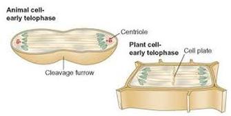
- Cytokinesis occurs concurrently with the final stage of mitosis (telophase) and is different in plant and animal cells
- In animal cells, cytokinesis results when a fiber ring composed of a protein called actin around the center of the cell contracts pinching the cell into two daughter cells, each with one nucleus.
- In plant cells, the rigid wall requires that a cell plate be synthesized between the two daughter cells.
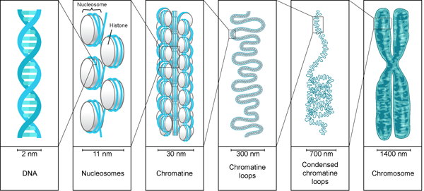
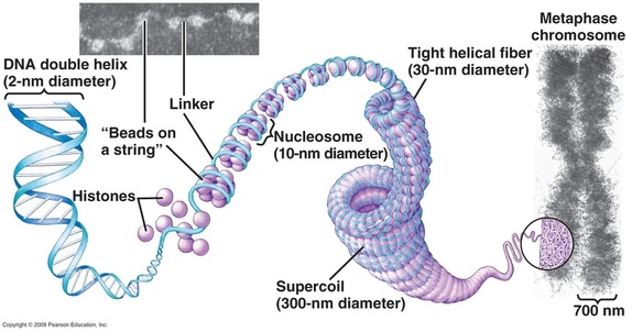
1.6.U.2 Chromosomes condense by supercoiling during mitosis (Oxford Biology Course Companion page 52).
- Describe the structure of a replicated chromosome, include the centromere and sister chromatids.
- Explain why chromosomes must condense during mitosis.
Chromatins are loosely packed DNA within the nucleus. In this unravelled form, the DNA is accessible to transcriptional machinery and so genetic information can be translated DNA is organised as chromatin in all non-dividing cells and throughout the process of interphase.
During mitosis chromosomes (tightly wounded strands of DNA) condense prior to division in a process called supercoiling. In this condensed form, the DNA is able to be easily segregated however is inaccessible to transcriptional machinery. Since a nucleus is generally less than 5 µm in diameter and some of the DNA molecules are over 50,000 µm in length.
The chromosomes have to condense and coil around histone proteins making the chromosome much shorter and fatter.. The nucleosomes (made of histones) will interact further with each other causing the chromosomes to supercoil.. This supercoiling helps regulate transcription because only certain areas of the DNA are accessible for the production of mRNA by transcription. This regulates the production of a polypeptide
DNA is organised as chromosomes during the process of mitosis (condense in prophase, decondense in telophase). A chromosome is visible during mitosis (via microscopy)
As the DNA is replicated during the S phase of interphase, the chromosome will initially contain two identical DNA strands. These genetically identical strands are called sister chromatids and are held together by a central region called the centromere. When these chromatids separate during mitosis, they become independent chromosomes, each made of a single DNA strand.
1.6.U.3 Interphase is a very active phase of the cell cycle with many processes occurring in the nucleus and cytoplasm
- Define cytokinesis.
- List example metabolic reactions occurring during cell interphase.
- Outline events of G1, S, G2 and G0 phases of interphase.
Interphase is an active period in the cell cycle when many metabolic reations occur. It is the longest part of the cell cycle which consists of 3 stages G1, S, G2
Many events need to occur in interphase to prepare the cell for successful division
These key processes include:
- DNA replication – DNA is copied during the S phase of interphase
- Organelle duplication – Organelles must be duplicated for twin daughter cells
- Cell growth – Cytoplasmic volume must increase prior to division
- Transcription / translation – Key proteins and enzymes must be synthesised
- Obtain nutrients – Vital cellular materials must be present before division
- Respiration (cellular) – ATP production is needed to drive the division process
1.6.U.4 Cytokinesis occurs after mitosis and is different in plant and animal cells
- Define cytokinesis.
- State the difference between mitosis and cytokinesis..
- Contrast cytokinesis in plant and animal cells.
- Describe the formation of the cleavage furrow in animal cell cytokinesis.
- Describe the formation of the middle lamella and cell wall in plant cell cytokinesis

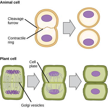
Cytokinesis is the process of cytoplasmic division, whereby the cell splits into two identical daughter cells. Cytokinesis occurs concurrently with the final stage of mitosis (telophase) and is different in plant and animal cells
Animal Cells
- After anaphase, microtubule filaments form a concentric ring around the centre of the cell
- The microfilaments constrict to form a cleavage furrow, which deepens from the periphery towards the centre
- When the furrow meets in the centre, the cell becomes completely pinched off and two cells are formed
- Because this separation occurs from the outside and moves towards the centre, it is described as centripetal
Plant Cells
- In plant cells tubular structures are formed by vesicles along the equator of the cell
- This continues until two layers of membrane exist across the equator, which develop into the plasma membrane of the two new cells
- Vesicles bring pectin and other substances and deposit these between the two membranes through exocytosis forming the middle lamella
- Cellulose is then brought and deposited by exocytosis between the membranes as well, forming the new cell walls
1.6.U.5 Cyclins are involved in the control of the cell cycle
- Explain the role of cyclin and cyclin-CDK complexes in controlling the cell cycle.
- State the role of cyclins D, B, A and E in the cell cycle.
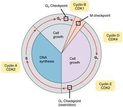
Cyclins are a family of regulatory proteins that control the progression of the cell cycle. Cyclins activate cyclin dependent kinases (CDKs), which control cell cycle processes through phosphorylation
When a cyclin and CDK form a complex, the complex will bind to a target protein and modify it via phosphorylation
The phosphorylated target protein will trigger some specific event within the cell cycle (e.g. centrosome duplication, etc.). After the event has occurred, the cyclin is degraded and the CDK is rendered inactive again.
There are 4 main types of cyclin in human cells.
- Cyclin D – causes G0 to move to G1 and G1 to move to S phase
- Cyclin E – causes the cell to prepare for replication in S phase
- Cyclin A – activates DNA replication in S phase
- Cyclin B – causes the mitotic spindle to begin to form and other tasks needed in the preparation of mitosis
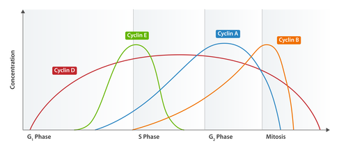
Cyclin Expression Patterns
Cyclin concentrations need to be tightly regulated in order to ensure the cell cycle progresses in a proper sequence. Different cyclins specifically bind to, and activate, different classes of cyclin dependent kinases
Cyclin levels will peak when their target protein is required for function and remain at lower levels at all other time
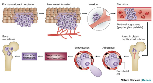
1.6.U.6 Mutagens, oncogenes and metastasis are involved in the development of primary and secondary tumours
- Define tumor, benign, malignant, metastasis, cancer, mutagen and carcinogen.
- Describe why mutagens are not necessarily carcinogens.
- Describe how cancer arises, referring to accumulation of mutations over time.
- Explain the relationship between oncogenes, tumor suppressor genes and cancer.
Tumors are the result of uncontrolled cell division, which can occur in any organ or tissue. Many of these cells can commonly avoid immune detection as they are not foreign bodies but abnormally functioning body cells. These abnormal growths can either be localized (primary tumours), meaning they do not move to other part of your body. These tumours are benign. If the cancer cells detach and move elsewhere into the body (secondary tumours), they are called malignant and are more life-threatening. Diseases due to malignant tumours are known as cancer.
Cancer is usually caused by genetic abnormalities due to a variety of different sources called carcinogens or due to inheritance or errors in DNA replication. Carcinogens are agents that can cause cancer, such as viruses, X-Rays, UV Radiation and many chemical agents. Mutagens are agents that can cause mutations in one’s DNA which can lead to cancer. Mutagens and carcinogens are strongly correlated and many mutagens can be carcinogens
Mutagens may be physical, chemical or biological in origin:
- Physical – Sources of radiation including X-rays (ionising), ultraviolet (UV) light and radioactive decay
- Chemical – DNA interacting substances including reactive oxygen species (ROS) and metals (e.g. arsenic)
- Biological – Viruses, certain bacteria and mobile genetic elements (transposons)
In cancer two types of genes are usually affected, oncogenes and tumor suppressor genes.
- Oncogenes are mutated forms of proto-oncogenes (which typically control synthesis of proteins involved in cell signaling or cell division). These cells with activated oncogenes cause uncontrolled growth and cell division, prevent the cancer cell from dying and allow them to invade other tissues.
- Tumor suppressor genes usually control replication and the cell cycle. In cancer cells these genes are generally inactivated causing a loss of normal function
Tumour cells may either remain in their original location (benign) or spread and invade neighbouring tissue (malignant). Metastasis is the spread of cancer from one location (primary tumour) to another, forming a secondary tumour. Secondary tumours are made up of the same type of cell as the primary tumour – this affects the type of treatment required.
E.g. If breast cancer spread to the liver, the patient has secondary breast cancer of the liver (treat with breast cancer drugs).
1.6.A.1 The correlation between smoking and incidence of cancers (Oxford Biology Course Companion page 57).
- Explain the use of correlations to determine the relationship between two variables (inclusive of positive and negative correlations).
- Explain why the existence of a correlation does not necessitate a causal relationship between two variables.
- Calculate a correlation coefficient using Pearson’s R.
- Determine if a correlation coefficient value is significant.
- Define significant as related to the relationship between two variables.
- Use epidemiological case study information to outline the relationships between smoking and cancer.
A significant body of scientific literature exists which provides a strong link between smoking and the incidence of cancers. Cigarette smoke contains over 4,000 chemical compounds, over 60 of which are known to be carcinogenic
There is a strong positive correlation between smoking and cancer (A correlation is a relationship between two variable factors). Surveys have shown that the more cigarettes that one smokes per day, the higher the death rate due to cancer. The main cancers involved are cancer of the mouth, pharynx, larynx, esophagus and lungs
What do the graphs indicate regarding the correlation between smoking and lung cancer?
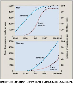
There is a positive correlation. However, correlation does not mean causation. Increase in lung cancer is observed in both men and women.
An increase in the number of cigarettes smoked per year leads to an increase in the incidence of lung cancer. Avoiding exposure to cigarette smoke reduces the chance of getting cancer.
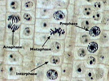
1.6.S.1 Identification of phases of mitosis in cells viewed with a microscope or in a micrograph (Oxford Biology Course Companion page 52). [Preparation of temporary mounts of root squashes is recommended but phases in mitosis can also be viewed using permanent slides.]
- Determine the phase of mitosis of a cell viewed in a micrograph or with a microscope.
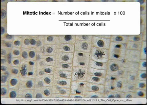
1.6.S 2 Determination of a mitotic index from a micrograph
- State the formula for calculation of a mitotic index.
- Calculate the mitotic index of a tissue as seen in a micrograph.
- Outline the use of mitotic index calculations in diagnosis and treatment of cancer.
The Mitotic index = number of cells containing visible chromosomes (in mitosis) divided by the total number of cells in field of view.
The mitotic index is a measure of the proliferation status of a cell population (i.e. the proportion of dividing cells). The mitotic index may be elevated during processes that promote division, such as normal growth or cellular repair
Identifying Mitotic Cells
Cells undergoing mitosis will lack a clearly defined nucleus and possess visibly condensed chromosomes
Prophase – Chromosomes condensed but still confined to a nuclear region
Metaphase – Chromosomes aligned along the equator of the cell
Anaphase – Two distinct clusters of chromosomes apparent at poles of the cell
Telophase – Two nuclear regions present within a single cell (difficult to see as cytokinesis occurs concurrently)