Meiosis
Also, known as Reductional Division because it reduces the number of chromosomes to half the parent cell. It is of two types meiosis I and meiosis II. Gametes such as sperm or egg under meiosis.
Meiosis I
It is divided into following phases:
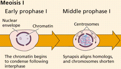
Prophase I
It is the longest phase of meiosis I. During this phase, homologous chromosomes pairs and DNA segments are exchanged (known as recombination). It is further divided into following-
Leptotene
- It is the first stage of meiosis.
- Each individual chromosome exists with two sister chromatids.
- Elements of synaptonemal complex assemble.
- Condensation and coiling of chromosomes occur.
Zygotene
- Chromosomes align as homologous pair of chromosomes.
- Synapsis of homologous chromosomes occur.
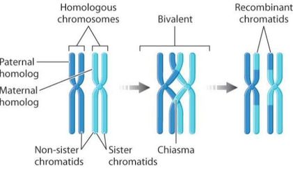
- The paired chromosomes are known as Bivalent or Tetrad.
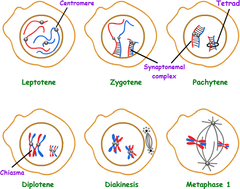
Fig. 7. Different stages of prophase I of meiosis I
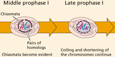
Pachytene
- During this stage, crossing over and homologous recombination occurs.
- Chiamata is formed where homologous chromosomes exchange there segments.
Diplotene
- Homologous chromosomes start to separate.
- Synaptonemal complex disassemble.
- Chromosomes remain bound at the chiasmata.
Diakinesis
- Chromosomes condenses further, so that four parts of the tetrads are visible.
- The nucleoli disappear, nuclear membrane disintegrates.
- Mitotic spindle starts to form.
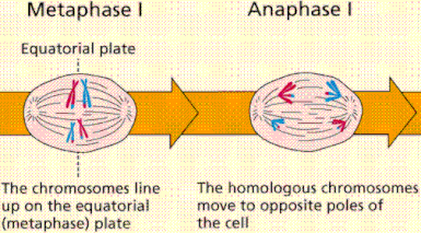
Metaphase I
Homologous chromosomes are aligned on the metaphasic plate. The replicated chromosomes are joined together via protein known as Cohesin.
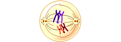
Fig. 8. Metaphase I
Anaphase I
Kinetochore microtubules shorten, pulling homologous chromosomes to opposite poles. The cohesin (protein complex) from the chromosome arms is degraded while the cohesin surrounding the centromere remains safe. This allows the sister chromatids to attach together while homologs are segregated.
Fig. 9. Metaphase I
Telophase I
Each daughter cell now has half the number of chromosomes as compared to parent cell. The microtubules that forms the spindle starts to disappear. The chromosomes again forms chromatin. Sister chromatids remain attached during telophase I.
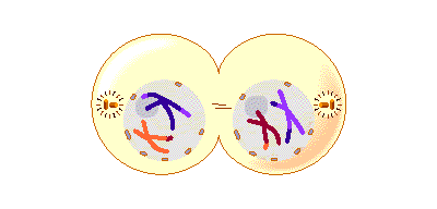
Fig. 10. Telophase I
Meiosis II
Meiosis II is the second meiotic division. It is similar to mitosis, but the genetic results are different. It results in the formation of four haploid cells from the two haploid cells produced in meiosis I.
The four main steps of Meiosis II are as follows: Prophase II, Metaphase II, Anaphase II, and Telophase II.
In prophase II nucleoli and nuclear membrane disappears. Shortening and thickening of the chromatids also occur during this phase. Centrosomes move to the poles and spindle fibers are formed for the second meiotic division.
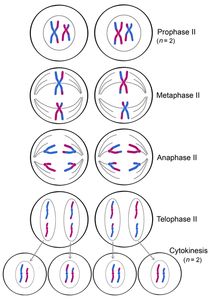
Fig. 11. Different stages of meiosis II
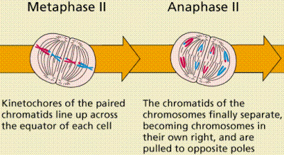
In metaphase II, the centromeres are present with two kinetochores that are attached to spindle fibers from the centrosomes at opposite poles. The metaphasic plate is rotated by 90 degrees when compared to meiosis I, perpendicular to the previous plate.
Anaphase II is accompanied by sister chromatid segregation. The remain of the cohesin is degraded to allow sister chromatid segregation.
Telophase II, which is like telophase I, de-condensation of chromosomes occurs. Nuclear envelopes reorganize and cleavage or cell plate formation starts to produce four daughter cells, each with a haploid set of chromosomes.
Topic 3.3: meiosis
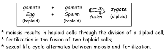
Meiosis is a type of cell division that reduces the number of chromosomes in the parent cell by half and produces four gamete cells. This process is required to produce egg and sperm cells for sexual reproduction.
This unit will take 2 school days
Essential idea:
- Alleles segregate during meiosis allowing new combinations to be formed by the fusion of gametes.
Nature of science:
- Making careful observations—meiosis was discovered by microscope examination of dividing germ-line cells. (1.8)
- Discuss difficulties in microscopic examination of dividing cells.
- Describe the discovery of meiosis.
3.3.U1 One diploid nucleus divides by meiosis to produce four haploid nuclei.
- Compare divisions of meiosis I and meiosis II.
Meiosis is the process by which sex cells (gametes) are made in the reproductive organs. It involves the reduction division of a diploid germline cell into four genetically distinct haploid nuclei
The process of meiosis consists of two cellular divisions:
- The first meiotic division separates pairs of homologous chromosomes to halve the chromosome number (diploid → haploid)
- The second meiotic division separates sister chromatids (created by the replication of DNA during interphase)
3.3.U2 The halving of the chromosome number allows a sexual life cycle with fusion of gametes.
- Compare sexual and asexual life cycles.
- Explain why meiosis must occur as part of a sexual life cycle.
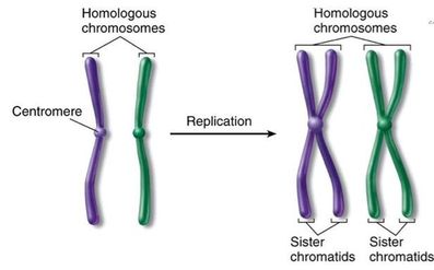
3.3.U3 DNA is replicated before meiosis so that all chromosomes consist of two sister chromatids.
- State that DNA is replicated in interphase before meiosis.
- Given a diploid number (for example 2n=4), outline the movement and structure of DNA through the stages of meiosis.
DNA is replicated in interphase, which is not a part of meiosis, but must precede it
- replication = formation of sister chromatids
- during S phase of interphase, DNA replication produces chromosomes made of two chromatids each, joined at the centromere
Meiosis I: prophase I
- chromosomes condense
- nucleolus disappears
- spindle forms
- synapsis of bivalents: pairing of homologous chromosomes
- crossing over occurs between non-sister chromatids
- nuclear membrane disappears
At the start of prophase I, the chromosomes have already duplicated. During prophase I, they coil and become shorter and thicker and visible under the light microscope.
The duplicated homologous chromosomes pair, and crossing-over (the physical exchange of chromosome parts) occurs. Crossing-over is the process that can give rise to genetic recombination. At this point, each homologous chromosome pair is visible as a bivalent (tetrad), a tight grouping of two chromosomes, each consisting of two sister chromatids. The sites of crossing-over are seen as crisscrossed nonsister chromatids and are called chiasmata (singular: chiasma)
|  |
Meiosis I: metaphase I
- chromosomes continue to condense
- spindle microtubules attach to centromeres
- bivalents line up at equator
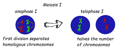
Meiosis I: anaphase I
- separation of homologous pairs;
- chromosomes moved to poles by spindle
Meiosis I: telophase I
- chromosomes at poles
- nuclear envelope reappears
- spindle disappears
- chromosomes partially uncoil

Meiosis II: prophase II
- chromosomes condense again
- nuclear envelope disappears
- new spindle forms (at right angles to previous spindle)
Meiosis II: metaphase II
- spindle microtubules attach to centromeres
- chromosomes move to the equator
Meiosis II: anaphase II
- sister chromatids separate
- spindle microtubules move chromatids to opposite poles
Meiosis II: telophase II
- chromosomes (=chromatids) arrive at poles
- spindle disappears
- nuclear membrane reappears
- nucleolus reappears
- chromosomes decondense into chromatin
cytokinesis not part of meiosis cell division
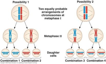
3.3.U5 Orientation of pairs of homologous chromosomes prior to separation is random
- Describe the attachment of spindle microtubules to chromosomes during meiosis I.
- Describe random orientation of chromosomes during meiosis I.
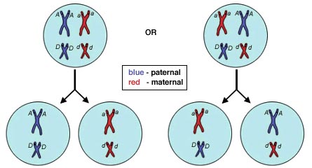
Independent assortment:
- Variety produced by recombination of maternal and paternal chromosomes
- For each pair of homologous chromosomes, maternal and paternal chromosomes assort to daughter cells randomly
- Possible arrangements of chromosomes in haploid daughter cells = (2)nth, where n = number of homologous pairs
- In humans, n = 23, and possible arrangements = (2)23 = about 8 million
- Mendel’s law of independent assortment applies only to traits carried on different chromosomes, i.e.unlinked genes
- Independent assortment occurs as a result of the alignment of homologs during metaphase I, determining which maternal and paternal chromosomes assort to each daughter cell
- Each pair of alleles separates independently of every other pair of unlinked alleles
3.3.U6 Separation of pairs of homologous chromosomes in the first division of meiosis halves the chromosome number.
- Explain why meiosis I is a reductive division.
- State that cells are haploid at the end of meiosis I.
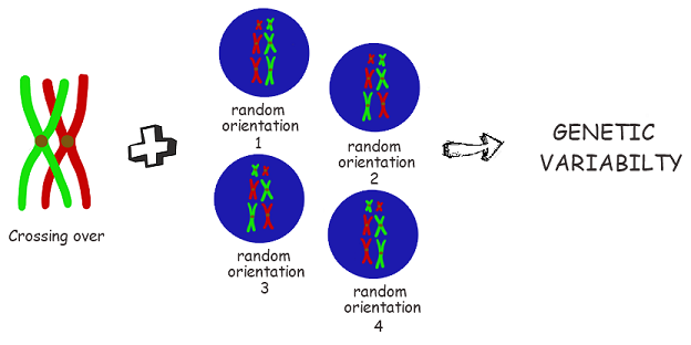
3.3.U7 Crossing over and random orientation promotes genetic variation.
- Explain how meiosis leads to genetic variation in gametes.
- State that the number of chromosome combinations possible due to random orientation is 2n.
The advantage of meiotic division and sexual reproduction is that it promotes genetic variation in offspring
The three main sources of genetic variation arising from sexual reproduction are:
- Crossing over (in prophase I)
- Random assortment of chromosomes (in metaphase I)
- Random fusion of gametes from different parents
Crossing over involves the exchange of segments of DNA between homologous chromosomes during prophase I
Because it mixes differents sets of DNA basically.
The possible combinations for random orientation in human are quite large, in fact, there are 2^23 possible combinations or 107,3741,824 possible orientations. (2 to the power of haploid number of chromosomes)
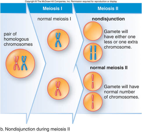
Meiosis is the process of creating haploid gametes from a diploid cell. If everything goes smoothing during meiosis, chromosomes will be separated and distributed evenly to produce four haploid gametes. However, sometimes chromosomes do not separate properly. This is called nondisjunction and results in gametes with either too many or too few chromosomes. In humans, nondisjunction becomes more common the older one gets.
If nondisjunction occurs during anaphase I of meiosis I, this means that at least one pair of homologous chromosomes did not separate. The end result is two cells that have an extra copy of one chromosome and two cells that are missing that chromosome. In humans, n + 1 designates a cell with 23 chromosomes plus an extra copy of one for a total of 24 chromosomes. n – 1 designates a cell missing a chromosome for a total of only 22 chromosomes in humans.
If one of these abnormal gametes undergoes fertilization, then a baby with an abnormal number of chromosomes in its cells could be born. Trisomy is the condition of having 3 copies of one chromosome type. It is designated as 2n + 1 because the cell has the normal two sets of each 23 types of chromosomes plus an extra copy of one chromosome. Monosomy is the condition of having only 1 copy of a chromosome and is designated as 2n – 1.
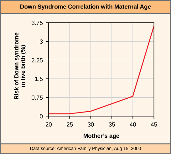
Studies show that the chances of non-disjunction increase as the age of the parents increase
There is a particularly strong correlation between maternal age and the occurrence of non-disjunction events
This may be due to developing oocytes being arrested in prophase I until ovulation as part of the process of oogenesis
Other studies also suggest that:
- The risk of chromosomal abnormalities in offspring increase significantly after a maternal age of 30
- There is a higher incidence of chromosomal errors in offspring as a result of non-disjunction in meiosis I
- Mean maternal age is increasing, leading to an increase in the number of Down syndrome offspring
3.3.A3 Description of methods used to obtain cells for karyotype analysis e.g. chorionic villus sampling and amniocentesis and the associated risks. (Oxford Biology Course Companion page 163).
- Describe the two procedures for obtaining fetal cells for production of a karyotype.
diagnosis of chromosomal abnormalities, such as Down syndrome, identifying extra chromosome 21 (trisomy 21)
3.3.S1 Drawing diagrams to show the stages of meiosis resulting in the formation of four haploid cells.
- Outline the events of prophase, metaphase, anaphase and telophase in meiosis I and meiosis II.
- Draw diagrams of cells in prophase, metaphase, anaphase and telophase in meiosis I and meiosis II.
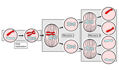
Meiosis consists of two divisions, both of which follow the same stages as mitosis (prophase, metaphase, anaphase, telophase). Meiosis is preceded by interphase, in which DNA is replicated to produce chromosomes consisting of two sister chromatids. A second growth phase called interkinesis may occur between meiosis I and II, however no DNA replication occurs in this stage.
Meiosis I
The first meiotic division is a reduction division (diploid → haploid) in which homologous chromosomes are separated
- P-I: Chromosomes condense, nuclear membrane dissolves, homologous chromosomes form bivalents, crossing over occurs
- M-I: Spindle fibres from opposing centrosomes connect to bivalents (at centromeres) and align them along the middle of the cell
- A-I: Spindle fibres contract and split the bivalent, homologous chromosomes move to opposite poles of the cell
- T-I: Chromosomes decondense, nuclear membrane may reform, cell divides (cytokinesis) to form two haploid daughter cells
Meiosis II
The second division separates sister chromatids (these chromatids may not be identical due to crossing over in prophase I)
- P-II: Chromosomes condense, nuclear membrane dissolves, centrosomes move to opposite poles (perpendicular to before)
- M-II: Spindle fibres from opposing centrosomes attach to chromosomes (at centromere) and align them along the cell equator
- A-II: Spindle fibres contract and separate the sister chromatids, chromatids (now called chromosomes) move to opposite poles
- T-II: Chromosomes decondense, nuclear membrane reforms, cells divide (cytokinesis) to form four haploid daughter cells