A level Biology 8.3 The heart – Exam style question – Paper 2

Question

Mammals have closed, double circulatory systems.
(a) Explain what are meant by the terms closed and double as applied to mammalian
circulatory systems.
closed ………………………………………………………………………………………………………………
double ………………………………………………………………………………………………………………
Fig. 5.1 shows a longitudinal section through a mammalian heart.

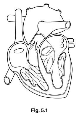
(b) Use label lines and the letters P, Q, R and S to label the following on Fig. 5.1:
P the right atrium
Q a semilunar valve
R a blood vessel that carries deoxygenated blood
S the position of Purkyne tissue
Catheters are small tubes that are inserted into blood vessels. A catheter was inserted into
an artery in the arm and then moved into the aorta and then into the left ventricle during a
diagnostic investigation. The catheter contained a device to measure the blood pressure in
the aorta and in the left ventricle. The results are shown in Fig. 5.2.

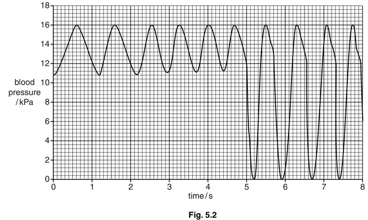
(c) (i) Calculate the heart rate during the period of the investigation.
Show your working.
answer …………………………………………..

(ii) Describe and explain the differences in pressure as the catheter moves from the aorta into the left ventricle.
Fig. 5.3 is an X-ray showing narrowing in the blood vessels supplying muscles in the heart.
A catheter is used to insert a dye into the blood vessels so that they appear clearly in the
X-ray. The arrows indicate where there is narrowing of the blood vessels.

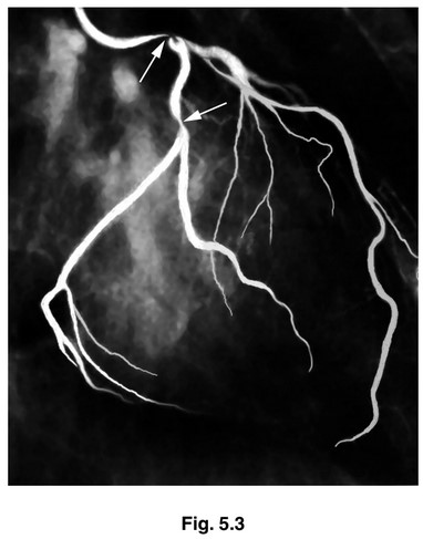
(d) (i) Name the blood vessels shown in Fig. 5.3.
(ii) State the likely effect of narrowing of these blood vessels.
(e) Suggest ways in which the condition shown in Fig. 5.3 may be treated.

Answer/Explanation

Answer:

(a) closed blood travels, inside blood vessels / AW ;
double blood travels through the heart twice during one, complete circuit / circulation
of the body ; AW
A pulmonary and systemic, systems / circuits

(b) P to right atrium ;
Q to (semilunar) pulmonary or aortic valve ;
R to, vena cava / pulmonary artery ;
S to, septum / wall(s) of ventricles ;

(c) (i) 75 (beats per minute) ;;
if incorrect answer or no answer allow one mark for extraction from Fig. 5.2 or for correct
working
e.g.10 beats in 8 seconds
10/8 × 60
(ii) max 3 if only description or only explanation given
lowest pressure in aorta, is 10.8 kPa / varies between 10.8-11.2 kPa v in left ventricle
is 0 KPa ;
difference between highest and lowest is greater in the ventricle / AW ;
4.8 – 5.2 kPa for aorta, 16.0 kPa in left ventricle ;
reference pressure differences (in left ventricle) as a direct result of ventricular systole
and diastole ;
semilunar / aortic, valve prevents backflow from aorta into ventricle ;
(so) no / little, blood in ventricle, when fully contracted / AW ;
elastic recoil of artery maintains (diastolic) blood pressure ;
AVP ;

(d) (i) coronary arteries ;
(ii) insufficient, glucose / oxygen (to, cardiac / heart, muscle) ;
angina ;
heart attack / myocardial infarction / cardiac arrest ;
description of anaerobic conditions in muscle ;

(e) coronary (artery) by-pass (graft) operation ;
R by-pass unless qualified
A described
insertion of a (coronary) stent ; A described
heart transplant ;
angioplasty ; A described
AVP ; e.g. calcium-channel blockers / named
further detail of treatments e.g. anticoagulants after angioplasty

Question

Fig. 5.1 shows a vertical section of the left side of the heart of a mammal.

   (a) Explain the difference in the thickness of the left ventricle and the left atrium.[2]

   (b) Explain how the structures labelled on Fig. 5.1 ensure that blood flows in the correct direction.[3]

   (c) During one cardiac cycle, blood is pumped from the heart into the pulmonary and systemic circulations.

         Explain how the contraction of the four chambers of the heart are coordinated and controlled to enable blood to be pumped simultaneously into both the pulmonary and systemic circulations. [4] [Total: 9]

Answer/Explanation

Ans:

5 (a) more/ thicker, (cardiac) muscle (tissue) in left ventricle ; A ora
                                                                                                             A thicker muscular wall

           either
           atrium pumps blood at lower pressure/ against less resistance/ to ventricle/ short(er) distance/with less force ;

           or
           ventricle pumps blood to the body / into systemic circulation/ long(er) distance/against
           greater resistance/ at higher pressure/with more force ;
           R ventricle wall withstands high pressure

     (b) valve opens to allow blood flow from atrium into ventricle/when pressure in atrium is greater than pressure in ventricle/ during atrial systole ;

           valve closes when ventricle contracts /when pressure in ventricle is greater than pressure in atrium/ during ventricle systole ;

           during contraction of ventricles
           papillary muscles contract to ‘pull on’ tendons ; R if tendons are said to open the valve tendons prevent valve, inverting/ going inside out/ everting/AW ;

     (c) 1 sino-atrial node/SAN sends out, waves of excitation /waves of depolarisation/ (electrical) impulses /action potential(s) ;
                 R nervous impulses / signal/ message penalise once only
           2 wave of excitation/AW/SAN stimulates, (both) atria to contract/atrial systole ;
           3 fibrous ring/ non-conducting tissue/insulating tissue (between atria and ventricles), prevents impulse reaching the ventricles / prevents atria and ventricles contracting at the same time ;
           4 atrio-ventricular node/AVN delays impulse (by 0.1s) / prevents ventricles contracting at the same time as atria ;
           5 allows, atria to empty / ventricles to fill ;
           6 AVN sends out, waves of excitation/impulses to Purkyne tissue/Bundle of His (in septum);
           7 causes ventricles to contract together/ at the same time/ simultaneously /AW ;

Question

There are many types of amino acids, but only twenty that are polymerised to make polypeptides and proteins in animals.

    (a) Name the type of chemical reaction that occurs when two amino acids form a dipeptide.[1]

    (b) Fig. 3.1 shows two amino acids, glycine and valine. Use the space below to make a drawing to show what happens when these two molecules join together to form a dipeptide.[4]

    (c) Angiotensinogen is an inactive protein molecule. When blood pressure decreases, part of angiotensinogen is removed to form a short polypeptide, angiotensin that stimulates an increase in blood pressure.

          Fig. 3.2 shows the base sequence within the gene for angiotensinogen that codes for this short polypeptide, the RNA codons and the primary structure of angiotensin.

          (i) Complete Fig. 3.2 to show the missing DNA triplet and the RNA codons. [1]

          (ii) State the full name of the type of RNA shown in Fig. 3.2.[1]

               Table 3.1 shows the blood pressure in the right ventricle and in the pulmonary artery of a person who is in good health.

(d) Use the information in Table 3.1 to explain why the blood pressure in the pulmonary artery is the same as the pressure in the right ventricle during systole, but higher during diastole.[3]

(e) People with long-term chronic obstructive pulmonary disease (COPD) usually have blood which is poorly oxygenated during its passage through the lungs. This leads to a constriction of blood vessels in the lungs.
               Suggest the likely effect of this on the heart.[2]

    (f) Describe the signs and symptoms of COPD that help doctors make an early diagnosis of this condition.[2] [Total: 14]

Answer/Explanation

Ans:

3 (a) condensation ; A dehydration

   (b) accept glycine-valine or valine-glycine

         peptide bond drawn correctly ;
         amino and carboxylic acid ends shown ;
         correct R-groups ;
         water eliminated ;

   (c)


         (ii) messenger ;

   (d) during systole semi-lunar valve is open ;
          during diastole semi-lunar valve is closed ;
          proximity/AW pulmonary artery to (right) ventricle (so no pressure lost) ;
          elastic recoil of pulmonary artery maintains blood pressure/AW ;
          no/little blood in (right) ventricle, after contraction/during diastole ;
          fills with blood at low pressure ;

   (e) increase in power of contraction ; AW
          increase in (systolic) blood pressure ;
          strain on right ventricle/right ventricle does not function efficiently ;
          growth of muscle in/right ventricle increases in thickness ;
          insufficient oxygen to, heart/cardiac, muscle ;
          heart failure/heart attack ;

   (f) persistent/AW, cough ;
          cough produces much mucus ;
          wheezing ;
          rapid breathing/difficulty breathing/breathlessness ;
          bluish colour to the skin ;
          recurrent chest infections/frequent colds or ‘flu/AW ;
          barrel-shaped chest ;
          chest pains ; R heart pains
          fatigue/weakness, (with exercise) ;

Scroll to Top