Question
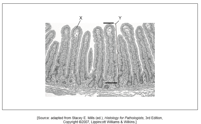
The micrograph shows a section of an organ in the human body.

State from which organ the section was taken.
Identify the layer of tissue found at X.
The actual length of the structure labelled Y is 0.8 mm between the two black lines. Calculate the magnification of the micrograph. Working should be shown.
The actual length of the structure labelled Y is 0.8 mm between the two black lines. Calculate the magnification of the micrograph. Working should be shown.
Answer/Explanation
Markscheme
small intestine
Do not accept villi/villus or intestine alone.
epithelium
Do not accept microvilli/brush border.
a. calculation shown with accurate measurement of length of villus
OR
For the first marking point to be awarded, the measurement must be between 53 and 55 mm.
b. 67 or 68 or 68
Allow any value between 67 and 69 inclusive.
Accept decimals e.g. 68.75.
Allow ECF from first marking point.
a. calculation shown with accurate measurement of length of villus
OR
53/0.8
or
54/0.8
or
55/0.8
«mm»
For the first marking point to be awarded, the measurement must be between 53 and 55 mm.
b. 67 or 68 or 68
Allow any value between 67 and 69 inclusive.
Accept decimals e.g. 68.75.
Allow ECF from first marking point.
Examiners report
Question
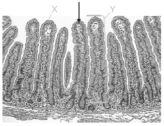
The micrograph shows a section of an organ in the human body.
One of the functions of this organ is absorption. On the micrograph, draw an arrow showing the direction of absorption.
Answer/Explanation
Markscheme
Question
The micrograph shows a transverse section through blood vessels of a mammal.
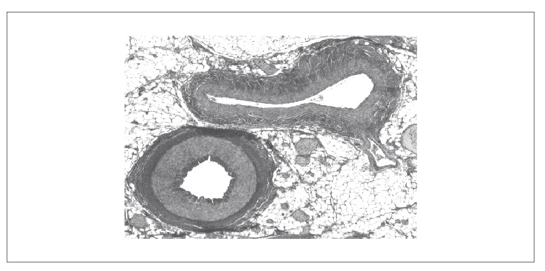
[Source: This book was originally published by OpenStax College, released under the CC-By license: https://creativecommons.org The eBook was adapted by Frank Lee.]
Identify the vein by labelling it with the letter V.
Distinguish between the vein and the artery with reference to structures visible in the micrograph.
Answer/Explanation
Markscheme
label pointing to the upper of the two blood vessels in the micrograph
Note: check the answer carefully as the scan of the diagram is not always clear for candidates writing in pencil
a. vein has larger lumen
b. vein has less elastic tissue
c. vein has less muscular/thinner walls/tunica media
OR
ratio of wall thickness to lumen is less in the vein
d. vein less rounded/squashed more easily
Accept inverse for artery
Do not accept non-visible differences such as valves
No ECF
[Max 2 Marks]