IB DP Biology 1.3 Membrane structure Question Bank SL Paper 2

Question

Draw a labelled diagram to show the structure of a membrane.

[5]
a.

Outline how vesicles are used to transport materials secreted by a cell.

[6]
b.

Explain how the structure of a villus in the small intestine is related to its function.

[7]
c.
▶️Answer/Explanation

Markscheme

Award [1] for each of the following clearly drawn and correctly labelled.
phospholipid bilayer – double row of opposing phospholipids, tails to inside;
phospholipid – with head and two tails;
hydrophilic/phosphate/polar (heads) and hydrophobic/hydrocarbon/fatty acid/non-polar (tails) labelled;
integral protein – embedded in the phospholipid bilayer;
protein channel – integral protein showing clear channel/pore;
peripheral protein – on the surface;
glycoprotein – with carbohydrate attached on one side;
cholesterol – shown embedded in bilayer;

a.

vesicles formed from rER transport proteins to Golgi apparatus;
these vesicles fuse with membranes of Golgi apparatus;
proteins are processed as they move through Golgi apparatus;
(transport) vesicles bud off/leave Golgi apparatus;
vesicles move through cytoplasm;
(vesicles) fuse with plasma membrane;
contents released to outside of cell / exocytosis;
cells use vesicles to secrete substances such as hormones/digestive enzymes/other appropriate example;
vesicles may contain cell products other than proteins;
Credit drawings which fully explain the points above.

b.

To achieve [1] answer must name the structure and relate it to its function.
the villus has a large surface area to volume ratio;
microvilli increase surface area for absorption;
thin surface (epithelial) layer so products of digestion can pass easily through;
channel proteins located in plasma membrane used for facilitated diffusion;
network of capillaries inside each villus (so only short distance) for movement of absorbed products;
capillaries transport absorbed nutrients/sugars and amino acids away from small intestine;
blood flow in capillaries maintains concentration gradient;
central lymph vessel/lacteal to transport absorbed fats/fatty acids away from small intestine;
large number of mitochondria provide ATP needed for active transport;
protein pumps in membrane (of microvilli) carry out active transport;
pinocytosis occurs at surface (epithelial) layer;
Accept any of the points above shown in a drawing.

c.


Question

Draw a labelled diagram to show the structure of membranes.

[6]
a.

Explain passive transport and active transport across membranes.

[8]
c.
▶️Answer/Explanation

Markscheme

Award [1] for each structure clearly drawn and correctly labelled.
phospholipid bilayer – with head and tails;
hydrophilic / phosphate / polar heads and hydrophobic / hydrocarbon / fatty acid / non-polar tails labelled;
integral/intrinsic protein – embedded in the phospholipid bilayer;
protein channel – integral protein showing clear channel/pore;
peripheral/extrinsic protein – on the surface;
glycoprotein with carbohydrate attached; (carbohydrate should project outwardly from membrane protein)
cholesterol – shown embedded in bilayer; (must appear in hydrophobic region)
thickness indicated (10 nm); (allow answers in the range of 7 nm to 13 nm)

a.

diffusion/facilitated diffusion and osmosis are passive;
do not require energy/ATP;
diffusion is movement from high to low concentration/down a (concentration) gradient;
facilitated diffusion uses (protein) channels/carrier proteins;
osmosis is water movement from lower to higher solute concentration / from higher to lower water potential/concentration;
across a partially permeable membrane;
active transport/formation of vesicles require energy;
in the form of ATP;
active transport moves materials up/against the (concentration) gradient/from low to high concentration;
protein pumps required;
endocytosis into cells / exocytosis out of cells;

example of active or passive transport; (e.g. sodium potassium pump for active transport / oxygen exchange in alveoli for passive transport)

c.


Question

List two functions of membrane proteins.

[2]
a.

Explain why digestion of large food molecules is essential.

[1]
b.

Outline why antibiotics are effective against bacteria but not against viruses.

[2]
c.

Outline the use of polymerase chain reaction (PCR) to copy and amplify minute quantities of DNA.

[2]
d.
▶️Answer/Explanation

Markscheme

a. hormone binding sites / receptors;

b. (immobilized) enzymes;

c. cell adhesion;

d. cell (to cell) communication;

e. passive transport/channels;

f. active transport/pumps;

g. facilitate diffusion;

h. carry electrons;

a.

a. many molecules are too large to be absorbed (by the villi) / small molecules are soluble and can be absorbed;

b. large food molecules are broken down so they can be reorganized/rearranged;

b.

a. antibiotics block/inhibit specific metabolic pathways/cell functions found in bacteria;

Accept specific examples of inhibition such as cell protein synthesis, cell wall formation

b. viruses must use host/eukaryotic cell metabolism / viruses do not have their own metabolic pathways;

c. host/eukaryotic cell metabolism/pathways not blocked/inhibited by antibiotics;

c.

a. strands of DNA (fragments) split/denatured with heat;

b. complementary nucleotides added to split stands (when cooling);

c. with the use of (DNA) polymerase (and primers);

d. process/heating and cooling cycle is repeated (until enough DNA is obtained);

Accept example of PCR application e.g. paternity cases or forensic investigations.

d.
 
.
.
 

Question

The diagram shows how vesicles are used to transport materials in a cell.

State the name of organelle A.

[1]
a (i).

State the process occurring at B.

[1]
a (ii).

Describe how the structure of the membrane allows the formation of vesicles.

[2]
b.

Explain active transport across membranes.

[3]
c.
▶️Answer/Explanation

Markscheme

Golgi apparatus/complex/body
Reject Golgi vesicle and Golgi unqualified.

a (i).

endocytosis/phagocytosis/pinocytosis
Reject exocytosis.

a (ii).

a. fluidity of membrane allows change of shape/invagination/formation of vesicles;
b. phospholipids can move / phospholipid bilayer makes membrane fluid/flexible;
c. weak bonding between phospholipid tails;
d. bends/kinks in the phospholipid tails prevent close packing;
e. cholesterol affects membrane fluidity;

b.

a. moves substances up/against a concentration gradient / from lower to higher concentration;
b. protein/pump (in membrane) that moves material; (reject channels)
c. ATP is used; (reject energy alone)
d. example/labeled diagram showing mechanism;

c.

Question

Draw a labelled diagram to show the molecular structure of a membrane.

[4]
a.

Some proteins in membranes act as enzymes. Outline enzyme-substrate specificity.

[6]
b.

Membranes of pre-synaptic and post-synaptic neurons play an important role in transmission of nerve impulses. Explain the principles of synaptic transmission.

[8]
c.
▶️Answer/Explanation

Markscheme

Award [1] for each of the following clearly drawn and correctly labelled.

phospholipid bilayer; (double row of opposing phospholipids, tails to inside)

hydrophilic/phosphate/polar (heads) and hydrophobic/hydrocarbon/fatty acid/nonpolar (tails) labeled;

integral protein; (embedded in the phospholipid bilayer)

protein channel/channel protein; (integral protein showing clear channel/pore)

peripheral protein; (shown on surface or slightly embedded on either side)

glycoprotein; (with carbohydrate attached on outer side)

cholesterol; (shown embedded in bilayer and smaller than the hydrophobic tail)

a.

enzyme shape is specific to (particular) substrate;
lock and key analogy/model;
example of specific enzyme and substrate;
has specific 3-D/tertiary configuration/3-D/tertiary shape essential to functioning;
active site on enzyme binds to substrate;
substrate and active site complementary/fit together;
(substrate and active site) are complementary due to structure/chemical attraction;
enzyme-substrate complex forms;
denaturation changes enzyme’s binding ability (to specific substrate);
Award [6] for the above points clearly shown in an annotated diagram.

b.

synapse is gap between adjacent neurons;
(arriving) action potential depolarizes pre-synaptic membrane;
opens (voltage-gated) calcium channels in membrane;
causes influx of calcium ions;
causes synaptic vesicles to fuse with pre-synaptic membrane;
vesicles release/exocytose neurotransmitter into the synaptic cleft;
neurotransmitter diffuses/moves across synaptic cleft;
neurotransmitter binds to receptors on post-synaptic membrane;
opens channels allowing sodium ions/potassium ions to diffuse;
initiation of action potential/depolarization in post-synaptic membrane;
removal/breakdown of neurotransmitter stops effect on post-synaptic membrane;
Award any of the above points for a clearly drawn correctly annotated diagram.
(Plus up to [2] for quality)

c.


Question

The image shows a phospholipid bilayer that is a component of the cell membrane.

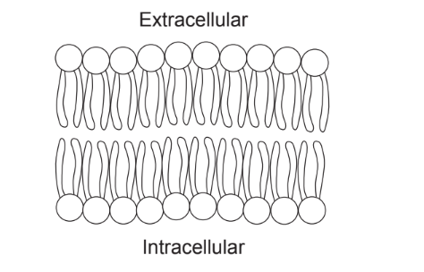
(a) Annotate the diagram to illustrate the amphipathic nature of phospholipids. [2]
(b) Outline a function of cholesterol in cell membranes. [1]

(c) Describe two pieces of evidence that show that eukaryotic cells originated by endosymbiosis. [2]

▶️Answer/Explanation

a a. line to circle labelled phosphate (head) and (tail) labelled fatty acid/hydrocarbon/lipid (tail);

  b. label hydrophilic/polar/attracted to water/ and hydrophobic/non polar/not attracted to water;

b reduces fluidity of membrane / reduces permeability of membrane (to some molecules);

c a. mitochondria/chloroplasts have their own DNA;

b. mitochondria can self-replicate/undergo a process like binary fission;
c. mitochondria/chloroplasts have double membranes;
d. mitochondria/chloroplasts have(70s) ribosomes;
e. mitochondria/chloroplasts are sensitive to antibiotics;
f. similar in size to bacteria

Scroll to Top