Home / IB DP Biology-B2.3 Cell specialization -FA 2025- IB Style Questions For HL Paper 1

IB DP Biology-B2.3 Cell specialization -FA 2025- IB Style Questions For HL Paper 1

Question

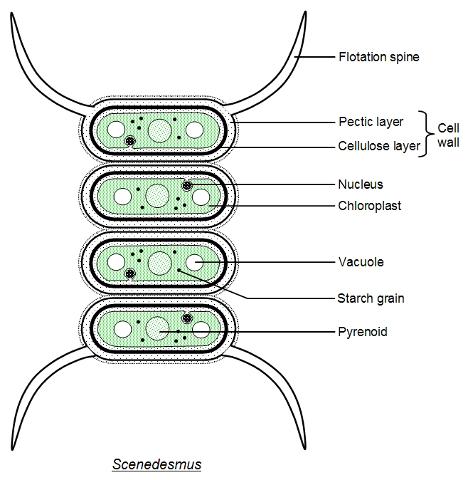
Scenedesmus is a microscopic, unicellular green alga. However, it often exists as multicellular colonies of cells.

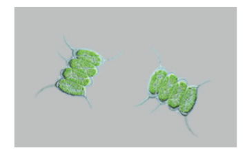
The magnification of the image is 500×. What is the length of one cell?
A. 10nm
B. 50 μm
C. 20 μm
D. 10mm

▶️Answer/Explanation

Answer: C. 20 μm

Explanation:

What is Magnification and How is Actual Size Determined?
Magnification is the number of times an object’s size is increased in an image. The actual size of the object can be calculated using the formula:

\text{Actual size} = \frac{\text{Image size}}{\text{Magnification}}

If we know the magnification but not the image size, we can still answer based on biological knowledge. Scenedesmus is a type of microscopic green alga. Individual cells typically measure between 10–30 micrometers (μm) in length.

Now, evaluate each option:

Option A. Incorrect – 10 nm
A nanometer (nm) is 1,000 times smaller than a micrometer (μm). 10 nm is the size of a virus or a protein, not a whole algal cell. This is far too small for any eukaryotic cell.

Option B. Incorrect – 50 μm
50 micrometers is possible for some large cells, but it is larger than the usual size of Scenedesmus cells. This value is slightly above the known typical range of 10–30 μm.

Option C. Correct – 20 μm
20 micrometers is within the normal size range for Scenedesmus cells. It is biologically accurate and fits the expected dimensions of these algae under 500× magnification.

Option D. Incorrect – 10 mm
10 millimeters equals 10,000 micrometers. This is far too large for any single algal cell and would be visible without a microscope. It is not realistic.

Question

The images of the radiolarian, a single-celled marine organism, were produced using a light microscope (left) and a scanning electron microscope (right).

What is a reason for the difference in quality of these images?
A. Light cannot pass through the specimen.
B. Higher magnification can be achieved with the electron microscope.
C. The resolution of the electron microscope is higher.
D. Samples are stained with methylene blue when viewed with the light microscope.

▶️Answer/Explanation

Answer: C. The resolution of the electron microscope is higher.

Explanation:

What affects microscope image quality?
Image quality depends mainly on resolution the ability to see fine detail not just magnification. Electron microscopes use electrons (not light), which gives them higher resolution than light microscopes.

Now let’s analyze the options:

Option A. Incorrect – Light cannot pass through the specimen.
This isn’t why the image is less clear. Light microscopes can still work with thin specimens.

Option B. Incorrect – Higher magnification can be achieved with the electron microscope.
True, but magnification without resolution still gives a blurry image. Resolution matters more.

Option C. Correct – The resolution of the electron microscope is higher.
This is the main reason SEM images are sharper. Electrons reveal much finer details.

Option D. Incorrect – Samples are stained with methylene blue when viewed with the light microscope.
Staining improves contrast, not resolution. It doesn’t explain the image quality difference.

Question

Which characteristic of stem cells makes them useful for treating Stargardt’s disease?

A. They can differentiate into retinal cells.
B. They are readily available from specially created embryos.
C. They transport white blood cells to the eyes.
D. They divide by binary fission so provide sufficient cells.

▶️ Answer/Explanation

Why are stem cells useful in treating diseases like Stargardt’s disease?
Stem cells are unspecialized cells that can differentiate into many types of specialized cells. In Stargardt’s disease, which damages retinal cells and causes vision loss, stem cells can become retinal pigment epithelial (RPE) cells to replace the damaged ones.

Now let’s analyze the options:

Option A. Correct – They can differentiate into retinal cells.
This is the main reason. Stem cells are used because they can become the exact type of cell needed — in this case, retinal cells.

Option B. Incorrect – They are readily available from specially created embryos.
The usefulness of stem cells depends on their ability to differentiate, not on where they come from.

Option C. Incorrect – They transport white blood cells to the eyes.
Stem cells do not transport immune cells. This has nothing to do with treating Stargardt’s disease.

Option D. Incorrect – They divide by binary fission so provide sufficient cells.
Stem cells divide by mitosis, not binary fission (which is how bacteria reproduce). This is incorrect.

Scroll to Top