▶️ Answer/Explanation
(a)
(b)
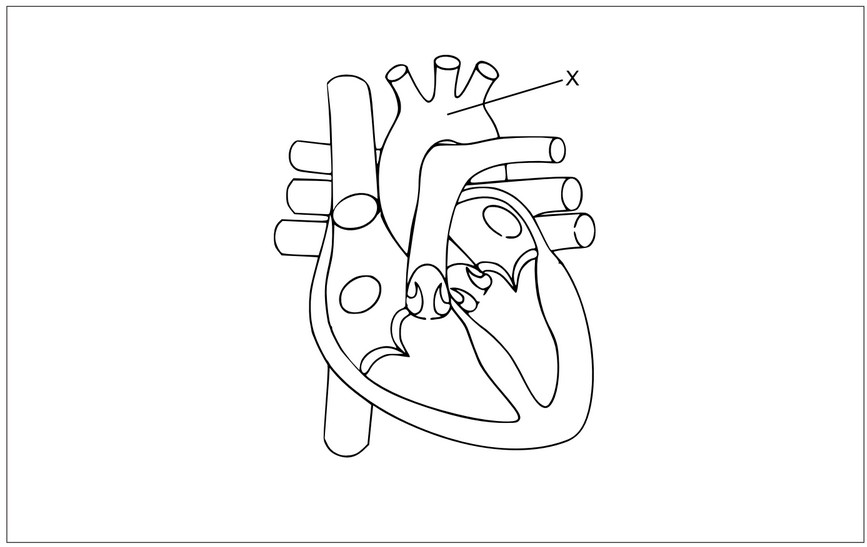
- X is the aorta.
- It is the largest artery in the body.
- It carries oxygenated blood from the left ventricle to the rest of the body.
(c)
- The left ventricle is responsible for pumping oxygen-rich blood to the entire body.
- When it contracts, it creates a high pressure that pushes blood through the aorta.
- It receives oxygenated blood from the left atrium, which gets it from the lungs via the pulmonary veins.
- The contraction of the left ventricle is controlled by electrical impulses from the atrioventricular (AV) node.
Markscheme:
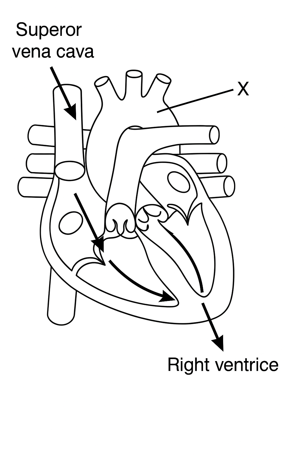
(a) Arrows added to the diagram to show how deoxygenated blood enters the heart: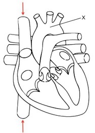
(b) Aorta;
(c)
a. Contracts to generate high pressure / pumps blood at high pressure;
b. Pump blood (through the aorta) to all parts of the body (apart from the lungs);
c. Receives blood from the left atrium;
d. Contraction is stimulated by the AV node;