Topic 1.3: membrane structure
In the Cell Membrane unit you will learn about cell size and the structure and function of cell membranes. Cell membranes protect and organize cells. All cells have an outer plasma membrane that regulates not only what enters the cell, but also how much of any given substance comes in.
Like all other cellular membranes, the plasma membrane consists of both lipids and proteins. The fundamental structure of the membrane is the phospholipid bilayer, which forms a stable barrier between the compartments inside and outside of the cell. Proteins embedded within the phospholipid bilayer carry out the specific functions of the plasma membrane, including selective transport of molecules and cell-cell recognition.
The unit is planned to take 2 school days
- The structure of biological membranes makes them fluid and dynamic.
- Using models as representations of the real world—there are alternative models of membrane structure. (1.11) Falsification of theories with one theory being superseded by another—evidence falsified the Davson-Danielli model. (1.9)
- Explain what models are and their purposes in science.
- Describe the observations and conclusions drawn by Gorter and Grendel in discovering the structure of cell membranes
- Describe why the understanding of cell membrane structure has changed over time.
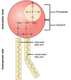
1.3.U.1 Phospholipids form bilayers in water due to the amphipathic properties of phospholipid molecules.
- Draw a simplified diagram of the structure of the phospholipid, including a phosphate-glycerol head and two fatty acid tails.
- Define hydrophilic and hydrophobic.
- Define amphipathic and outline the amphipathic properties of phospholipids.
- Explain why phospholipids form bilayers in water, with reference to hydrophilic phosphate heads and two hydrophobic hydrocarbon tails.
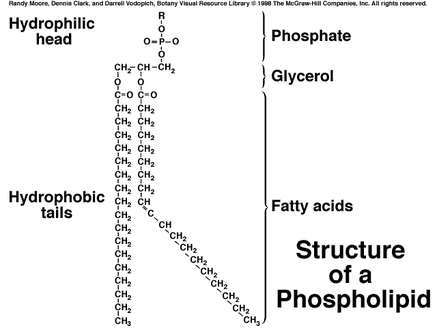
Phospholipids are one of the principal components of cell membranes (in conjunction with membrane proteins)
Phospholipids typically share a common basic structure that includes:
- A polar organic molecule (e.g. choline, serine)
- A phosphate group
- A glycerol molecule (replaced by sphingosine in sphingomyelin)
- Two fatty acid tails (may be saturated or unsaturated)
Hydrophilic and Hydrophobic Properties
- Cell membranes are composed of phospholipids that consist of a hydrophilic (attracted to water) head and a hydrophobic (repelled by water) tail
- This property is described as Amphipathic
- phospholipid-web
- The phospholipid head contains a negatively charged phosphate group which because of its charge is attracted water because of its polarity
- The fatty acid hydrocarbon tail has no charge and is therefore repelled by water
- When placed in water, the phospholipids naturally form a double layer with the heads facing outwards towards the water and the tails facing each other inwards
- This forms a very stable structure that surrounds the cell because of the attractions and bonds that are formed between the heads to the water and to each other, and the hydrophobic interactions between the tails
- Even though it is a very stable structure, it is still fluid, as the phospholipids can move along the horizontal plane
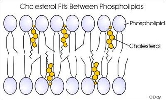
- To increase stability, many cells have cholesterol imbedded between the phospholipids
Phospholipids may vary in the length and relative saturation of the fatty acid tails
- Shorter fatty acid tails will increase fluidity as they are less viscous and more susceptible to changes in kinetic energy
- Lipid chains with double bonds (unsaturated fatty acids) have kinked hydrocarbon tails that are harder to pack together
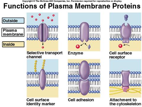
1.3.U2 Membrane proteins are diverse in terms of structure, position in the membrane and function.
- State the primary function of the cell membrane.
- Contrast the structure of integral and peripheral proteins.
- List at least four functions (with example) of membrane bound proteins.
- Contrast the two types of transport proteins: pumps and channels.
Hormone binding sites (receptor proteins)
- Proteins embedded in the membrane, which bind to specific hormones.
- When the hormone binds, it causes the receptor protein to undergo a conformational change, which signals the cell to perform a function.
- For example, insulin receptors.
Immobilized Enzymes
- Integral proteins that catalyze specific chemical reactions.
- Many of these enzymes catalyze metabolic reactions or are a part of a metabolic pathway, such as ATP Synthase in aerobic respiration.
Cell Adhesion
- Proteins that form tight bonds between adjacent cells in tissues and organs.
- For example, gap junctions.
Cell-to-cell communication
- Receptors for neurotransmitters at synapses between two nerve cells.
- Glycoproteins on the surface can also be used for cell identification purposes.
Channels for passive transport
- Integral proteins that span the membrane and provide a passageway for molecules to move from an area of high concentration to low concentration.
- Specific proteins are also used for facilitated diffusion.
Pumps for active transport
- Proteins that use ATP to move substances from a low concentration to a high concentration across the membrane.
- For example, Sodium/Potassium (Na+/K+) pumps and the proton (H+) pumps
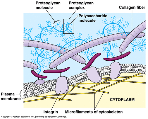
The extracellular matrix typically provides structural and biochemical support to surrounding cells, including:
- Providing sites for anchorage by cells within a tissue and segregating separate tissues from one another
- Sequestering and storing growth factors until receipt of a chemical signal (thereby regulating intercellular communication)
In plant cells the extracellular matrix includes cell wall components (like cellulose) and hence plays an important role in:
- Regulating water uptake (maintenance of cell turgor)
- Providing mechanical strength and rigidity to the cell (maintains cell shape)
1.3.U.3 Cholesterol is a component of animal cell membranes.
- Identify the structure of cholesterol in molecular diagrams.
- Describe the structural placement of cholesterol within the cell membrane.
Cholesterol is able to stop the hydrocarbon from crystalizing and behaving as a solid, but the cholesterol also retricts molecular motion, which reduces the fluidity of the membrane. Also reduces the permiability to the hydrophilic particles like sodium ions and hydrogen ions
It is absent in plant cells, as these plasma membranes are surrounded and supported by a rigid cell wall made of cellulose
Most of the cholesterol molecule is hydrophobic and therefore embeds within the tails of the bilayer. A small portion (hydroxyl –OH group) is hydrophilic and is attracted to the phospholipid head.
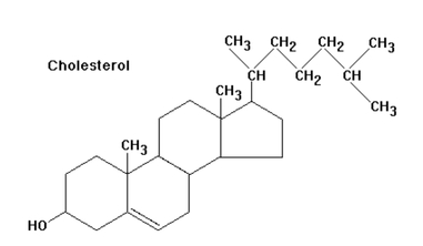
1.3.A.1 Cholesterol in mammalian membranes reduces membrane fluidity and permeability to some solutes.
- Describe the function of cholesterol molecules in the cell membrane.
At the high concentrations it is found in our cell’s plasma membranes (close to 50 percent, molecule for molecule) cholesterol helps separate the phospholipids so that the fatty acid chains can’t come together and crystallize.
Therefore, cholesterol helps prevent extremes– whether too fluid, or too firm– in the consistency of the cell membrane.
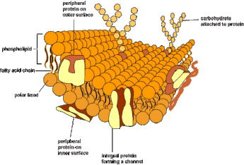
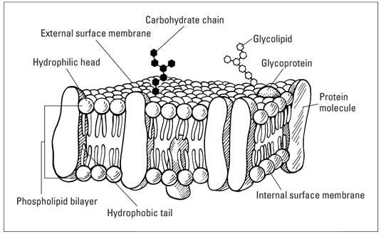
[Drawings of the fluid mosaic model of membrane structure can be two dimensional rather than three dimensional. Individual phospholipid molecules should be shown using the symbol of a circle with two parallel lines attached. A range of membrane proteins should be shown including glycoproteins.]
- Draw and label the structure of membranes. Include
- Phospholipid bilayer
- Integral proteins shown spanning the membrane
- Peripheral proteins on membrane surface
- Protein channels with a pore
- Glycoproteins with a carbohydrate side chain
- Cholesterol between phospholipids in the hydrophobic region
- An indication of thickness (10nm)
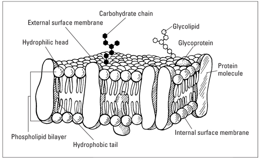
The diagram of the plasma membrane must show the
- phospholipid bilayer
- cholesterol
- glycoproteins
- integral (transmembrane) and peripheral proteins.
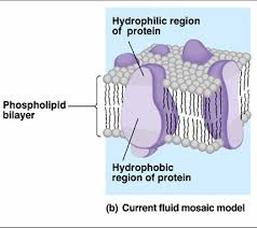
Integral proteins are embedded in the phospholipid of the membrane, whereas peripheral proteins are attached to its surface. Glycoproteins are carbohydrates attached to surface proteins.
NOTE: When you draw a peripheral protein for the IB exam, the peripheral protein must not be embedded into the membrane in order to score the point. Also, you should label the whole phospholipid bilayer not just the individual phospholipid on the majority of mark schemes
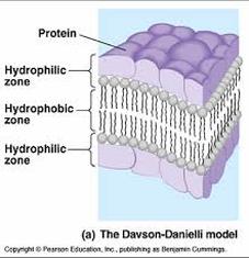
1.3.S.2 Analysis of evidence from electron microscopy that led to the proposal of the Davson-Danielli model.
- Describe the observations and conclusions drawn by Davson and Danielli in discovering the structure of cell membranes
The fluid-mosaic model was not the first scientifically accepted paradigm to describe membrane structure. The first model that attempted to describe the position of proteins within the bilayer was proposed by Hugh Davson and James Danielli in 1935. Davson and Danielli proposed that the lipid bilayer was coated on either side with a layer of globular proteins. The hydrophobic tails of the lipids are orientated towards each other, while the hydrophilic heads are oriented to the outside. Although the membrane composition is correct, there are some problems with the proposed model:
Membranes are not identical. The differ in thickness and the ratio of proteins:lipids.
- Membranes have distinct inside and outside layers (defined by the membrane proteins which are present on the surface of the membrane)
- Other than predicted by the model, the membrane proteins do not have a very good solubility in water – in fact they have hydrophilic and hydrophobic regions. The hydrophobic side is anchored inside the membrane.
- When the membrane proteins would cover the lipid bilayer, their hydrophobic regions would be in contact with water, which destabilizes this construct. Even if they would be oriented towards the membrane, they would face towards the hydrophilic heads of the phospholipids causing the same effect. Additionally the proteins would also separate the hydrophilic phospholipid heads from the water. So there is no real stable solution in embedding the membrane with proteins.
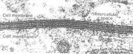
The model was described as a ‘lipo-protein sandwich’, as the lipid layer was sandwiched between two protein layers
The dark segments seen under electron microscope were identified (wrongly) as representing the two protein layers
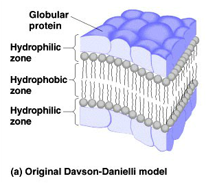
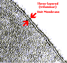
When viewed under a transmission electron microscope, membranes exhibit a characteristic ‘trilaminar’ appearance
Trilaminar = 3 layers (two dark outer layers and a lighter inner region)
1.3.S.3 Analysis of the falsification of the Davson-Danielli model that led to the Singer-Nicolson model.
- Describe conclusions about cell membrane structure drawn from freeze-etched electron micrograph images of the cell membrane.
- Describe conclusions about cell membrane structure drawn from cell fusion experiments.
- Describe conclusions about cell membrane structure drawn from improvements in techniques for determining the structure of membrane proteins..
- Compare the Davson-Danielli model of membrane structure with the Singer-Nicolson model.
There were a number of problems with the lipo-protein sandwich model proposed by Davson and Danielli:
- It assumed all membranes were of a uniform thickness and would have a constant lipid-protein ratio
- It assumed all membranes would have symmetrical internal and external surfaces (i.e. not bifacial)
- It did not account for the permeability of certain substances (did not recognise the need for hydrophilic pores)
- The temperatures at which membranes solidified did not correlate with those expected under the proposed model
Falsification Evidence:
- Membrane proteins were discovered to be insoluble in water (indicating hydrophobic surfaces) and varied in size
- Such proteins would not be able to form a uniform and continuous layer around the outer surface of a membrane
- Fluorescent antibody tagging of membrane proteins showed they were mobile and not fixed in place
- Membrane proteins from two different cells were tagged with red and green fluorescent markers respectively
- When the two cells were fused, the markers became mixed throughout the membrane of the fused cell
- This demonstrated that the membrane proteins could move and did not form a static layer (as per Davson-Danielli)
- Freeze fracturing was used to split open the membrane and revealed irregular rough surfaces within the membrane
- These rough surfaces were interpreted as being transmembrane proteins, demonstrating that proteins were not solely localised to the outside of the membrane structure
New Model:
- In light of these limitations, a new model was proposed by Seymour Singer and Garth Nicolson in 1972
- According to this model, proteins were embedded within the lipid bilayer rather than existing as separate layers
- This model, known as the fluid-mosaic model, remains the model preferred by scientists today (with refinements)