Topic 1.4: membrane transport
In the Cell Membrane unit we will learn that the cell membrane is one of the great multi-taskers of biology. It provides structure for the cell, protects cytosolic contents from the environment, and allows cells to act as specialized units. We will also learn that the membrane is the cell’s interface with the rest of the world – it’s gatekeeper. This phospholipid bilayer determines what molecules can move into or out of the cell, and so is in large part responsible for maintaining the delicate homeostasis of each cell.
The unit is planned to take 3 school days
- Membranes control the composition of cells by active and passive transport.
- Experimental design—accurate quantitative measurement in osmosis experiments are essential. (3.1) (covered by the practical and processing of data)
- Define quantitative and qualitative.
- Determine measurement uncertainty of a measurement tool.
- Explain the need for repeated measurements (multiple trials) in experimental design.
- Explain the need to control variables in experimental design.
1.4.U.1 Particles move across membranes by simple diffusion, facilitated diffusion, osmosis and active transport.
- Describe simple diffusion.
- Explain two examples of simple diffusion of molecules into and out of cells.
- Outline factors that regulate the rate of diffusion.
- Describe facilitated diffusion.
- Describe one example of facilitated diffusion through a protein channel.
- Define osmosis.
- Predict the direction of water movement based upon differences in solute concentration.
- Compare active transport and passive transport.
- Explain one example of active transport of molecules into and out of cells through protein pumps.
Cellular membranes possess two key qualities:
- They are semi-permeable (only certain materials may freely cross – large and charged substances are typically blocked)
- They are selective (membrane proteins may regulate the passage of material that cannot freely cross)
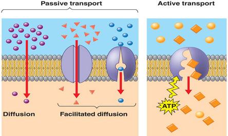
Movement of materials across a biological membrane may occur either actively or passively
Diffusion is the passive movement (does not require energy) of particles from a region of high concentration to a region of low concentration.
- Passive transport means there is no expenditure of energy (ATP).
- Passive transport requires the substance to move from an area of high concentration to low concentration.
Osmosis – the passive movement of water molecules, across a semi-permeable membrane, from a region of lower solute concentration to a region of higher solute concentration.
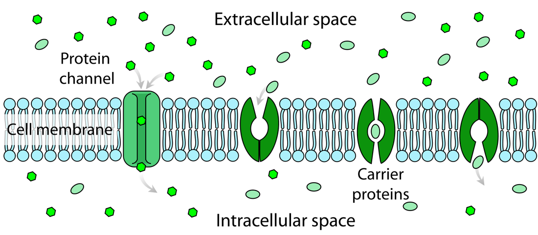
Simple diffusion – passive movement of particles from an area of high concentration to an area of low concentration (follows its concentration gradient). Simple diffusion across membranes occurs when substances other than water move across the phospholipid bilayer (between the phospholipids) or through protein channels.
- Substances that move across the membrane are usually small non-charged particles (i.e. Oxygen, Carbon Dioxide, and Nitrogen) or other lipids.
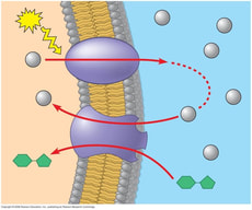
Facilitated Diffusion – specific ions and other particles that cannot move through the phospholipid bilayer sometimes move across protein channels
- During facilitated diffusion the membrane protein changes its shape to allow a specific substance to move across the membrane.
- Each protein channel structure allows only one specific molecule to pass through the channel. For example, magnesium ions pass through a channel protein specific to magnesium ions.
Active transport – movement of substances across membranes using energy from ATP.
- Active transport generally moves substances against their concentration gradient (low to high concentration).
- Many different protein pumps are used for active transport. Each pump only transports a particular substance; therefore cells can control what is absorbed and what is expelled.
- Pumps work in a specific direction; substances enter only on one side and exit through the other side.
- Substances enter the pump from the side with a lower concentration.
- Energy from ATP is used to change the conformational shape of the pump.
- The specific particle is released on the side with a higher concentration and the pump returns to its original shape.
1.4.U 2 The fluidity of membranes allows materials to be taken into cells by endocytosis or released by exocytosis.
- Describe the fluid properties of the cell membrane and vesicles.
- Explain vesicle formation via endocytosis.
- Outline two examples of materials brought into the cell via endocytosis.
- Explain release of materials from cells via exocytosis.
- Outline two examples of materials released from a cell via exocytosis.
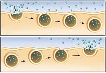
Endocytosis
- Plasma membrane is pinched as a result of the membrane changing shape.
- External material (i.e. Fluid droplets) are engulfed and enclosed by the membrane.
- A vesicle is formed that contains the enclosed particles or fluid droplets, now moves into the cytoplasm.
- The plasma membrane easily reattaches at the ends that were pinched because of the fluidity of the membrane.
- Vesicles that move through the cytoplasm are broken down and dissolve into the cytoplasm.
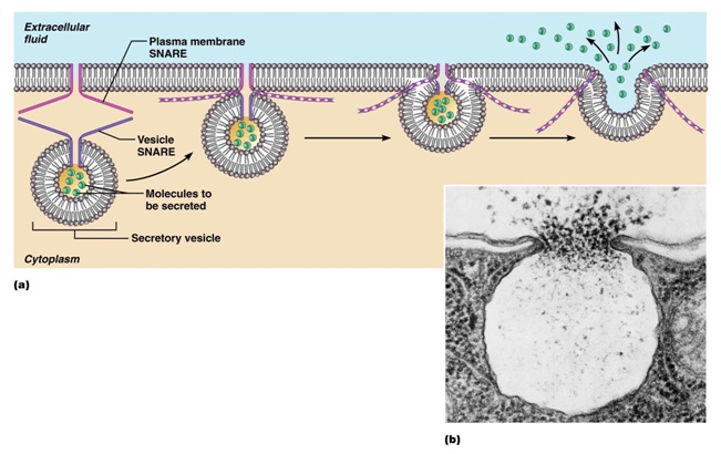
Exocytosis
- After a vesicle created by the rough ER enters the Golgi apparatus, it is again modified, and another vesicle is budded from the end of the Golgi apparatus, which moves towards the cell membrane.
- This vesicle migrates to the plasma membrane and fuses with the membrane, releasing the protein outside the cell through a process called exocytosis.
- The fluidity of the hydrophilic and hydrophobic properties of the phospholipids and the fluidity of the membrane allows the phospholipids from the vesicle to combine to the plasma membrane to form a new membrane that includes the phospholipids from the vesicle.
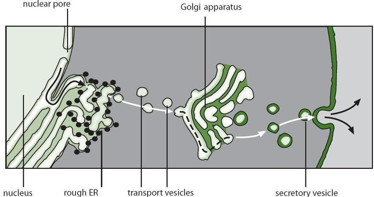
1.4.U3: Vesicles move materials within cells
- List two reasons for vesicle movement.
- Describe how organelles of the endomembrane system function together to produce and secrete proteins (rough ER, smooth ER, Golgi and vesicles).
- Outline how phospholipids and membrane bound proteins are synthesized and transported to the cell membrane.
1.4.A.1 Structure and function of sodium–potassium pumps for active transport and potassium channels for facilitated diffusion in axons.
- Describe the structure of the sodium-potassium pump.
- Describe the role of the sodium-potassium pump in maintaining neuronal resting potential.
- Outline the six steps of sodium-potassium pump action.
- Describe the structure of the potassium channel.
- Describe the mechanism of potassium movement through the potassium channel.
- Explain the specificity of the potassium channel.
- Describe the action of the “voltage gate” of the potassium channel.
The axons in nerve cells transmit electrical impulses by translocating ions to create a voltage difference across the membrane. In the resting phase, the sodium-potassium pump releases sodium ions from the nerve cell, while potassium ions are accumulated in. When the neuron fires, these ions swap locations through facilitated diffusion using the sodium and potassium channels
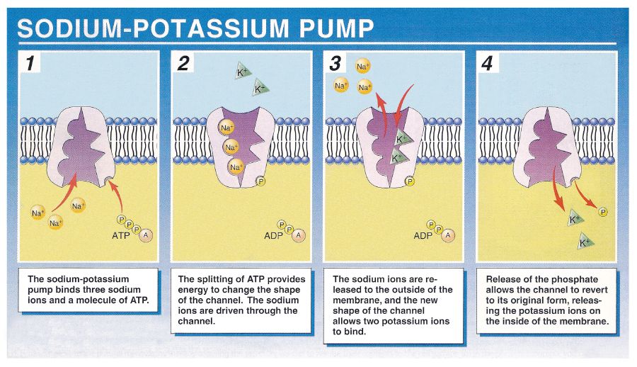
Sodium-Potassium Pump
An integral protein that exchanges 3 sodium ions out of cell with two potassium ions into the cell.
The process of ion exchange against the gradient is energy-dependent and involves a number of key steps:
Three sodium ions bind to intracellular sites on the sodium-potassium pump
- A phosphate group is transferred to the pump via the hydrolysis of ATP
- The pump undergoes a conformational change, translocating sodium across the membrane
- The conformational change exposes two potassium binding sites on the extracellular surface of the pump
- The phosphate group is released which causes the pump to return to its original conformation
- This translocates the potassium across the membrane, completing the ion exchange
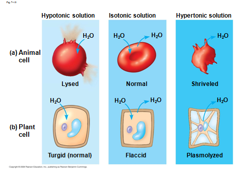
1.4.A.2 Tissues or organs to be used in medical procedures must be bathed in a solution with the same osmolarity as the cytoplasm to prevent osmosis
- Explain what happens to cells when placed in solutions of the same osmolarity, higher osmolarity and lower osmolarity.
- Outline the use of normal saline in medical procedures.
Hypertonic solution – Is a solution with a higher osmolarity (higher solute concentration) then the other solution. If cells are placed into a hypertonic solution, water will leave the cell causing the cytoplasm’s volume to shrink and thereby forming indentations in the cell membrane.
Hypotonic solution – Is a solution with a lower osmolarity (lower solute concentration) then the other solution. If cells are placed in a hypotonic solution, the water will rush into the cell causing them to swell and possibly burst.
Both of the above solutions would damage cells, therefore isotonic solutions are used (same osmolarity as inside the cell)
Isotonic solution: A solution that has the same salt concentration as cells and blood.
Organs placed in hypotonic solution or hypertonic solution would damage cells, therefore isotonic solutions are used (same osmolarity as inside the cell)
In medical procedures, isotonic solutions are commonly used as
- Intravenously infused fluids in hospitalized patients.
- Used to rinse wounds and skin abrasions
- Saline eye drops
- Packing donor organs for transport (frozen to slush)
- During skin grafts, used to keep damaged area moist (normal saline is approx. 300 mOsm)