Topic 11.2: Movement
 image from Better Movement
This unit will last 4 school days.
Essential idea:
- The roles of the musculoskeletal system are movement, support and protection.
Nature of science:
- Developments in scientific research follow improvements in apparatus—fluorescent calcium ions have been used to study the cyclic interactions in muscle contraction. (1.8)
11.2 U 1 Bones and exoskeletons provide anchorage for muscles and act as levers.
- State the function of bones and exoskeletons.
- Contrast bones with exoskeletons.
- Identify the fulcrum, effort force and resultant force in the motion of the spine and the grasshopper leg.
- Determine the class of motion of a lever
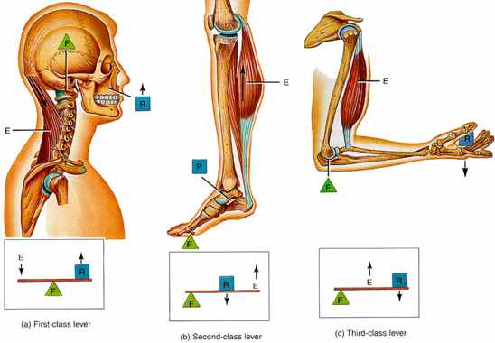
Muscles and bones act together to form levers. A lever is a rigid rod (usually a length of bone) that turns about a pivot (usually a joint). Levers can be used so that a small force can move a much bigger force. This is called mechanical advantage.
- Bones act as levers so the body can move and provide structural support (skeleton).
- Ligaments are strong bands that connect bone to bone strengthening the joint during movement.
- Tendons have dense connective tissue that connects muscles to bones, allowing movement of the bone when a muscle contracts.
- Muscles provide the force for movement by contracting (shortens the muscle fibers)
- The joint acts as a pivot point or a fulcrum
- The force applied (when the muscle contracts) is called the effort
- The force or load needed to overcome for movement to take place is called the resistance
Levers can be used so that a small force can move a much bigger force. This is called mechanical advantage. In our bodies bones act as lever arms, joints act as pivots, and muscles provide the effort forces to move loads
- Levers are classified by first, second, and third class, depending upon the positions among the fulcrum, the effort, and the resistance.
- First-class levers have the fulcrum in the middle, like a seesaw. An example of a first class lever is when a human nods their head (top of the spinal column is the fulcrum, the effort force is provided by the muscles in the back of the neck, and the resistance is weight of the head).
- Second-class levers have a resistance in the middle, like a load in a wheel-barrow. The body acts as second class lever when engaged in push up or calf raise. During a calf raise ball of foot is fulcrum, the body’s mass is the resistance and the effort is applied by calf muscle.
- Third-class levers have the effort from the muscle in the middle of the lever. The majority of the human body’s musculoskeletal levers are third class. These levers are built for speed and range of motion. Muscle attachments are usually close to the fulcrum. In the example of the arm, the effort force is provided by the contraction of the biceps, the fulcrum is the elbow joint and the resistance would be provided by whatever weight is being lifted.
- Exoskeletons in insects and crustaceans can facilitate the movement by providing an anchorage for muscles; similarly to how bones provide anchorage for animals with internal skeletons.
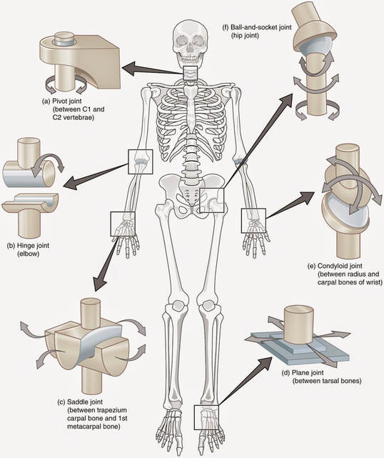
11.2 U 2 Synovial joints allow certain movements but not others.
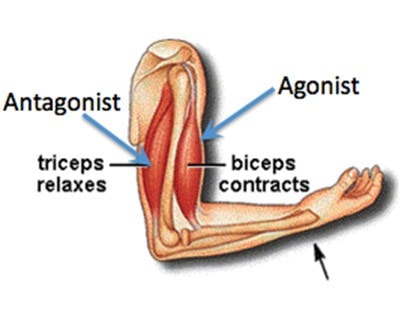
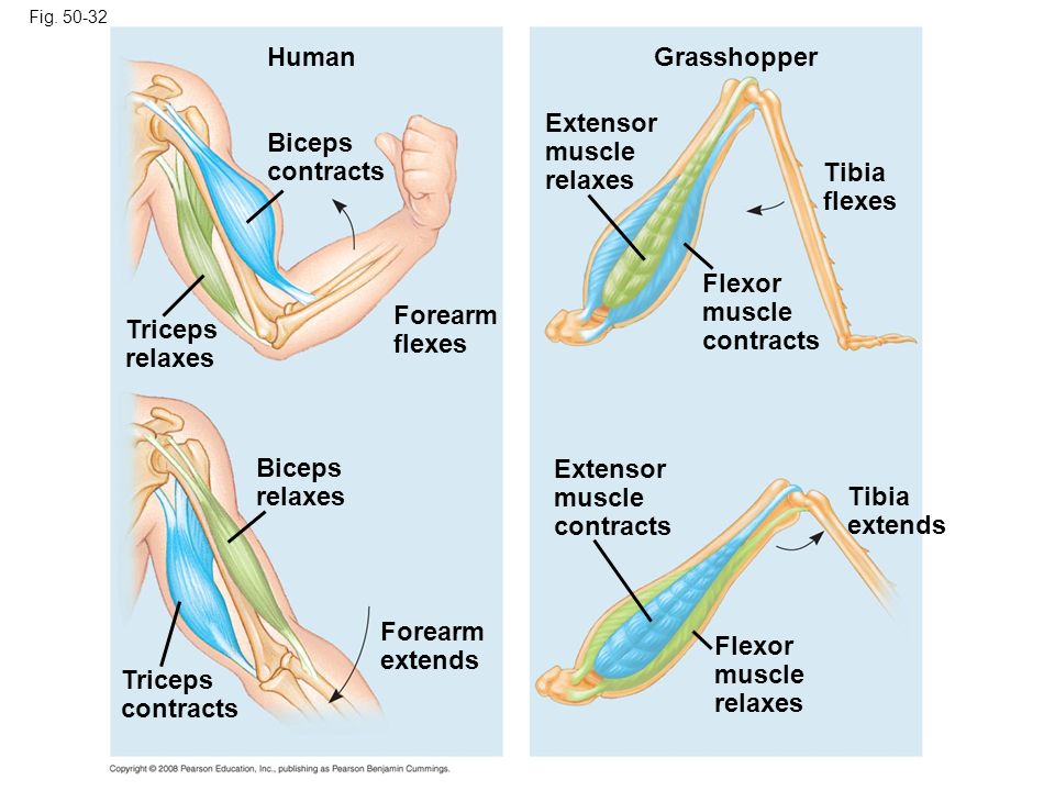
- Define antagonistic pairs in relation to muscle movement.
- State an example of an antagonistic pair of muscles.
11.2 U 3 Movement of the body requires muscles to work in antagonistic pairs.
- Compare the motion of hinge joints with the motion of a ball and socket joint.
- Outline motion of the human knee, shoulder and hip in terms of flexion, extension, rotation, abduction and adduction.
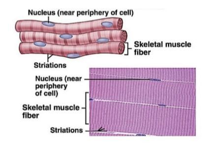
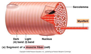
11.2 U 4 Skeletal muscle fibres are multinucleate and contain specialized endoplasmic reticulum.
- List three types of muscle tissue found in the human body.
- Label a diagram of a muscle fibre cell, including the sacrolemma, nuclei, sacroplasmic reticulum and mitochondria.
The 3 types of muscle tissue are cardiac, smooth, and skeletal. Cardiac muscle cells are located in the walls of the heart, appear striated, and are under involuntary control. Smooth muscle fibers are located in walls of hollow visceral organs, except the heart, appear spindle-shaped, and are also under involuntary control. Skeletal muscle fibers occur in muscles which are attached to the skeleton. They are striated in appearance and are under voluntary control.
- Skeletal muscles are composed of bundles of muscle fibers and have a striped appearance because of areas of thick and thin filaments (myosin and actin)
- Muscle cells have many nuclei and are long because the embryonic muscle cells fuse together
- Muscle fibers are composed of many parallel elongated fibers called myofibrils
- A modified endoplasmic reticulum, called the sarcoplasmic reticulum (fluid-filled membranous sacs), extends throughout the muscle fibre, wrapping around each myofibril, sending a signal to the all parts of the muscle fibre to contract at the same time
- The sarcoplasmic reticulum stores calcium – between myofibrils are large numbers of mitochondria, which provide ATP needed for contractions.
11.2 U 5 Muscle fibres contain many myofibrils.
- Outline the relationship between muscles, muscle fibre cells and myofibrils
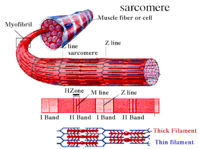
A skeletal muscle is made up of many multinucleate muscle fibers (cells), each of which runs the entire length of the muscle and is connected to at least one motor neuron. Each muscle fiber contains multiple bundles of contractile protein called myofibrils. Myofibrils made up units called sacromere, strung out in single file along the length of myofibrils.
Within each muscle fibre there are many parallel, elongated structures called myofibrils. These have alternating light and dark bands, which give striated muscle its strips. In the centre of each light band is a disc-shaped structure, referred to as the Z-line.
11.2 U 6 Each myofibril is made up of contractile sarcomeres.
- Outline the relationship between myofibrils and sacromeres.
- Describe a structure of a sarcomere., including the Z line, thin actin filaments, thick myosin filaments, light band and dark band.
Myofibrils – rod-shaped parallel bodies consisting of actin and myosin filaments
- Sarcolemma – plasma membrane of the muscle cell
- Mitochondria – large numbers; found dispersed around individual myofibrils
Sarcomere
- Lies between two Z-lines which are dense protein discs
- Contains the thick filament (myosin) and thin filament (actin)
- Myosin contains a head which binds to the binding site on the actin; interaction between myosin and actin (cross-bridge) is responsible for muscle contraction
- Myosin is seen as dark bands while actin is seen as light bands
- Actin filaments are attached to a Z-line at one end
- Myosin filaments are interdigitated with actin filaments at both ends and occupy the centre of the sarcomere
- Each mysosin filament is surrounded by six actin filaments
- A bands contain a full length of myosin and some of the actin filaments
- I bands contain only actin filaments
11.2 U 7 The contraction of the skeletal muscle is achieved by the sliding of actin and myosin filaments.
- Explain the sliding-filament mechanism of muscle contraction, including the role of myosin heads, cross bridges and ATP
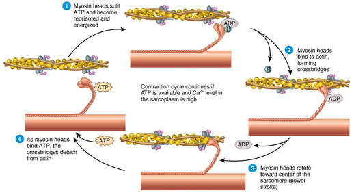
11.2 U 8 ATP hydrolysis and cross bridge formation are necessary for the filaments to slide.
- Explain the exposure of myosin head binding sites on actin, including the role of the sarcoplasmic reticulum, calcium, troponin and tropomyosin.
- ATP attaches to the myosin heads breaking the cross-bridges between the myosin heads and actin binding sites
- The ATP undergoes a hydrolysis reaction forming ADP + Pi.
- This causes a positional change in the myosin head (cocked back).
- The myosin heads bind to actin filaments forming cross-bridges at a site one position further from the centre of the sarcomere
- When the ADP + Pi are released the myosin heads change conformational position, sliding the actin filaments towards the center of the Sarcomere.
- This is called the “power stroke”.
- After the power stroke ATP again binds to the myosin head, causing it to detach from the actin filament ready for another cycle.
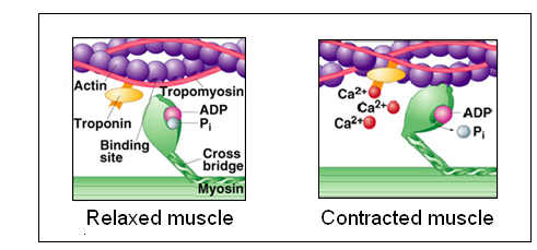
11.2 U 9 Calcium ions and the proteins tropomyosin and troponin control muscle contractions.
- List the events that occur during cross-bridge cycles.
- Describe the role of ATP in muscle contraction.
- An action potential propagated along a motor neuron arrives at the neuromuscular junction.
- This causes the release of the neurotransmitter acetylcholine into the synapse between the terminal axon of the motor neuron and the sarcolemma of the skeletal muscle.
- The acetylcholine binds to receptors on the sarcolemma, causing voltage-gated channels to open and Na+ ions to flow into the muscle cells.
- This creates an action potential in the striated muscle.
- The action potential is further propagated along the sarcolemma of the skeletal muscle.
- The action potential moves into the interior of the muscle cell through folds called t tubules.
- The depolarization of the t tubules causes voltage-gated Ca+ channels on the sarcoplasmic reticulum to open, causing an influx of Ca+ ions into the sarcoplasm.
- Ca+ ions bind to troponin which causes tropomyosin to move exposing the myosin binding sites (troponin and tropomyosin are regulatory proteins blocking the myosin binding sites).
11.2 A 1 Antagonistic pairs of muscles in an insect leg (Oxford Biology Course Companion page 478).
- Label the tibia, femur, tarsus, flexor muscle and extensor muscle on a diagram of a grasshopper hindlimb.
- Describe the contraction of muscles and movement of hindlimb structures that produces a grasshopper jump.
- The hind limbs of grasshoppers are specialized for jumping
- It has a jointed appendage with three parts
- Below the joint is the tibia, and at the base of the tibia is another joint called the tarsus
- Above the joint is the femur
- When the grasshopper jumps, the flexor muscles contract, and the femur and tibia are brought closer together (flexing)(extensor muscles are relaxed)
- As the grasshopper jumps the extensor muscles contract, extending the tibia, creating a powerful jump force
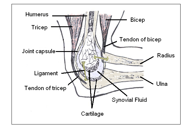
11.2 S 1 Annotation of a diagram of the human elbow. include cartilage, synovial fluid, joint capsule, named bones and named antagonistic muscles.
- Label a diagram of the human elbow inclusive of: humerus, triceps, biceps, joint capsule, synovial fluid, radius, cartilage and ulna.
- State the function of structures found in the human elbow, including: humerus, triceps, biceps, joint capsule, synovial fluid, radius, cartilage and ulna.
11.2 S 2 Drawing labelled diagrams of the structure of a sarcomere. Include Z lines, actin filaments, myosin filaments with heads, and the resultant light and dark bands.
- Draw a diagram of the structure of a sarcomere.
- Label a sarcomere diagram, including Z lines, actin filaments, myosin filaments with heads and the resultant light and dark bands.
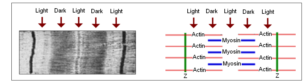
11.2 S 3 Analysis of electron micrographs to find the state of contraction of muscle fibres. Measurement of the length of sarcomeres will require calibration of the eyepiece scale of the microscope. (Oxford Biology Course Companion page 482)
- Compare a relaxed sarcomere to a contracted sarcomere, referring to Z line distance and size of light bands.
- Determine of a sarcomere is contracted or relaxed given an electron micrograph image.
