B3.1.1 – Gas Exchange as a Vital Function in All Organisms
Needed for respiration
Small organisms: Diffusion
Large organisms: Specialized systems
All living organisms need energy to perform essential functions like movement, growth, and repair. This energy is released through aerobic respiration, which requires oxygen and produces carbon dioxide as a waste product.
Gas exchange is the biological process by which oxygen is taken in and carbon dioxide is removed. In most organisms, this happens by diffusion across membranes or specialized surfaces.
Gas Exchange in Different Organisms
- Plants: Take in CO₂ for photosynthesis and release O₂. They also respire all the time, using O₂ and producing CO₂. Gases move through stomata on leaves.
- Unicellular organisms: Use their whole-body surface for gas exchange due to a high surface area-to-volume ratio.
Surface Area-to-Volume Ratio (SA:V)
| Organism Size | SA:V Ratio | Gas Exchange Efficiency |
|---|---|---|
| Small | Large | Efficient by diffusion |
| Large | Small | Needs specialized systems |
As organisms grow, their volume increases faster than surface area, reducing their SA:V ratio. This makes diffusion alone too slow for effective gas exchange.
Why Specialized Systems Are Needed
Larger organisms have internal cells that are far from the external environment. They need:
- Special exchange surfaces (lungs, gills, tracheae)
- Transport systems (blood, haemolymph)
Why the need for a gas exchange system inside large organisms?


Summary
- Gas exchange is essential for aerobic respiration in all organisms.
- Small organisms use simple diffusion due to high SA:V ratios.
- Larger organisms need specialized systems to ensure efficient gas exchange over longer distances.
B3.1.2 – Properties of Gas Exchange Surfaces
Thin • Permeable • Moist • Large surface area
To ensure efficient gas exchange, all organisms have evolved specialized surfaces that allow oxygen to enter and carbon dioxide to exit rapidly. Despite structural differences, these surfaces share four essential features:
1. Thin Tissue Layer
Gas exchange surfaces are typically only one cell thick, minimizing the diffusion distance and speeding up gas exchange.
2. Permeability
Gas exchange surfaces must allow oxygen and carbon dioxide to diffuse freely. These gases are small and nonpolar, so they move easily through cell membranes by simple diffusion.
No barriers = faster diffusion
3. Moisture
Gases must dissolve in water before they can diffuse across membranes. That’s why gas exchange surfaces are always kept moist.
4. Large Surface Area
A larger surface area increases the amount of gas that can diffuse at any one time, compensating for the naturally slow process of diffusion.
Summary of Key Features
| Feature | Function |
|---|---|
| Thin surface | Short diffusion distance ⟶ faster gas exchange |
| Permeable | Allows O₂ and CO₂ to diffuse freely |
| Moist | Enables gases to dissolve before diffusing |
| Large surface area | Maximizes total gas exchange capacity |
B3.1.3 – Maintenance of Concentration Gradients at Exchange Surfaces in Animals
Gas exchange only works efficiently if a steep concentration gradient is maintained between oxygen and carbon dioxide.
Oxygen and carbon dioxide move by diffusion. For diffusion to stay rapid and effective, there must be a constant concentration gradient across the gas exchange surface.
1. Dense Network of Blood Vessels
Capillaries are packed closely around the alveoli (lungs) or gills (fish). They quickly:
- Carry away oxygen after it diffuses in
- Deliver carbon dioxide from body tissues

2. Continuous Blood Flow
Blood is constantly moving, which prevents buildup of oxygen or slow removal of carbon dioxide.
3. Ventilation (Air or Water Movement)
Ventilation ensures that the external side of the surface (air or water) stays high in O₂ and low in CO₂:
- In lungs → breathing brings in fresh oxygen-rich air
- In gills → water flows over the surface constantly
🔑 Summary of Gradient Maintenance
| Mechanism | Function |
|---|---|
| Dense capillary network | Quickly removes O₂ and brings CO₂ ⟶ keeps blood side steep |
| Continuous blood flow | Delivers fresh blood ⟶ prevents oxygen saturation |
| Ventilation | Maintains external O₂/CO₂ levels ⟶ gradient across the surface |
B3.1.4 – Adaptations of Mammalian Lungs for Gas Exchange
✔ Oxygen fuels aerobic respiration
✔ CO₂ must be removed to avoid toxicity and maintain pH
The lungs are adapted to allow rapid and efficient exchange of gases between air and blood. Several structural features support this essential function.

1. Alveoli – The Main Site of Gas Exchange
Alveoli are tiny air sacs found at the end of bronchioles. Each lung has ~300 million alveoli, providing a vast surface area for diffusion.
2. Very Thin Walls
Alveolar and capillary walls are one cell thick, reducing the diffusion distance to less than 1 µm.
3. Surfactant Production
Type II pneumocytes inside alveoli secrete surfactant – a fatty fluid that reduces surface tension.
- Prevents alveoli from collapsing
- Keeps alveoli moist for efficient gas diffusion
4. Dense Network of Capillaries
Each alveolus is wrapped in a rich bed of capillaries, which:
- Deliver CO₂ from tissues
- Carry away O₂ to the body
5. Highly Branched Bronchial Tree
Air enters the lungs through a system of branching tubes:
Trachea → Bronchi → Bronchioles → Alveoli
This branching structure increases surface area and ensures air reaches deep into the lungs.
6. Ventilation Maintains Gradients
Breathing movements bring in fresh air and remove used air:
- Inhalation: Brings in O₂-rich air
- Exhalation: Removes CO₂-rich air
✅ Summary of Lung Adaptations
| Adaptation | Function |
|---|---|
| Thin walls | Short diffusion distance |
| Large surface area (alveoli) | More gas exchange per breath |
| Rich capillary network | Maintains steep gradients |
| Surfactant | Prevents collapse & helps diffusion |
| Ventilation | Refreshes air to maintain gradients |
B3.1.5 – Ventilation of the Lungs
Inhalation: breathing in
Exhalation: breathing out
Maintains steep O₂ and CO₂ gradients for efficient gas exchange
Ventilation is the physical movement of air into and out of the lungs. It involves two processes:
- Inhalation (Inspiration): Brings in fresh air rich in oxygen
- Exhalation (Expiration): Removes air rich in carbon dioxide
🫧 Inhalation (Breathing In)
During inhalation:
- Diaphragm contracts and moves downward (flattens)
- External intercostal muscles contract → ribs move up & out
- Thoracic cavity volume increases
- Pressure inside lungs drops below atmospheric pressure
- Air flows into the lungs
💨 Exhalation (Breathing Out)
During exhalation:
- Diaphragm relaxes and moves upward (dome shape)
- External intercostal muscles relax → ribs move down & in
- Abdominal muscles may contract (especially during forced exhalation)
- Thoracic cavity volume decreases
- Pressure inside lungs increases above atmospheric pressure
- Air flows out of the lungs
🧠 Summary: Muscles Involved in Ventilation
| Muscle Group | During Inhalation | During Exhalation |
|---|---|---|
| Diaphragm | Contracts ↓ | Relaxes ↑ |
| External Intercostals | Contract ↑ & → | Relax ↓ & ← |
| Ribs | Move up and out | Move down and in |
| Abdominal Muscles | Relax | May contract |
B3.1.6 – Measurement of Lung Volumes
Vital Capacity = TV + IRV + ERV
✅ You can measure:
– Tidal Volume (TV)
– Inspiratory Reserve (IRV)
– Expiratory Reserve (ERV)
❌ You can’t directly measure:
– Residual Volume (RV)
– Total Lung Capacity (TLC)
📊 Key Lung Volume Terms
| Term | Description |
|---|---|
| Tidal Volume (TV) | Air breathed in or out in a normal breath (at rest) |
| Inspiratory Reserve (IRV) | Extra air you can forcefully inhale after a normal breath |
| Expiratory Reserve (ERV) | Extra air you can forcefully exhale after a normal breath |
| Vital Capacity (VC) | Maximum air exhaled after a full inhalation (TV + IRV + ERV) |
| Residual Volume (RV) | Air left in lungs after forced exhalation (not measurable directly) |
| Total Lung Capacity (TLC) | Vital Capacity + Residual Volume |
🧪 How Do We Measure These?
Using a spirometer: a machine that records breathing patterns on a graph.
- Breathe normally → record Tidal Volume (TV)
- Inhale deeply → record Inspiratory Reserve (IRV)
- Exhale forcefully → record Expiratory Reserve (ERV)
- Calculate Vital Capacity = TV + IRV + ERV
📐 Example Values (Average Adult)
- Tidal Volume (TV): ~500 mL
- Inspiratory Reserve (IRV): ~3000 mL
- Expiratory Reserve (ERV): ~1100 mL
- Vital Capacity (VC): ~4600 mL
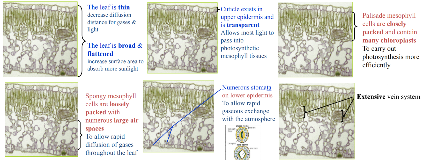
B3.1.7 – Adaptations for Gas Exchange in Leaves
📌 Leaf Features
– Large surface area → more air contact
– Thin → short diffusion distance
– Air spaces → fast gas movement
– Stomata + guard cells → regulate flow
– Moist inner surface → gases dissolve
– Veins → water in, sugars out
Leaves are adapted for efficient gas exchange, which is essential for photosynthesis and respiration.
🌬️ Why Do Leaves Need Gas Exchange?
- Photosynthesis needs CO₂ and releases O₂
- Respiration (in all cells) uses O₂ and produces CO₂
- Leaves must allow gas exchange without losing too much water
🧬 Key Adaptations in Leaf Structure
| Structure | Function (How it helps gas exchange) |
|---|---|
| Waxy Cuticle | Thin, waterproof layer that reduces water loss but allows light through |
| Upper Epidermis | Transparent, protective layer that lets light reach photosynthetic cells |
| Spongy Mesophyll | Loosely packed cells with air spaces to allow easy gas diffusion |
| Air Spaces | Allow gases like CO₂ and O₂ to diffuse quickly between cells and stomata |
| Stomata (pores) | Let CO₂ enter and O₂ exit the leaf |
| Guard Cells | Surround each stoma and control opening/closing |
| Veins (xylem & phloem) | Xylem brings water; Phloem carries sugars away |
🌡️ How Do Guard Cells Work?
- Location: Mostly on underside of the leaf
- Only epidermal cells with chloroplasts
Daytime (light):
- Photosynthesis → sugar made
- Guard cells absorb K⁺ ions
- Water enters by osmosis → cells swell
- Stoma opens to let CO₂ in

Night / Water stress:
- K⁺ pumped out → water exits
- Guard cells shrink → stoma closes
B3.1.8 – Distribution of Tissues in a Leaf
📚 Basic Structure of a Dicot Leaf (Transverse Section)
Below is a simplified order of tissue layers from top to bottom:
- Cuticle
- Upper Epidermis
- Palisade Mesophyll
- Spongy Mesophyll
- Lower Epidermis (with stomata and guard cells)
- Cuticle
🔬 Tissue Distribution & Functions
| Tissue / Layer | Key Features & Function |
|---|---|
| Cuticle | Waxy outer layer (top and bottom) Prevents water loss |
| Upper Epidermis | Transparent cells Protect internal tissues from damage & microbes |
| Palisade Mesophyll | Tightly packed, column-shaped cells Rich in chloroplasts → main site of photosynthesis |
| Spongy Mesophyll | Loosely packed with air spaces Allows gas diffusion between cells |
| Vein (Vascular Bundle) | Contains xylem (for water) & phloem (for sugars) Leaf’s transport system |
| Lower Epidermis | Has stomata and guard cells Regulates gas exchange and water loss |
| Guard Cells | Bean-shaped, contain chloroplasts Open/close stomata for gas control |
B3.1.9 – Transpiration as a Consequence of Gas Exchange in a Leaf
Halophytes → adapted to salty environments
Both minimize water loss through unique structural & physiological features.
Transpiration is the loss of water vapor from the surface of leaves, mainly through the stomata during gas exchange.

This process is an unavoidable side-effect of stomata opening to allow carbon dioxide in for photosynthesis. When water potential (Ψ) inside the leaf is higher than the surrounding air, water vapor exits.
🌞 When Does It Happen?
- Mostly during the daytime when stomata are open
- Nighttime transpiration is minimal (stomata are usually closed)
- Plants can lose up to 98% of absorbed water through transpiration!
📊 Factors Affecting Rate of Transpiration
| Factor | Effect on Transpiration |
|---|---|
| Light Intensity | More light → stomata open wider → more water loss |
| Temperature | Warmer air holds more water vapor → increases evaporation rate |
| Humidity | High humidity → reduces water potential gradient → slows transpiration |
| Wind / Air Movement | Wind removes humid air near stomata → maintains steep gradient |
| Soil Water Availability | Dry soil → limits water uptake → reduces transpiration |
🌱 Link with Gas Exchange
Stomata open to allow CO₂ to enter for photosynthesis. At the same time, oxygen and water vapor exit the leaf. Thus, transpiration and gas exchange are directly linked.
B3.1.10 – Stomatal Density
Stomatal density is the number of stomata per mm² of leaf surface. These pores regulate gas exchange and water loss.
More stomata = greater potential for photosynthesis and transpiration.
🧪 How Do We Measure It?
- Leaf Casts: Apply clear nail polish to leaf, peel it off, and place it under a microscope.
- Micrographs: Take clear images of the leaf surface and count stomata using a grid or scale.
📏 Steps for Accurate Measurement
- Select a known field of view under high magnification.
- Count stomata in that area.
- Repeat for multiple areas on the same leaf.
- Calculate the average number of stomata per mm².
🔍 Why Repeat Counts?
“Biological samples are variable”
→ Stomatal number may vary across a single leaf
→ Repeating measurements gives more reliable, unbiased data
→ Always calculate an average from multiple fields of view
🌿 Link to Environmental Conditions
| Environment | Stomatal Density Trend |
|---|---|
| Dry Conditions | Fewer stomata to reduce water loss |
| Humid Conditions | More stomata to allow maximum gas exchange |
Additional Higher Level
B3.1.11 – Adaptations of Foetal and Adult Haemoglobin for Oxygen Transport
Foetal Hb → left shift → higher O₂ affinity
Bohr effect → right shift → more O₂ unloading in active tissues
Haemoglobin (Hb) is the protein in red blood cells that carries oxygen. It’s made of 4 polypeptide chains and 4 haem groups, each binding one molecule of oxygen.
🧷 Cooperative Binding of Oxygen
When the first O₂ molecule binds, it causes a conformational change, making it easier for the next three to bind. This is called cooperative binding, and it leads to an S-shaped (sigmoidal) oxygen dissociation curve.
Foetal vs. Adult Haemoglobin
| Feature | Foetal Hb | Adult Hb |
|---|---|---|
| Oxygen affinity | Higher | Lower |
| Reason | Takes O₂ from maternal blood | Already exposed to high O₂ in lungs |
| Dissociation curve | Shifted to the left | Normal or slightly right-shifted |
👉 Why left shift for foetus?
Higher O₂ affinity → binds oxygen more readily in placenta, even at low O₂ concentration.


The Bohr Effect – CO₂’s Role
CO₂ binds to haemoglobin at a different site (not the haem group). This is allosteric binding, which changes Hb’s shape and reduces its oxygen affinity.
High CO₂ → more H⁺ ions → more acidic → promotes O₂ unloading in tissues. This is known as the Bohr Shift – the oxygen dissociation curve shifts to the right.
🔁 Summary of Adaptations
| Adaptation | Purpose |
|---|---|
| Cooperative binding | Efficient oxygen loading in lungs |
| Foetal Hb higher affinity | Takes O₂ from mother’s blood |
| Bohr Effect (CO₂ binding) | Promotes O₂ unloading where it’s needed |
B3.1.12 – Bohr Shift
🧠 Tip:
Right shift = Release of O₂
Bohr shift helps muscles get more oxygen during exercise or stress!
The Bohr Shift explains how increased carbon dioxide (CO₂) in tissues causes haemoglobin (Hb) to release more oxygen (O₂). This effect is crucial in active tissues that produce lots of CO₂, such as during exercise.
🔁 How It Works (Step-by-Step)
- Active cells respire → produce more CO₂.
- CO₂ dissolves in blood and reacts with water to form carbonic acid (H₂CO₃):
CO₂ + H₂O→ H₂CO₃ - Carbonic acid releases H⁺ ions → lowers blood pH.

- H⁺ binds to haemoglobin → changes its shape.
- This reduces Hb’s affinity for oxygen → O₂ is released more easily.
📊 Effect on the Oxygen Dissociation Curve
The curve shifts to the right
More O₂ is unloaded at higher CO₂ levels
Tissues get more oxygen when they need it most
💪 Benefits of the Bohr Shift
| Feature | Benefit to Body |
|---|---|
| More O₂ released in active tissues | Supports high respiration rate |
| Responds to ↑ CO₂ and ↓ pH | Helps maintain homeostasis |
| Curve shifts right | Efficient O₂ delivery under stress |
🧪 Summary
More CO₂ → More H⁺ → Lower pH → Hb releases O₂
The Bohr effect allows faster and more targeted oxygen delivery to where it’s needed most – like working muscles and organs.
B3.1.13 – Oxygen Dissociation Curves & Haemoglobin’s Affinity for Oxygen
💡Tip:
Cooperative binding makes oxygen load quickly in lungs and unload fast in tissues.
The sigmoid curve reflects haemoglobin’s smart oxygen delivery!
The Oxygen Dissociation Curve is a graph that shows how much oxygen (O₂) is bound to haemoglobin (Hb) at different oxygen pressures.
- Y-axis: % saturation of haemoglobin with O₂
- X-axis: Partial pressure of oxygen (pO₂)
🧬 Why is the Curve S-Shaped (Sigmoidal)?
- Cooperative binding causes the S-shape.
- Binding of first O₂ makes it easier for the next ones.
- Curve steepens in middle, then flattens at top as Hb gets saturated.
Haemoglobin has 4 haem groups. As each O₂ binds, it alters the shape of Hb slightly, increasing its affinity for the next O₂ molecule. However, once nearly full, it becomes harder for the last O₂ to bind – causing the curve to plateau.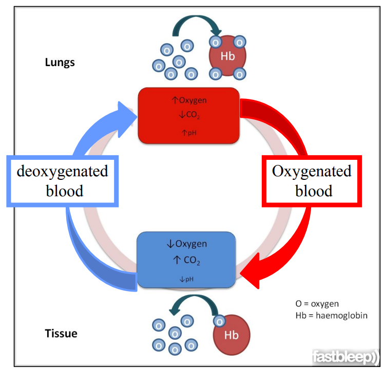
In the Lungs: High Affinity
At high pO₂ (in lungs), Hb has high oxygen affinity and binds O₂ easily, forming oxyhaemoglobin (HbO₂). Blood becomes highly saturated with oxygen.
In Tissues: Low Affinity
At low pO₂ (in tissues), Hb’s affinity drops, causing it to release oxygen where it’s needed for respiration.
🔄 Cooperative Binding Summary
| Binding Stage | Affinity for O₂ | Curve Behavior |
|---|---|---|
| 1st O₂ | Low | Curve starts shallow |
| 2nd & 3rd O₂ | Higher | Curve steepens |
| 4th O₂ | Lower again | Curve levels off |
Haemoglobin structure
Haemoglobin = Respiratory pigment
- Haemoglobin has quaternary structure
- Four subunits
- Two alpha chains
- Two beta chains
- Each contains a haeme group that can bind oxygen
Haemoglobin binding oxygen

📊 What the Curve Tells Us
Right shift: Hb releases more O₂ (e.g. Bohr shift)
Left shift: Hb holds onto O₂ more (e.g. fetal haemoglobin)