C2.2.1 – Neurons as Cells That Carry Electrical Impulses
🧬 Structure of a Neuron
Neurons are specialized cells in the nervous system that transmit electrical signals.
Each neuron consists of:
- Cell body (soma): Contains the nucleus and cytoplasm.
- Axon: A single, long fibre that carries impulses away from the cell body.
- Dendrites: Multiple short fibres that carry impulses towards the cell body.
🔌 How Neurons Work
Neurons conduct electrical impulses along their fibres to communicate with other neurons, muscles, or glands.
The direction of the impulse is typically:
Dendrites → Cell body → Axon → Axon terminals
🧠 Example: Motor Neuron
| Part | Function |
|---|---|
| Dendrites | Receive signals from other neurons |
| Cell body | Processes signals; contains the nucleus |
| Axon | Transmits the signal over long distances |
| Axon terminals | Pass the signal to another cell (e.g. a muscle or another neuron) |
– Neurons are cells specialized for transmitting electrical impulses.
– Axons carry signals away from the cell body, while dendrites bring them in.
– The cell body holds the nucleus and processes information before sending signals down the axon.
C2.2.2 – Generation of the Resting Potential in Neurons
🔋 What Is the Resting Potential?
The resting potential is the electrical charge difference across the plasma membrane of a neuron when it is not transmitting a signal.
It’s typically around –70 mV, meaning the inside of the neuron is more negative than the outside.
🔄 How the Resting Potential Is Generated
The sodium-potassium pump (Na⁺/K⁺ pump) is a protein in the neuron membrane that uses ATP to move ions:
- 3 Na⁺ (sodium ions) out
- 2 K⁺ (potassium ions) in
This active transport process creates a concentration gradient:
- High Na⁺ outside
- High K⁺ inside
⚡ Why the Inside Is More Negative
Although K⁺ is brought in, many potassium channels are open, so K⁺ leaks back out.
Large negative proteins remain inside the neuron and can’t cross the membrane.
This leads to a net negative charge inside → membrane polarization.
🧠 Key Concepts
| Term | Explanation |
|---|---|
| Resting potential | Electrical charge across the membrane at rest (~–70 mV) |
| Membrane potential | Difference in charge between inside and outside of the neuron |
| Polarization | Inside of neuron is more negative than the outside |
| ATP use | Na⁺/K⁺ pump needs ATP to work against concentration gradients |
– The resting potential is around –70 mV.
– The Na⁺/K⁺ pump moves 3 sodium ions out and 2 potassium ions in using ATP.
– The resting potential is negative due to ion gradients and leaky potassium channels.
C2.2.3 – Nerve Impulses as Action Potentials
⚡ What Is a Nerve Impulse?
A nerve impulse is an action potential – a temporary reversal of the electrical charge across a neuron’s membrane.
It’s a rapid, electrical signal that travels along the axon or nerve fibre.
⚙️ How Is It Electrical?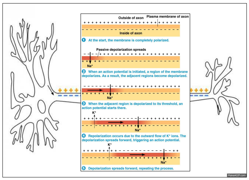
It’s caused by the movement of positively charged ions (Na⁺ and K⁺).
When the neuron is stimulated:
- Sodium (Na⁺) channels open
- Na⁺ rushes into the cell, making the inside less negative → depolarization
Then:
- Potassium (K⁺) channels open
- K⁺ flows out, restoring negativity inside → repolarization
🔁 Propagation of the Impulse
The action potential moves along the axon like a wave:
- One part depolarizes
- It triggers the next section to depolarize
This is called propagation.
🧠 Key Concepts Table
| Term | Explanation |
|---|---|
| Action potential | Rapid change in membrane potential (inside becomes positive briefly) |
| Depolarization | Na⁺ enters → inside becomes less negative |
| Repolarization | K⁺ exits → inside becomes more negative again |
| Propagation | Action potential spreads along the neuron like a domino effect |
📊 Real Example
A typical action potential lasts ~3–5 ms and peaks at +30 to +40 mV.
Impulses can travel up to 120 m/s in myelinated axons!
– A nerve impulse is an action potential, involving ion movement.
– Na⁺ and K⁺ ions cause depolarization and repolarization of the membrane.
– The action potential is electrical due to movement of positive ions.
– It propagates along the nerve fibre in one direction.
C2.2.4 – Variation in the Speed of Nerve Impulses

🚀 What Affects the Speed of a Nerve Impulse?
The speed of a nerve impulse can vary depending on:
- Axon diameter
- Presence or absence of a myelin sheath
- Animal size
🔬 Comparisons of Nerve Impulse Speed
| Fibre Type | Speed | Reason |
|---|---|---|
| Giant axons (e.g. squid) | Fast | Large diameter → less resistance to ion flow |
| Small, non-myelinated fibres | Slow | Narrow → more resistance; no insulation |
| Myelinated fibres (e.g. humans) | Very fast (up to 120 m/s) | Saltatory conduction – impulse jumps between nodes |
| Non-myelinated fibres | Slower | Impulse travels continuously → slower conduction |
💡 Key Terms
- Myelin sheath: Fatty insulating layer around axons.
- Saltatory conduction: The “jumping” of impulses between nodes of Ranvier in myelinated axons.
- Axon diameter: Wider axons have less resistance → faster impulse.
📉Understanding Correlations
| Correlation Type | Description |
|---|---|
| Negative correlation | As one variable increases, the other decreases |
| Positive correlation | As one variable increases, the other also increases |
| Correlation coefficient (r) | Value between -1 and +1 showing strength and direction of correlation |
| Coefficient of determination (R²) | Indicates how much variation in one variable is explained by another (0 to 1) |
📊 Example Correlations
| Variables | Type of Correlation |
|---|---|
| Animal size vs conduction speed | Negative |
| Axon diameter vs conduction speed | Positive |
If R² = 0.85 for axon diameter vs speed, it means 85% of the variation in speed is explained by axon diameter.
– Nerve impulses travel faster in giant or myelinated axons.
– Axon diameter and myelination increase conduction speed.
– Speed is negatively correlated with animal size, but positively correlated with axon diameter.
– Correlation coefficients (r) and R² values help analyse these relationships mathematically.
C2.2.5 – Synapses as Junctions Between Neurons and Effector Cells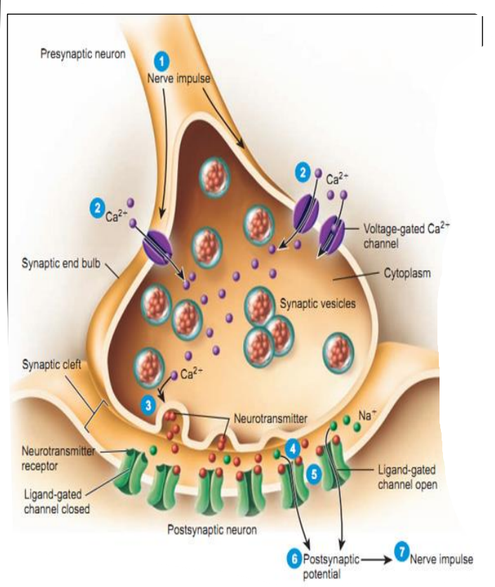
🧬 What Are Synapses?
Synapses are tiny gaps (junctions) between:
- Two neurons
- A neuron and an effector cell (e.g. muscle or gland)
Only chemical synapses are covered here (not electrical).
📩 How Do Synapses Work?
| Step | Process |
|---|---|
| 1 | An electrical impulse reaches the axon terminal. |
| 2 | It triggers release of neurotransmitters from vesicles. |
| 3 | Neurotransmitters diffuse across the synaptic cleft. |
| 4 | They bind to receptors on the postsynaptic membrane. |
| 5 | This generates a new impulse in the next neuron or activates an effector. |
🛑 Why Does the Signal Only Go One Way?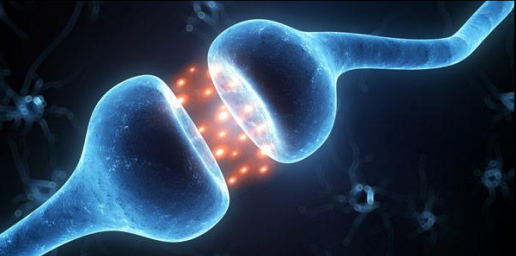
- Vesicles with neurotransmitters are only in the presynaptic neuron.
- Receptors for neurotransmitters are only on the postsynaptic membrane.
This ensures that signals only pass in one direction across synapses.
📘 Key Terms
| Term | Meaning |
|---|---|
| Synaptic cleft | Gap between neurons at a synapse |
| Neurotransmitter | Chemical messenger (e.g. acetylcholine) |
| Presynaptic neuron | Sends the signal |
| Postsynaptic cell | Receives the signal |
| Effector cell | Muscle or gland cell that responds to a signal |
– Synapses connect neurons or neurons to effectors (muscles/glands).
– Only chemical synapses are considered.
– Signal transmission is one-way only due to vesicle and receptor placement.
– Neurotransmitters carry the signal across the synaptic cleft.
Additional Higher Level
C2.2.8 – Depolarization and Repolarization During Action Potentials
⚙️ What is an Action Potential?
An action potential is a rapid, temporary change in membrane potential that travels along a neuron to transmit electrical signals.
To generate an action potential, the membrane must reach a threshold potential (around –55 mV) to trigger the opening of voltage-gated ion channels.
⚡ Depolarization (Making the Inside More Positive)
| Event | What Happens |
|---|---|
| 1. Threshold reached | A small stimulus causes the membrane to depolarize to –55 mV. |
| 2. Voltage-gated sodium (Na⁺) channels open | Sodium ions flood into the cell due to the electrochemical gradient. |
| 3. Membrane potential spikes | Inside becomes more positive, rising up to +30 mV. |
🔋 Repolarization (Resetting the Charge)
| Event | What Happens |
|---|---|
| 4. Na⁺ channels close | After peaking, the sodium channels shut. |
| 5. Voltage-gated potassium (K⁺) channels open | K⁺ ions move out of the neuron. |
| 6. Inside becomes negative again | The membrane repolarizes, dropping back below 0 mV. |
⬇️ After-Hyperpolarization (Optional Extra Dip)
- Too much K⁺ leaves the cell, briefly making the membrane more negative than resting potential (e.g. –75 mV).
- This is called the after-hyperpolarization or undershoot.
- It quickly returns to normal via the Na⁺/K⁺ pump and leak channels.
📊 Graph: Action Potential Stages
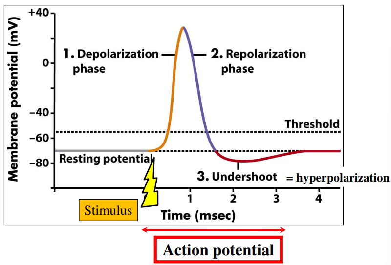
🔑 Key Channels Involved
| Ion Channel | When It Opens | Ion Movement |
|---|---|---|
| Voltage-gated Na⁺ channels | When threshold is reached | Na⁺ in (depolarizes) |
| Voltage-gated K⁺ channels | After Na⁺ peak | K⁺ out (repolarizes) |
– Action potentials occur only if the threshold potential (~–55 mV) is reached.
– Depolarization: Na⁺ channels open → Na⁺ enters → inside becomes positive.
– Repolarization: Na⁺ channels close, K⁺ channels open → K⁺ exits → inside becomes negative again.
– Action potentials allow rapid and unidirectional transmission of nerve signals.
C2.2.9 – Propagation of an Action Potential Along a Nerve Fibre/Axon
🚀 What is Propagation?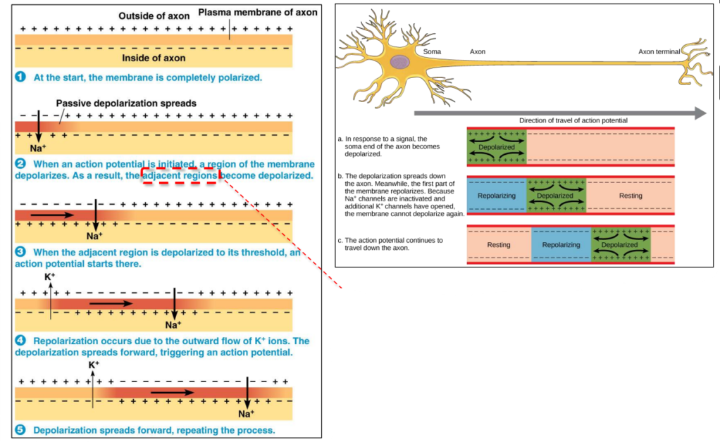
Propagation = how an action potential travels along an axon. It doesn’t move all at once – it’s regenerated at each point along the membrane. This is possible due to local currents caused by ion diffusion.
🌊 What Are Local Currents?
Local currents are small movements of Na⁺ ions that:
- Spread along the inside of the axon (cytoplasm), and
- Attract ions on the outside of the membrane.
This movement depolarizes nearby membrane regions, helping them reach the threshold potential (≈ –55 mV).
⚙️ Step-by-Step: How an Action Potential Moves
| Step | What Happens |
|---|---|
| 1 | A section of axon is depolarized (Na⁺ channels open). |
| 2 | Na⁺ floods in → inside becomes more positive. |
| 3 | Na⁺ ions diffuse along the inside of the axon to the next region. |
| 4 | This local current raises the voltage of the next segment. |
| 5 | If it reaches threshold, new Na⁺ channels open and another action potential starts. |
| 🔁 | This continues all the way down the axon. |
🔄 Why Can’t It Go Backward?
After firing, that region of the axon enters a refractory period. During this time, Na⁺ channels are inactivated, so a new action potential can’t start there. This ensures one-way direction of impulse travel.
🧠 Analogy: Mexican Wave in a Stadium
Just like a wave of people stands up and sits down in sequence during a game, an action potential travels as each part of the axon “fires” and then resets.
– Action potentials move due to local currents caused by sodium ion diffusion.
– These currents cause nearby areas to depolarize, triggering new action potentials.
– The process ensures fast, one-way signal transmission along the axon.
C2.2.10 – Oscilloscope Traces Showing Resting Potentials and Action Potentials
🧪 What is an Oscilloscope Trace?
A graphical display of voltage changes over time used to visualize nerve impulses (action potentials).
- X-axis = Time
- Y-axis = Membrane potential (mV)
🔋 Key Features of an Action Potential on a Trace
| Phase | What Happens | Typical Voltage (mV) |
|---|---|---|
| Resting Potential | Na⁺/K⁺ pumps maintain –70 mV | –70 mV |
| Depolarization | Voltage-gated Na⁺ channels open → Na⁺ in | Up to +30 mV |
| Repolarization | Voltage-gated K⁺ channels open → K⁺ out | Falls toward –70 mV |
| Hyperpolarization | Too much K⁺ leaves temporarily | Around –80 mV |
| Return to Resting | Pumps restore original gradient | Back to –70 mV |
📊 Interpreting the Trace: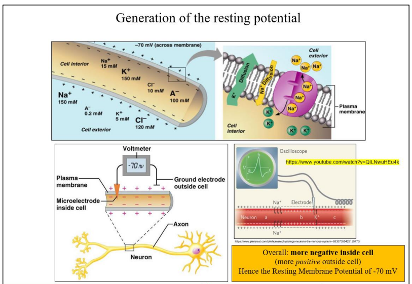
- Before spike = resting (–70 mV)
- Rising curve = depolarization (Na⁺ in)
- Peak = maximum voltage (+30 mV)
- Falling curve = repolarization (K⁺ out)
- Dip below baseline = hyperpolarization
- Flat line return = back to resting potential
⏱️ Measuring Impulse Frequency (Hz or impulses/sec)
To calculate:
- Measure time between peaks on the oscilloscope.
- Use the formula:
Frequency (Hz) = 1 / time interval (s)
More frequent impulses = stronger stimulus or higher intensity.
– Oscilloscopes show voltage changes during action potentials.
– Each phase on the trace corresponds to ion movements across the membrane.
– Impulse frequency can be calculated from the time between peaks.
C2.2.11 – Saltatory Conduction in Myelinated Fibres
🧠 What is Saltatory Conduction?
- A fast method of transmitting nerve impulses along myelinated axons.
- The action potential “jumps” from one node of Ranvier to the next.
🧪 How It Works:
| Feature | Role in Saltatory Conduction |
|---|---|
| Myelin sheath | Insulates axon; prevents ion flow in these regions |
| Nodes of Ranvier | Gaps in the myelin where ion channels are concentrated |
| Na⁺/K⁺ pumps & ion channels | Only located at nodes – where depolarization happens |
| Jumping effect | Action potential only forms at the nodes, not along the whole axon |
➡️ This speeds up transmission because less of the membrane needs to depolarize!
🚀 Why Is It Faster Than Non-Myelinated Conduction?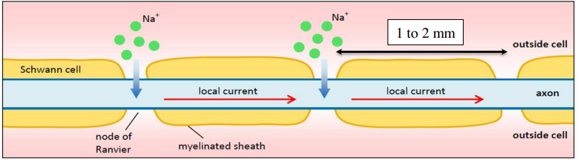
- Non-myelinated axons: Impulse moves continuously along entire axon
- Myelinated axons (saltatory): Impulse skips between nodes → less resistance, faster depolarization
- Saltatory conduction = up to 120 m/s
- Non-myelinated conduction = around 2 m/s
🧠 Real-World Analogy:
Imagine walking vs hopping:
- Non-myelinated = walking the whole path
- Myelinated = hopping between stones in a river = faster and more efficient
– Saltatory conduction = impulse “jumps” from node to node.
– Myelin prevents ion flow except at nodes of Ranvier.
– Increases impulse speed significantly compared to continuous conduction.
C2.2.15 – Perception of Pain by Neurons with Free Nerve Endings in the Skin
🩹 What Are Free Nerve Endings?
Free nerve endings are sensory neurons found in the skin.
They are unencapsulated-meaning they are exposed nerve endings that detect pain (nociception).
⚡ How Do They Work?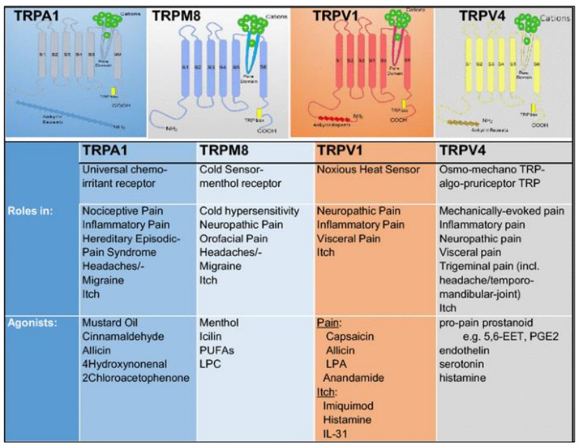
These neurons have ion channels that open in response to:
- High temperatures (e.g. >43°C)
- Acidic pH (e.g. lactic acid)
- Irritating chemicals like capsaicin (from chili peppers)
🧪 Triggering a Nerve Impulse
When the channels open, positively charged ions (like Na⁺ or Ca²⁺) enter the neuron.
This causes depolarization of the membrane.
If the threshold potential is reached (typically ~ -55 mV):
- An action potential is triggered.
- The signal travels along the neuron to the spinal cord and brain.
🧠 Where Is Pain Felt?
Although the receptors are in the skin, the sensation of pain is only experienced when the brain processes the signal.
Pain is a perception, not a direct property of the stimulus.
🌶️ Example: Capsaicin
| Stimulus | Effect on Free Nerve Endings |
|---|---|
| Capsaicin (from chilli) | Binds to TRPV1 ion channels, making them open |
| Result | Na⁺/Ca²⁺ ions enter → depolarization → pain signal sent |
– Free nerve endings detect painful stimuli like heat, acid, and chemicals.
– Pain perception starts when ion channels open and positive ions enter.
– If the threshold is reached, a nerve impulse travels to the brain.
– The brain interprets this signal as “pain”.