Neural Control and Coordination
Table of contents
- Neural System/Nervous System
- Human Nervous System
- Structure and the Function of Neuron
- Types of Neurons
- Generation and Conduction of Nerve Impulses
- Transmission of Impulses
- Central Nervous System
- Reflex Action and Reflex Arc
- Sensory Perception and Processing
The process by which two or more organs interact with each other and complements each other function is known as Coordination.
Neural System/Nervous System
The structural and functional unit of neural system are known as Neurons. They can detect, receive and transmit stimuli.
Human Nervous System
Human neural or nervous system is divided into- Central Nervous System (CNS) and Peripheral Nervous System (PNS). Central nervous system includes brain and spinal cord whereas peripheral nervous system includes all the nerves present in the brain and the spinal cord. The PNS has two types of nerve fibers- afferent nerve fibers and efferent nerve fibers.
Afferent nerve fibers carry impulses from organs/tissues to the CNS whereas efferent nerve fibers transmit nerve impulses from CNS to the target tissue/organ.
PNS is divided into somatic nervous system and autonomic nervous system. The somatic nervous system transmits impulses from the CNS to skeletal muscles while the autonomic nervous system transmits impulses from the CNS to the involuntary organs and smooth muscles of the body. The autonomic nervous system is classified into sympathetic nervous system and parasympathetic nervous system.
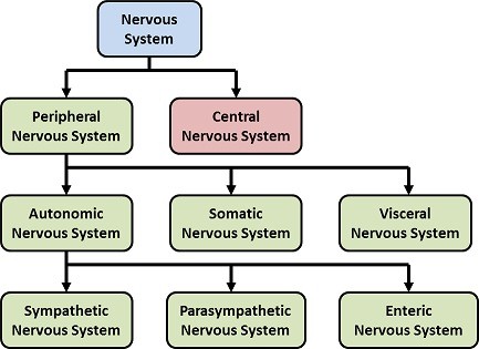
Fig. 1. Types of Nervous System
Structure and the Function of Neuron
The structural and the functional unit of nervous system is neuron. It consists of 3 parts- cell body, dendrites, and axon. Cell body contains cytoplasm that contain granular bodies known as Nissl’s granules. Finger-like projections that arises from cell body are known as Dendrites. Dendrites transmit nerve impulses towards the cell body.
From the cell body arises a long axon, whose distal end is branched. Each axon terminates into a bulb-like structure known as Synaptic Knob, which contains neurotransmitter. The cells of the axon which are covered by myelin sheath are known as Schwann Cells. The gap between the two-adjacent myelin sheath are known as Nodes of Ranvier.
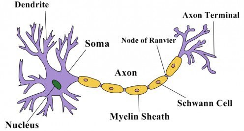
Fig. 2. Structure of the Neuron
Types of Neurons
Multipolar Neurons are those neurons that with one axon and two or more dendrites. For Example, in cerebral cortex.
Bipolar Neurons are with one axon and one dendrite. For Example, Retina.
Unipolar Neurons are with one axon, no dendrite. For Example, Embryonic Stage.

Fig. 3. Types of Neuron
Generation and Conduction of Nerve Impulses
Neurons are electrically excitable cells, that is, there membranes are in polarized state. Membrane of neurons contain ion channels that are selectively permeable to different ions. When no nerve impulse transmission is occurring, the membrane is more permeable to potassium ions and impermeable to sodium ions. This condition is known as Resting Membrane Potential. So, inside the membrane high concentration of potassium ions are present as compared to sodium ions. But outside the membrane, sodium concentration is high as compared to potassium ions. The difference in the concentration of these ions creates a concentration gradient. This gradient is maintained by sodium-potassium pump that actively transport 3 sodium ions outside and 2 potassium ions inside the neuronal membrane. As a result, outer surface of the axonal membrane possesses a positive charge and its inner surface becomes negatively charged. That is why the membrane is said to be polarized.
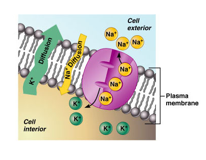
Fig. 4. Membrane Potential
When a stimulus reaches a neuronal membrane, the membrane becomes freely permeable to sodium ions. This will cause influx of sodium ions inside the neuronal membrane. Due to this the polarity of the membrane changes, the inner side of the membrane becomes positively charged whereas outside the membrane is negatively charged. The electrical potential difference created is known as Action Potential.
After some time, the permeability of sodium ions decreases and permeability of potassium ion decreases. The efflux of sodium ion occurs from inside to outside of the axonal membrane. Again, the polarity is reversed. This sequence of events is repeated along the full axon and as a result nerve impulse is conducted.
Transmission of Impulses
Nerve impulse transmission occurs from one neuron to another neuron through junctions known as Synapses. Synapse is formed by the membrane of pre-synaptic neuron and post-synaptic neuron separated by a gap known as Synaptic Cleft.
Synapses are of two types- Electrical Synapse and Chemical Synapse. During electrical synapse, pre- and post-synaptic neuron are in close proximity. Electric current can directly flow from one neuron to another neuron. This mode of nerve impulse transmission is faster than the chemical synapse.
During chemical synapses, pre- and post-synaptic neuron are separated by a fluid filled space known as Synaptic Cleft. Neurotransmitters are involved in chemical synapses. The axon terminals contain vesicles which are filled with neurotransmitter. When the nerve reaches the axon terminal, synaptic vesicles move towards the membrane and get fuses with it. This results in the release of the neurotransmitters in the synaptic cleft. The released neurotransmitters than bind the receptors on the post-synaptic neuron. This open ion channels as a result nerve impulse is transmitted.
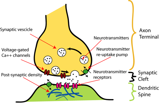
Fig. 5. Chemical Synapses
Central Nervous System
Brain acts as a command and control system. Human brain is protected inside the skull. Brain is covered by 3 layers known as meninges. They are as follows- outermost dura mater, middle arachnoid, and innermost pia mater. The brain is divided into forebrain, midbrain, and hindbrain.
Forebrain consists of three parts- Cerebrum, Thalamus and Hypothalamus. Cerebrum is the largest part of the brain. Cerebrum is divided into two halves known as Left and Right Cerebral Hemispheres. These hemispheres are connected by a tract on nerve fibers known as Corpus Callosum. Cerebral hemispheres are covered by a layer of cells known as Cerebral Cortex. The area present at the center of the forebrain is known as Thalamus. It is involved in sensory and motor signaling.
At the base of the thalamus lies the hypothalamus. It controls urge for eating, drinking and body temperature.
Fig. 6. Structure of human brain
Midbrain is located between the forebrain and hindbrain. It contains 4 lobes known as Corpora Quadrigemina.
Hind brain comprises of pons, cerebellum, and medulla oblongata. Cerebellum are the second largest part of the brain. The connection between the brain and spinal cord is known as Medulla Oblongata. Medulla controls gastric secretion, control respiration and cardiovascular functions.
Reflex Action and Reflex Arc
The involuntary response towards any stimulus is known as Reflex Action. The pathway of reflex action is known as Reflex Arch. Reflex arch or reflex pathway consist of afferent neuron and efferent neuron. The afferent neuron receives signal from a sensory organ and transmits the impulse via a dorsal nerve root into the CNS. The efferent neuron then carries signals from CNS to the effector organ or muscle.

Fig. 7. Reflex Arc
Sensory Perception and Processing
Sensory organs detect the changes in the environment and send signals to the central nervous system.
Human Eye
Eyes are situated in the sockets of the skull known as Orbits. Human eye is spherical in structure. It is composed of 3 layers- the outermost sclera (dense connective tissue), middle choroid (supplied with blood vessels) and innermost retina. Choroid becomes thick anteriorly and it forms ciliary body. The visible colored portion of the eye is known as Iris. A transparent, crystalline lens is held by ligaments which are attached to the ciliary body.
Retina consists of 3 layers of neuronal cells- ganglion cells, bipolar cells, and photoreceptor cells. Rods and cones are two types of photoreceptor cells. These cells contain light sensitive proteins. Cones are meant for bright light and color vision whereas rods are meant for dim light. Rods contain protein known as Rhodopsin. Cones respond to 3 different colors- red, green, and blue.
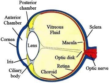
Fig. 8. Structure of human eye
Optic nerve is the nerve that connects eye to the brain. Photoreceptors are absent in this region, hence known as blind spot. Lateral to the blind spot lies the macula lutea, which is densely packed with only cones. Macula lutea is a place where visual resolution is highest. The fluid filled space between the cornea and the lens is known as Aqueous Chamber and fluid is known as Aqueous Humor. Similarly, the space between the lens and the retina is known as vitreous chamber and the fluid in it is known as Vitreous Humor.
Mechanism of vision
When light rays are focused on the retina, they generate action potential in rods and cones. Due to this, a photosensitive pigment separates into opsin (protein component) and retinal (aldehyde of vitamin A). This generates action potential which travels from optic nerve to the visual cortex of the brain. This leads to the image formation on the retina.
The Ear
Hearing and balancing are the two main functions of ears. Ear is divided into 3 parts- outer ear, middle ear, and external ear. The outer ear consists of pinna and external auditory meatus. Pinna collects the vibration from the air and produces sound. Wax secreting gland are present in pinna and meatus.
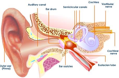
Fig. 9. Structure of ear
The middle ear consists of 3 ossicles- malleus, incus, and stapes. Middle ear is connected to pharynx by a eustachian tube. Inner ear comprises of bony labyrinth that contains 3 semicircular canals. The coiled part of the labyrinth is known as Cochlea. The space within the cochlea is filled with endolymph. Structure located on the basilar membrane is known as Organ of Corti. They contain hair cells that acts as auditory receptors.
Mechanism of hearing
The external ear receives sound waves and transfer it to ear drum. This in turn vibrates the ear drum which transfers the vibrations to the ear ossicles. The vibrations are further transferred to cochlea. This generates the nerve impulse which are transmitted via auditory nerve to the auditory cortex of the brain.