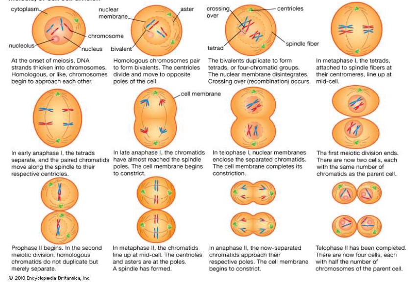
Prophase I:
- DNA supercoils and condenses. Chromosomes are visible under light microscope.
- Nuclear membrane begins to break down and disintegrate.
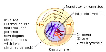
- The homologous chromosomes associate with each other to form
bivalent or tetrads. - Crossing over occurs: non-sister chromatids exchange genetic
information. The crossing over point is called chaisma (pl. chaismata) - Spindle fiber begins to form
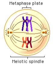
Metaphase I:
- Bivalents line up at the equator
- Random Orientation occurs: bivalents (homologous pairs) that come from the
mother or the father line up randomly on either side of the cell equator, independently of the other homologous pairs. Hence the daught nuclei get a different mix of chromosomes. - Spindle fibers (microtubules) from each of the centrosomes attach to the centromere of bivalents
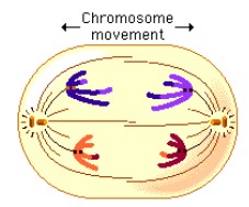
Anaphase I:
- Contraction of the spindle fibers pulls homologous chromosome pair apart.
- Chaismata breaks apart and separate.
- One chromosome of each pair move to opposite poles of the cell
Telophase I:
- Chromosome begins to uncoil and nuclear envelop reforms.
- Chromosome number reduces from 2n (diploid) to n (haploid); however each
chromatid still has the replicated sister chromatid still attached (not homologous
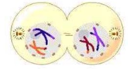
pairs anymore). - Cytokinesis occurs and the cell splits into two separate cells.
Meiosis, or sex cell division
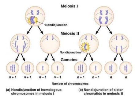
Non-disjunction:
- A non-disjunction is an error in meiosis, where the chromosome pairs fail to split during cell division.
- Non-disjunction can occur in anaphase I where the homologous pairs fail to split, or it can occur in anaphase II, where the sister
chromatids fail to split. - The result of this error is too many chromosomes in a gamete cell or too few chromosomes in the final gamete cell.
- One of the gamete cells could have 22 chromosomes and one could have 24 chromosomes. The resulting zygote will therefore have 47 or 45 chromosomes.
- An example of a non-disjunction is Down’s syndrome.
- Down syndrome occurs when chromosome 21 fails to separate, and one of the gametes ends up with an extra chromosome 21.
Therefore, a child that receives that gamete with an extra chromosome 21 will have 47 chromosomes in every cell. - Down syndrome is also called Trisomy 21.
- Some Down syndrome symptoms include impairment in cognitive ability and physical growth, hearing loss, oversized tongue, shorter limbs and social difficulties