Blood Vessel
⇒ Arteries:
- Take blood away from the heart to tissues around the body
- Because large volumes of blood are flowing directly out of the heart, arteries must be able to withstand the high pressure and high blood volume created when the ventricles contract.
- Very thick wall of smooth muscle tissue surrounding arteries makes them strong and elastic in nature with a narrow lumen (area where the blood flows).
- Elastic fibres store energy when they are stretched by the flow of blood. As they recoil the blood is further propelled through the artery.
- The thick smooth muscle layer in the arteries can be used to help regulate blood pressure by changing the diameter of the arteries.
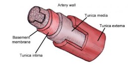
- Tunica externa – outer layer made from connective tissue and elastic fibre
- Tunica media – thick layer containing smooth muscle
- Tunica intima – endothelium layer that lines the inside of the artery
- When the ventricles of the heart contract (systole), the blood leaves the heart through the arteries at a very high pressure
- The blood pushes the walls of the arteries outwards, thus increasing the diameter of the lumen and creating potential energy within the elastic walls of the artery
- As the blood passes after the heart has contracted, the pressure drops and the stretched elastic walls snap back, squeezing the blood in the lumen to conserve energy and preventing the pressure from becoming too low inside the arteries
- However, since this pressure still relatively high, blood flow in the arteries is fairly consistent and steady, even though the heart pumps in pulsating manner
⇒ Capillaries:
- Capillaries have a very narrow diameter (one cell thick 10 μm) with thin surrounding endothelium cells to allow the shortest distance for \(O_2\) to diffuse into the blood from the alveoli in the lungs and from the blood into the body tissues. \(CO_2\) also can easily diffuse out of the blood into the alveoli in the lungs and from the tissue into the blood after respiration.
- The walls have pores, making them very permeable allowing plasma to leak out and form tissue fluid, which contains oxygen, glucose and all other substances contained in the blood plasma, except proteins (too large to fit through the pores in the capillary wall)
- Highly branched networks of capillaries increase the surface area, maximizing the amount of nutrients and gases that can move in and out of the capillaries.
- Because they are highly branched, the blood slows down to allow efficient transfer of \(O_2\) and \(CO_2\) into and out of the capillaries.
- Capillaries have small lumen and low pressured blood
⇒ Veins:
- Transport blood back to the heart from the capillary beds in tissues.
- Very low blood pressure and therefore the walls can be thin. Blood is pushed back to the heart through the contraction of skeletal muscles. As the muscles contract, the veins are squeezed, pushing the blood back towards the heart
- Large lumen allows large amounts of blood to slowly return to the heart because the blood has to slow down as it passes through the capillary beds.
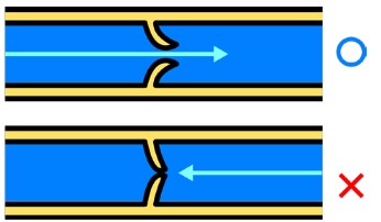
- Since the blood pressure in the veins is quite low because the blood slows down considerably when it reaches the capillary bed and there is not another pump like the heart to speed up the flow and increase the pressure, veins have a series of valves to prevent backflow
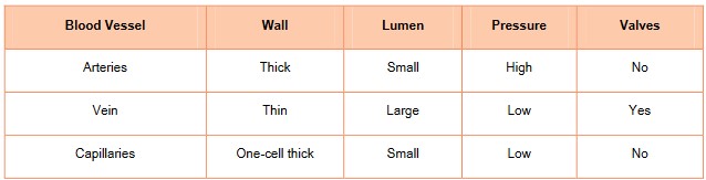
Comparison table between different types of blood vessels
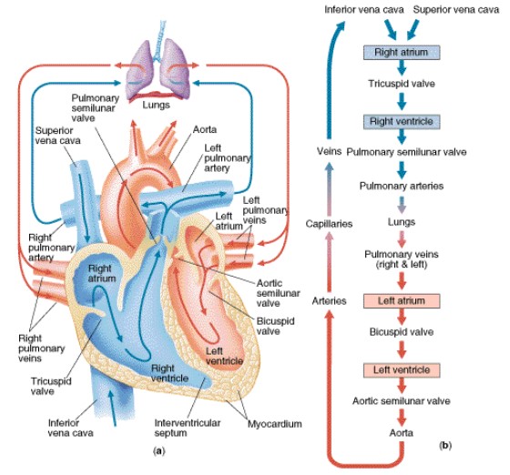
Heart structure