▶️ Answer/Explanation
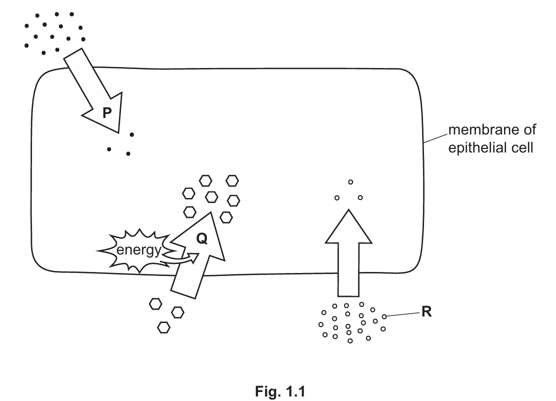
(a)(i)
Diffusion is the net movement of particles from a region of their higher concentration to a region of their lower concentration (down a concentration gradient), as a result of their random movement.
Explanation: Molecules are constantly in motion due to kinetic energy. While they move randomly, the statistical probability results in them spreading out until they are evenly distributed.
(a)(ii)
Kinetic energy.
Explanation: Diffusion is a passive process; it relies on the inherent motion (kinetic energy) of the particles themselves, not on metabolic energy (ATP) produced by the cell.
(b)(i)
Aerobic respiration.
Explanation: This is the chemical reaction in mitochondria where glucose reacts with oxygen to release energy, producing carbon dioxide and water as byproducts.
(b)(ii)
The arrow should be drawn pointing from the inside of the epithelial cell to the outside (towards the blood/tissue fluid).
Explanation: Since cells produce \(CO_2\) during respiration, the concentration is highest inside the cell. Therefore, \(CO_2\) diffuses out down its concentration gradient.
(c)
Type of movement: Active transport.
Explanation: The particles are moving from a region of lower concentration to a region of higher concentration (against the concentration gradient). The diagram also shows the use of energy, which is required for this process.
Additional Note: Active transport typically involves specific protein carriers in the cell membrane that use energy (from ATP) to pump substances against the gradient.
(d)
Particle R cannot be starch because:
1. Starch molecules are too large.
2. Starch is insoluble.
3. Starch molecules cannot pass through the cell membrane (they must be digested into simple sugars like glucose first).
Explanation: The cell membrane is partially permeable, allowing small molecules like glucose or amino acids to pass, but blocking large polymers like starch.
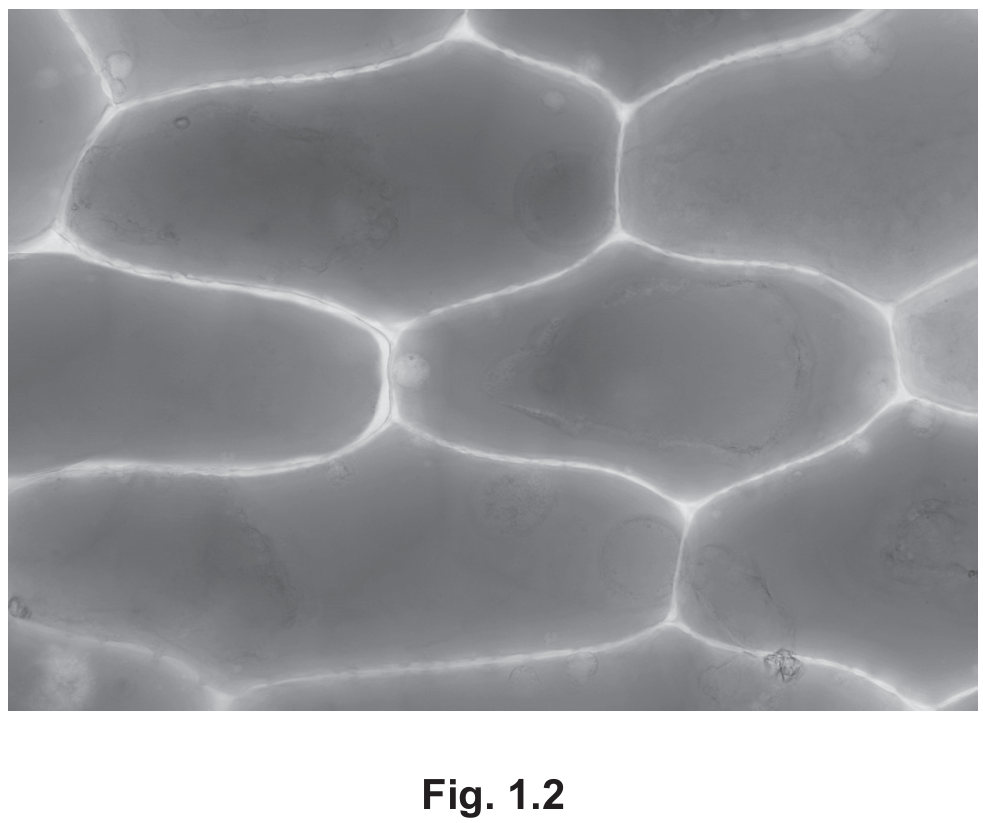
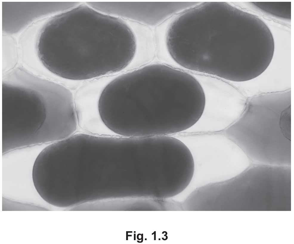
(e)
Description:
- Before immersion (Fig 1.2), the cells are turgid; the cytoplasm pushes fully against the cell wall.
- After immersion (Fig 1.3), the cells are plasmolysed or flaccid. The cell membrane and cytoplasm have pulled away from the cell wall.
- The vacuole/cytoplasm appears darker or more concentrated after immersion.
Explanation:
- This change is due to osmosis.
- The salt solution surrounding the cells is hypertonic (concentrated), meaning it has a lower water potential than the cell cytoplasm.
- Water moves out of the cell, from a region of higher water potential (inside) to a region of lower water potential (outside), through the partially permeable membrane.
- The loss of water causes a decrease in turgor pressure, leading to the cytoplasm shrinking.
▶️ Answer/Explanation
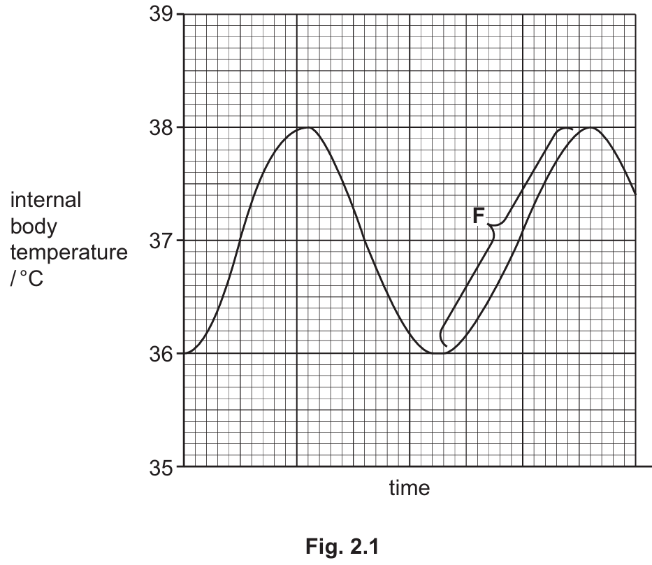
(a)(i) Temperature Range
Value: \(2\)
Units: \(^{\circ}\text{C}\)
Explanation: The graph shows the maximum temperature reaches \(38^{\circ}\text{C}\) and the minimum drops to \(36^{\circ}\text{C}\). The range is the difference between these two values: \(38 – 36 = 2^{\circ}\text{C}\).
(a)(ii) Set Point
A horizontal line should be drawn at \(37^{\circ}\text{C}\).
Explanation: The set point in homeostasis is the ideal value around which the variable fluctuates. In human body temperature, this is typically \(37^{\circ}\text{C}\) (the midpoint of the oscillation).
(a)(iii) Mechanism
Negative feedback.
Explanation: Homeostasis relies on negative feedback loops. When a deviation from the set point occurs (temperature goes too high or too low), mechanisms are triggered to oppose that change and bring the value back to the normal level.
(a)(iv) Explanation of Region F
Region F shows the temperature rising from a low point (\(36^{\circ}\text{C}\)) back towards the set point. This indicates the body is reacting to being too cold. The body raises temperature via:
1. Vasoconstriction: Arterioles supplying skin capillaries constrict (narrow), reducing blood flow to the skin surface to minimize heat loss.
2. Shivering: Rapid contraction of skeletal muscles generates heat through respiration.
3. Increased Metabolism: The rate of respiration increases to generate more metabolic heat.
4. Piloerection: Hair erector muscles contract, causing hairs to stand on end to trap an insulating layer of air.
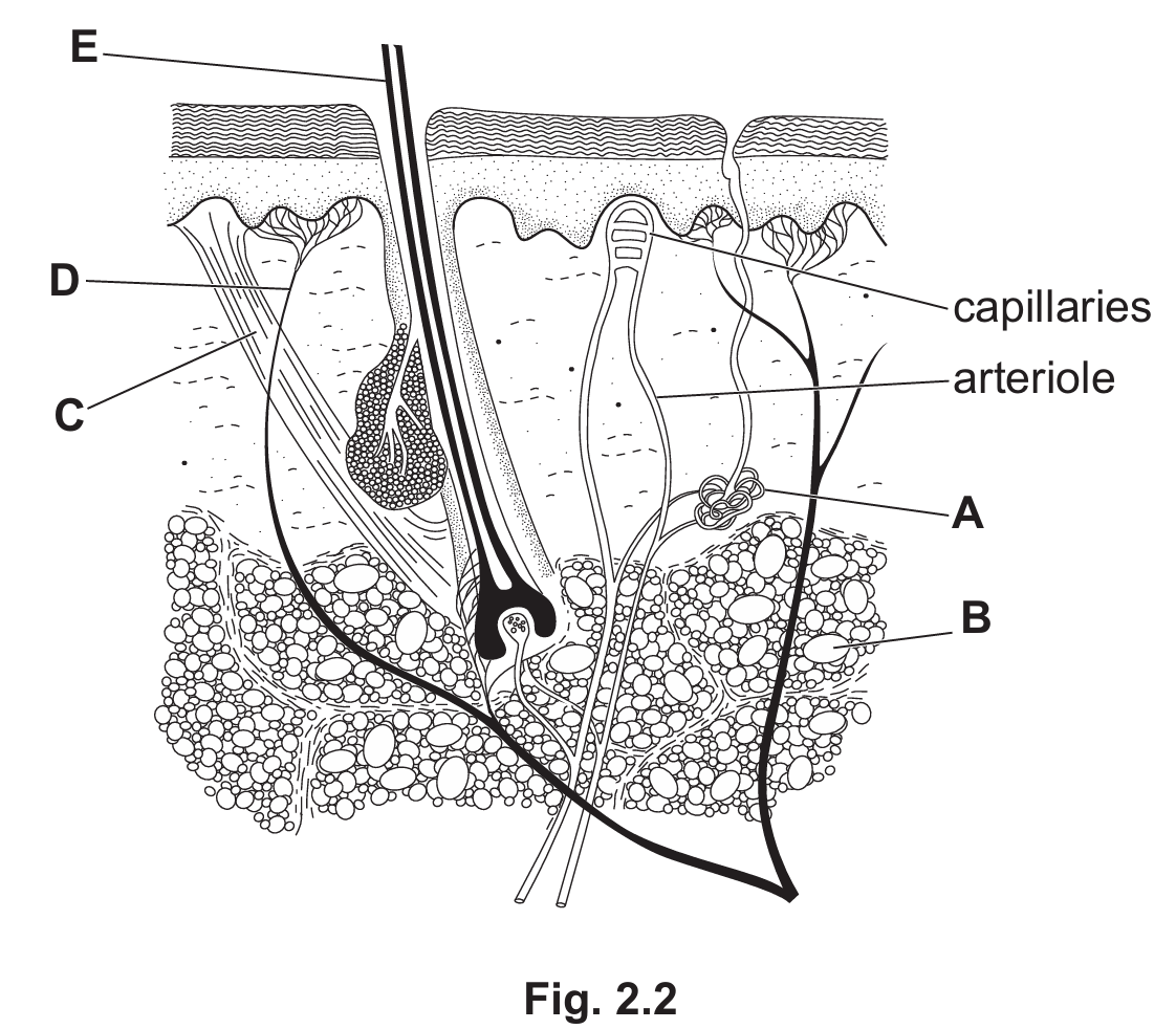
(b)(i) Structure producing sweat
A
Explanation: Structure A is a coiled tube located in the dermis with a duct leading to a pore on the surface. This is the characteristic structure of a sweat gland.
(b)(ii) Structures C and D
C: (Hair) erector muscle
D: Sensory neurone (or receptor)
Explanation: Structure C is the small muscle attached to the base of the hair follicle, responsible for pulling the hair upright (piloerection). Structure D acts as a receptor sensing stimuli (like temperature or touch) and connects to a nerve fiber.
▶️ Answer/Explanation
(a)(i)
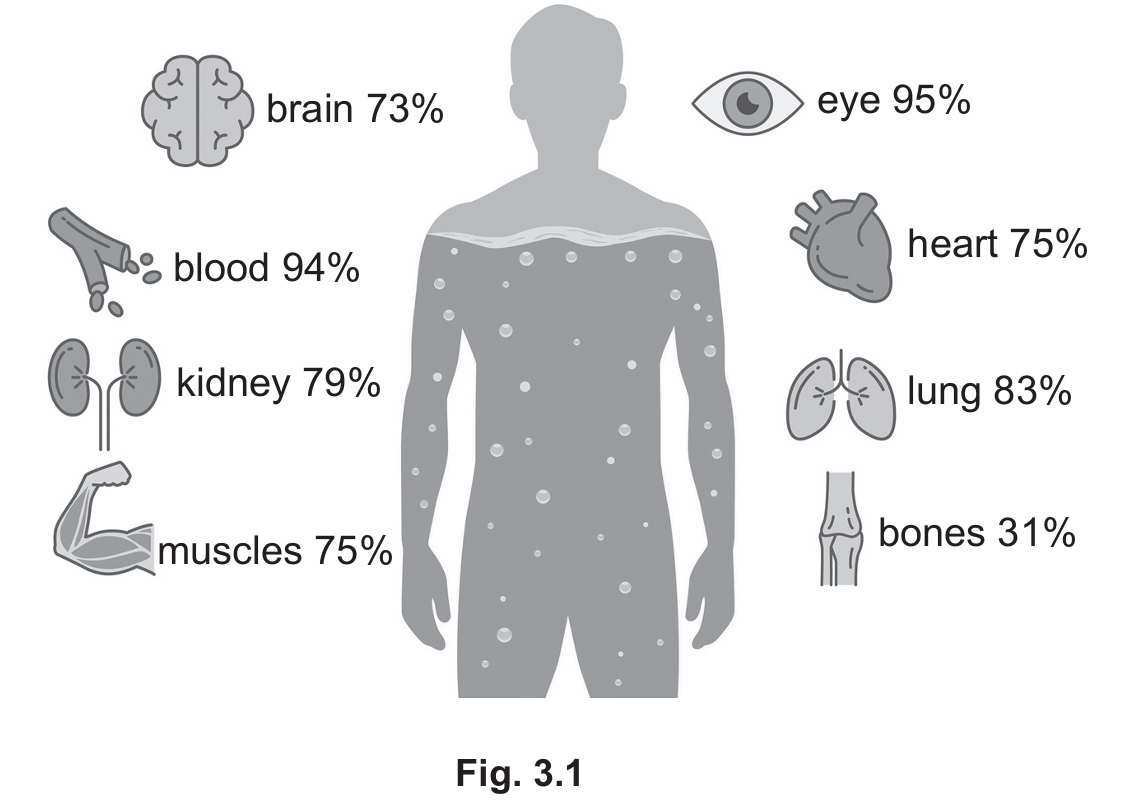
According to Fig 3.1, the eye is composed of $95\%$ water.
Mass of water = $28\,\text{g} \times 0.95 = 26.6\,\text{g}$.
Rounding to two significant figures, the answer is $27\,\text{g}$.
(a)(ii)
Water acts as a critical solvent in the body, allowing substances to dissolve for transport (e.g., in blood plasma) and for metabolic reactions (e.g., digestion/hydrolysis). It is also vital for excretion (urine) and temperature regulation (sweating).
(b)(i)
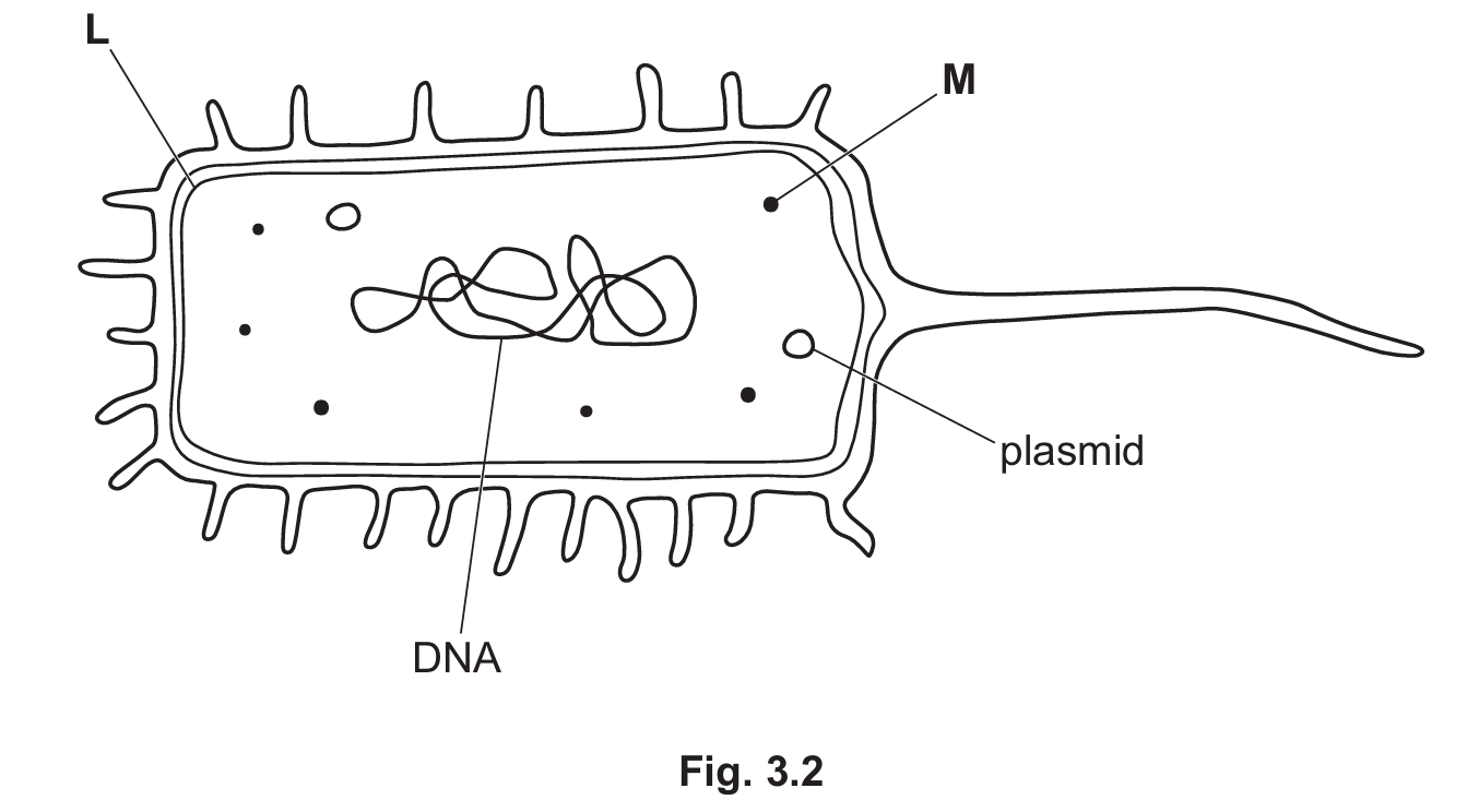
L: Cell membrane (the inner line beneath the cell wall).
M: Ribosome (the small dots in the cytoplasm).
(b)(ii)
Typical prokaryotic features visible in the diagram include:
1. Circular DNA (the looped genetic material, not enclosed in a nucleus).
2. Plasmids (small circular rings of DNA).
3. Cell wall (the outer layer; note: prokaryotes do not have a nucleus).
(b)(iii)
The type of pathogen is a bacterium.
(b)(iv)
The scientific name is Vibrio cholerae. The genus name is the first part: Vibrio.
(b)(v)
The cholera bacterium produces a toxin that causes the secretion of chloride ions ($\text{Cl}^-$) into the small intestine. This accumulation of ions lowers the water potential in the gut lumen. Consequently, water moves out of the cells and blood into the gut by osmosis, leading to watery diarrhoea and dehydration.
(b)(vi)
During genetic modification:
1. Restriction enzymes create sticky ends.
2. These are joined together using an enzyme called DNA ligase (or simply ligase).
3. The modified plasmid is called a recombinant plasmid.
▶️ Answer/Explanation
(a)(i)
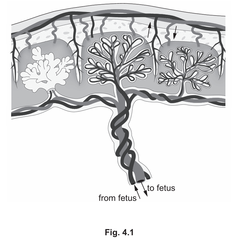
The functions of the placenta and umbilical cord include:
- Separation of blood: It keeps maternal and fetal blood separate to prevent immune rejection and damage from high maternal blood pressure.
- Exchange of substances: It facilitates the transfer of nutrients (e.g., glucose, amino acids) and oxygen from the mother to the fetus, and the transfer of metabolic wastes (e.g., carbon dioxide, urea) from the fetus to the mother.
- Passive Immunity: It allows the transfer of antibodies from mother to fetus.
- Protection: The placenta acts as a barrier to certain toxins and pathogens.
- Hormone Secretion: The placenta secretes hormones like progesterone and oestrogen to maintain the pregnancy.
(a)(ii)
The amniotic sac and fluid perform the following functions:
- Protection: It cushions the fetus, protecting it against mechanical damage or shocks.
- Temperature Regulation: It maintains a constant temperature suitable for development.
- Development: It allows the fetus to move freely, which is essential for proper muscle and bone development.
- Barrier: It helps protect the fetus from infection.
- Lung Development: It allows for lung development (fetal breathing movements).
(b)(i)
HIV (Human Immunodeficiency Virus) is another STI that can be passed from mother to fetus.
(b)(ii)
Ways to control the spread of STIs include:
- Barrier methods: Using condoms or femidoms during sexual intercourse.
- Abstinence: Refraining from sexual activity.
- Education: Increasing awareness about safe sex practices.
- Screening/Testing: Regular testing and tracing sexual partners of infected individuals.
- Vaccination: Using vaccines (e.g., for Hepatitis B or HPV).
- Medication: Using antibiotics to treat bacterial infections (preventing further spread).
Explanation:
Part (a): The placenta is a vital temporary organ. It provides a large surface area for the diffusion of materials between the maternal and fetal blood supplies without the blood actually mixing. Mixing would be dangerous because the mother’s immune system might attack the fetal cells (which contain foreign antigens from the father), and her higher blood pressure could damage delicate fetal vessels. The umbilical cord connects the fetus to the placenta; the umbilical artery carries deoxygenated, waste-laden blood away from the fetus, while the umbilical vein returns oxygenated, nutrient-rich blood to the fetus. The amniotic fluid creates a weightless environment where the fetus can exercise its muscles against the fluid resistance, crucial for orthopedic development.
Part (b): Syphilis is bacterial, whereas HIV is viral; both can cross the placental barrier or be transmitted during birth. Controlling STIs relies heavily on breaking the chain of transmission. Barrier methods physically prevent the exchange of bodily fluids which carry the pathogens. Contact tracing ensures that asymptomatic partners are treated so they do not unknowingly infect others.
▶️ Answer/Explanation
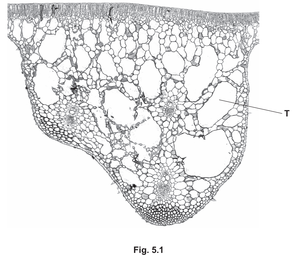
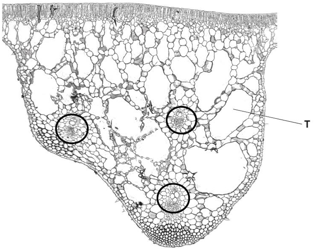
(a)(i) Vascular Bundle Location
To answer this correctly on the diagram, you would circle one of the distinct circular structures embedded in the spongy mesophyll layer (the veins). These structures contain the xylem and phloem.
(a)(ii) Palisade Mesophyll Location
The label line X should point to the layer of cells immediately below the upper epidermis. These cells are distinctive because they are elongated, rectangular, and packed tightly together to maximize light absorption.
(a)(iii)
Chloroplasts
Explanation: The palisade mesophyll is the primary site of photosynthesis. These cells contain a high density of chloroplasts, the organelles containing chlorophyll, which absorbs light energy.
(a)(iv)
The vascular bundle contains two main tissues with distinct functions:
- Phloem: Responsible for translocation, which is the transport of sucrose and amino acids from sources (leaves) to sinks (roots/fruits).
- Xylem: Responsible for the transport of water and dissolved mineral ions from the roots to the leaves. Xylem vessels also provide structural support to the plant due to their lignified walls.
(b)(i)
Hydrophyte
Explanation: This is the specific biological term for plants adapted to live in aquatic environments (either submerged or floating).
(b)(ii)
Feature T: Air space (or aerenchyma).
Explanation: These large air gaps reduce the overall density of the leaf. This creates buoyancy, allowing the leaf to float on the water surface to access sunlight for photosynthesis.
(b)(iii)
Acceptable answers include:
- Stomata on the upper epidermis: In land plants, stomata are usually on the bottom to prevent water loss. In floating plants, they must be on top to access the air for gas exchange ($CO_2$ intake and $O_2$ release).
- Thin or no cuticle: Since the plant is in water, there is no risk of desiccation (drying out), so the thick waxy cuticle found in terrestrial plants is unnecessary.
▶️ Answer/Explanation
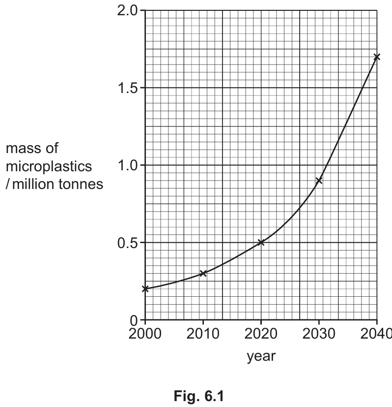
(a)(i)
The value is an estimate because it is impossible to count every piece of microplastic in the entire ocean. The ocean is too large, deep, and largely inaccessible to survey completely. Additionally, some microplastics may have been eaten by marine organisms or settled on the ocean floor where they cannot be collected. Scientists rely on samples taken from specific areas and extrapolate the data, rather than measuring the whole.
(a)(ii)
To calculate the percentage increase:
1. Identify the initial mass in 2000: \(0.2\) million tonnes.
2. Identify the final mass in 2040: \(1.7\) million tonnes.
3. Calculate the difference: \(1.7 – 0.2 = 1.5\).
4. Calculate the percentage increase: \(\frac{1.5}{0.2} \times 100 = 750\%\).
(b)(i)
A producer is an organism that makes its own organic nutrients (like glucose). It does this using energy from sunlight through the process of photosynthesis.
(b)(ii)
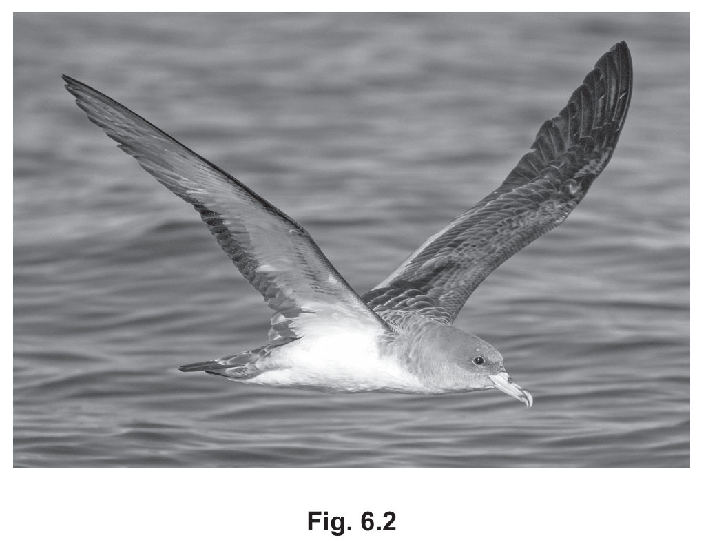
Microplastics are non-biodegradable, meaning they do not break down. They are first absorbed or ingested by producers like phytoplankton. These phytoplankton are then eaten by fish (primary/secondary consumers). Finally, the shearwater birds eat the fish. The microplastics are passed along the food chain and can accumulate in the bodies of the top consumers (bioaccumulation).
(b)(iii)
Ways to conserve the population include:
• Monitoring the species numbers to track population health.
• Protecting their habitats or nesting sites to ensure they have a safe place to reproduce.
• Implementing captive breeding programmes to increase numbers before releasing them back into the wild.
• Creating laws to ban hunting or poaching of the birds.
• Education and awareness campaigns to reduce human impact.
(b)(iv)
A decrease in population size reduces the genetic variation within the species. This leads to a higher chance of inbreeding, which increases the likelihood of harmful recessive alleles being expressed (genetic diseases). With less genetic variation, the population is less able to adapt to environmental changes (like new diseases or climate change), significantly increasing the risk of extinction. It also becomes more difficult for individuals to find mates.