▶️ Answer/Explanation
(a)
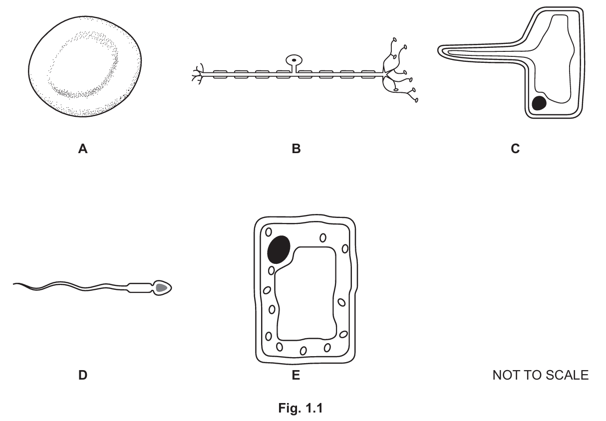
• D (contain an acrosome)
• A (contain haemoglobin)
• C and E (are found in plants)
• B (are found in the peripheral nervous system)
Explanation:
Cell D is a sperm cell; the acrosome is a vesicle containing enzymes. Cell A is a red blood cell containing haemoglobin. Cell C (root hair cell) and Cell E (palisade mesophyll cell) are plant cells; both have cell walls (E also has chloroplasts). Cell B is a neurone (nerve cell).
(b)(i)
Controls the activities of the cell / Contains genetic material (DNA/chromosomes)
(b)(ii)
Any two from:
• Cell membrane
• Cytoplasm
• Ribosomes
Note: Cell A (Red Blood Cell) lacks a nucleus and mitochondria, and animal cells (A, B, D) lack cell walls. Therefore, only the cell membrane and cytoplasm are universally present in these mature cells (ribosomes are present in developing RBCs but lost in mature ones, though often accepted in broad mark schemes; cell membrane and cytoplasm are the safest answers).
(c)
• Has a long extension / projection / hair-like structure
• Increases the surface area
• For the absorption of water and mineral ions
(d)
• Magnification
• Image size (or length of the cell in the image)
Formula: \( \text{Actual size} = \frac{\text{Image size}}{\text{Magnification}} \)
▶️ Answer/Explanation
(a)
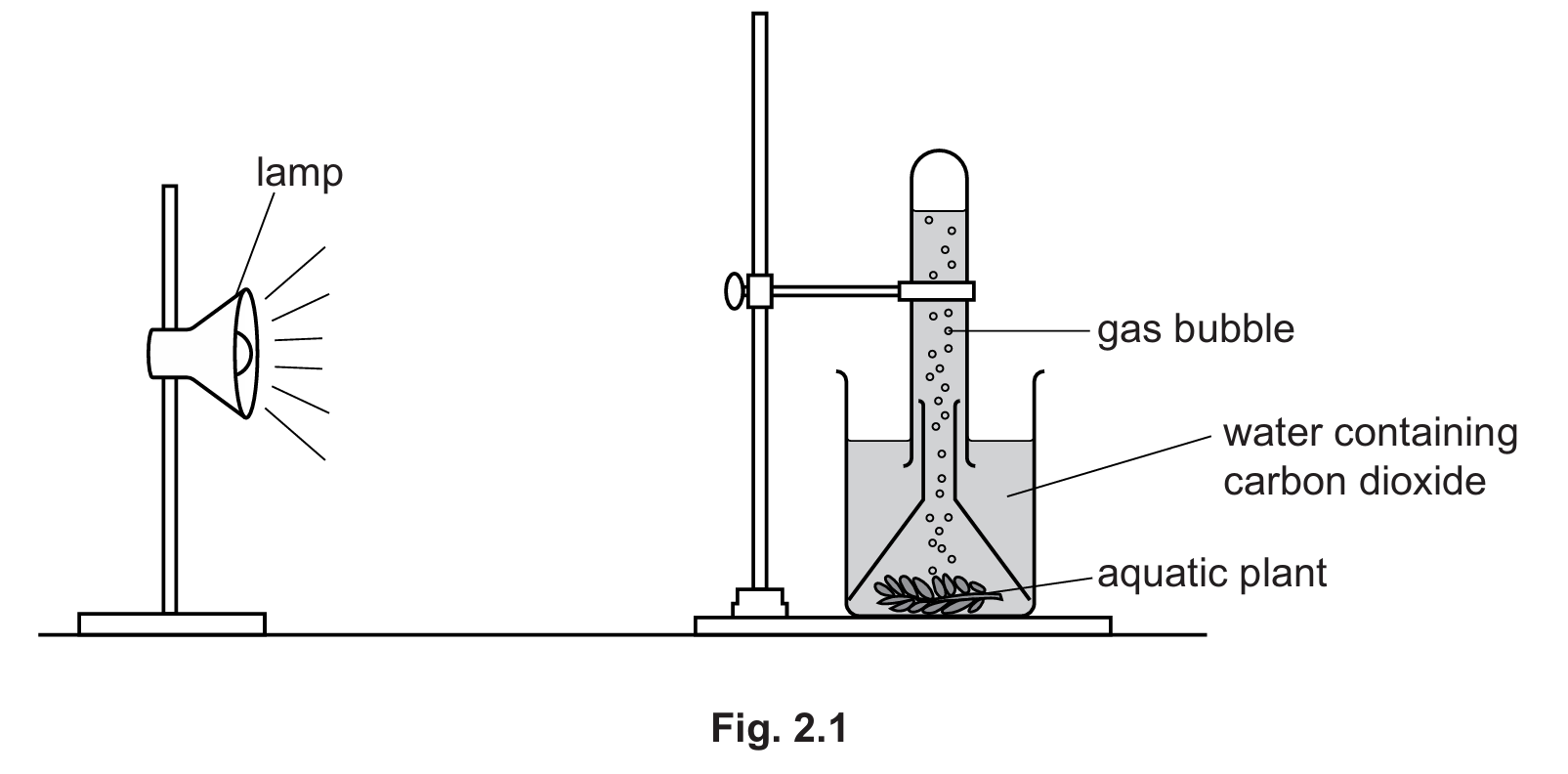
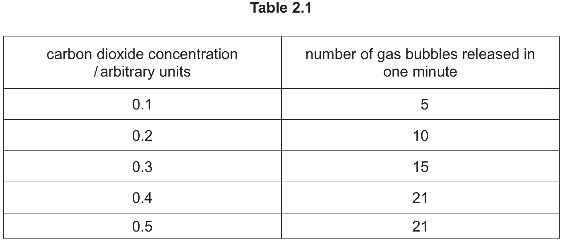
As the carbon dioxide concentration increases, the number of gas bubbles released in one minute increases and then remains the same.
This is because carbon dioxide is a raw material required for photosynthesis.
The number of gas bubbles released remains the same between the carbon dioxide concentrations of \(0.4\) and \(0.5\) arbitrary units.
Photosynthesis produces oxygen gas which is released as bubbles.
The distance between the lamp and the aquatic plant is kept constant during the investigation so that the light intensity remains the same.
Explanation:
The experiment measures the rate of photosynthesis by counting oxygen bubbles. Initially, as the concentration of the reactant ($CO_2$) rises from \(0.1\) to \(0.4\), the rate of reaction rises, showing that \(CO_2\) was the limiting factor. However, between \(0.4\) and \(0.5\), the rate stays constant at \(21\) bubbles per minute. This indicates that \(CO_2\) is no longer the limiting factor; another factor (such as light intensity or temperature) has become limiting, preventing the rate from increasing further.
(b)
The green pigment is chlorophyll, which is located in the chloroplasts. Its primary importance is that it absorbs light energy (from the sun). It then transfers this energy into chemical energy to synthesize carbohydrates (specifically glucose) from carbon dioxide and water.
(c)
Respiration (specifically aerobic respiration).
Explanation:
While photosynthesis consumes carbon dioxide to produce oxygen, respiration is the metabolic process occurring in all living cells (including plants) that breaks down glucose using oxygen to produce energy, releasing carbon dioxide as a waste product.
▶️ Answer/Explanation
(a)
- Physical digestion occurs: stomach. (The muscular walls of the stomach churn food).
- Releases hydrochloric acid: stomach. (Secreted by the gastric glands in the stomach wall).
- Secretes protease and amylase: pancreas. (The pancreas secretes pancreatic juice containing these enzymes into the small intestine).
- Secretes insulin: pancreas. (Insulin is a hormone produced by the pancreas to regulate blood glucose).
(b)
Hydrochloric acid has two primary functions in the stomach:
- It kills harmful microorganisms (pathogens) present in food.
- It provides an acidic pH (optimum pH) for the enzyme pepsin (a protease) to work effectively.
(c)
Protease is important because it breaks down large, insoluble protein molecules into small, soluble amino acids. This allows the nutrients to be absorbed through the wall of the small intestine into the blood for use by the body (e.g., for growth and repair).
(d)(i)
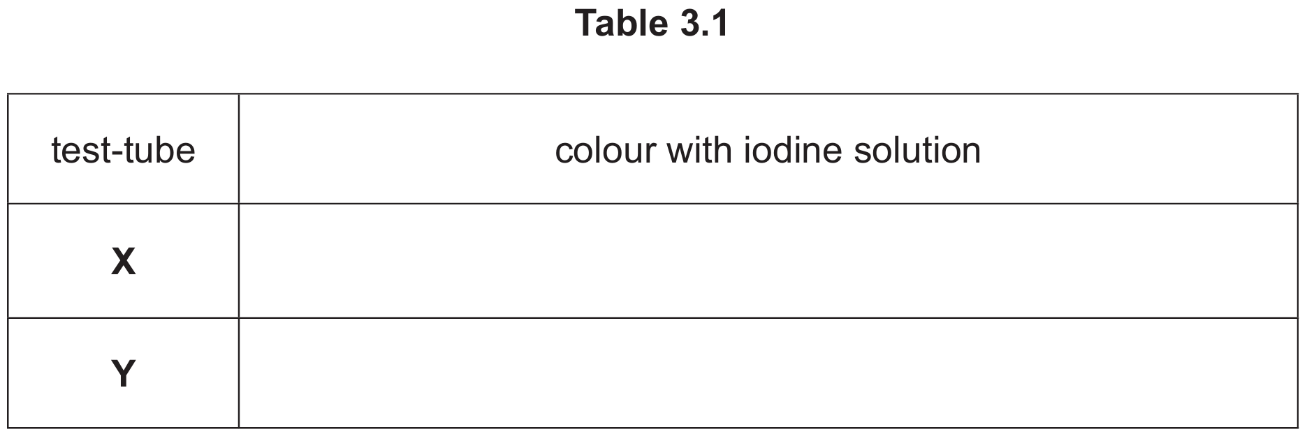
| test-tube | colour with iodine solution |
| X | blue-black |
| Y | yellow-brown (or orange/brown) |
(d)(ii)
The colours are different because in test-tube Y, the enzyme amylase has broken down (digested) the starch into simple sugars (maltose). Since starch is no longer present, the iodine solution remains its original yellow-brown colour. In test-tube X, there is no amylase, so the starch remains undigested, causing the iodine to turn blue-black.
▶️ Answer/Explanation
(a) Completion of the sentence:
Sense organs are groups of receptor cells responding to specific stimuli such as light, sound, touch, temperature and chemicals.
(b) Description and Explanation:
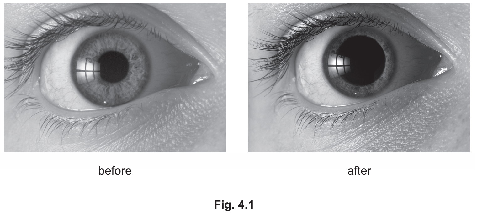
• Description: The pupil becomes larger (dilates) in the “after” diagram.
• Explanation: The light intensity in the environment decreased (became dimmer). The pupil dilated to allow more light to enter the eye to reach the retina.
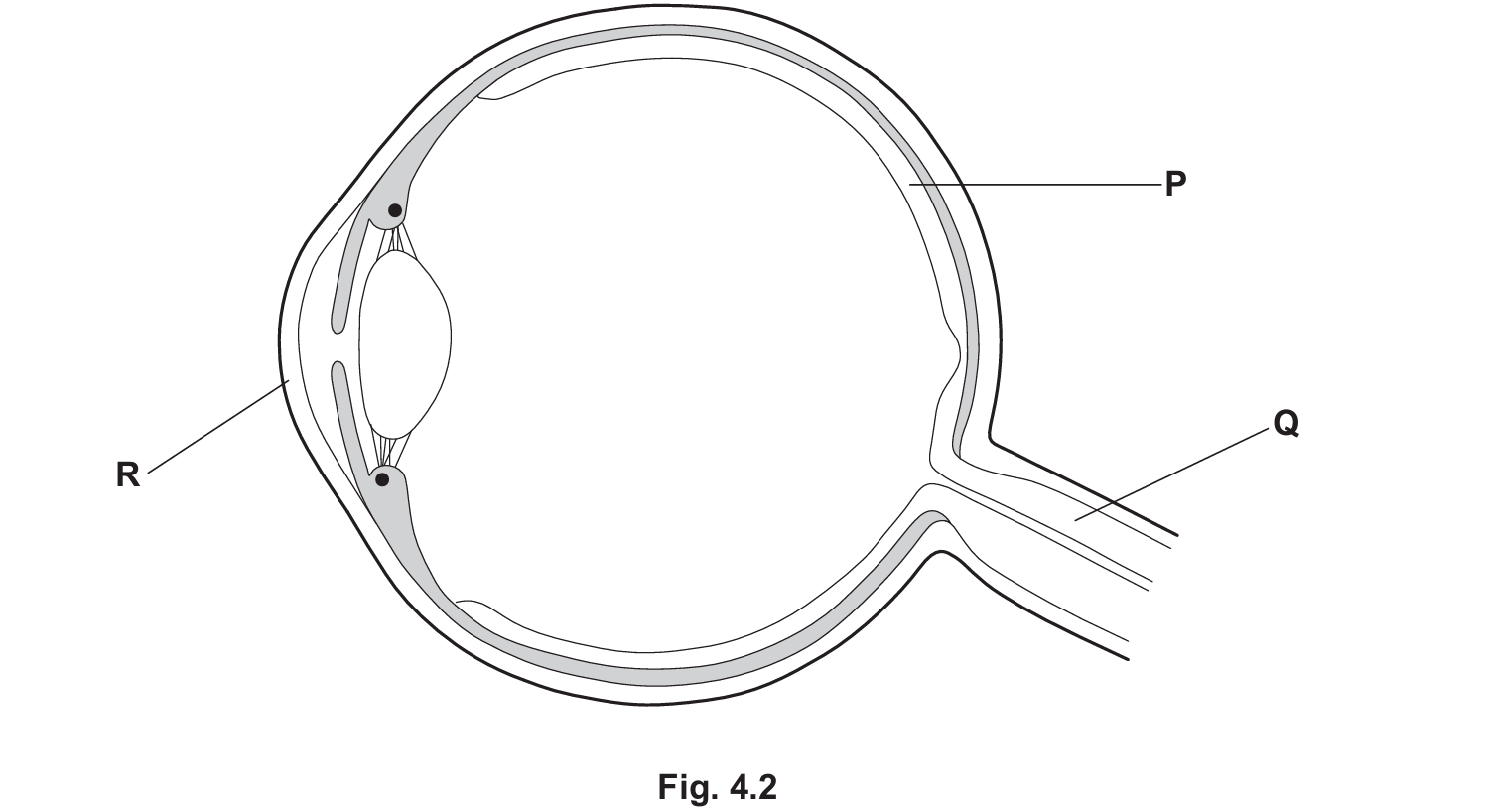
(c)(i) Blind Spot Identification:
The X should be marked at the point where the optic nerve (Q) leaves the eye (the optic disc), where there are no receptor cells.
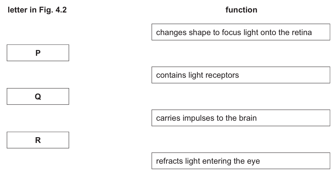
(c)(ii) Matching functions:
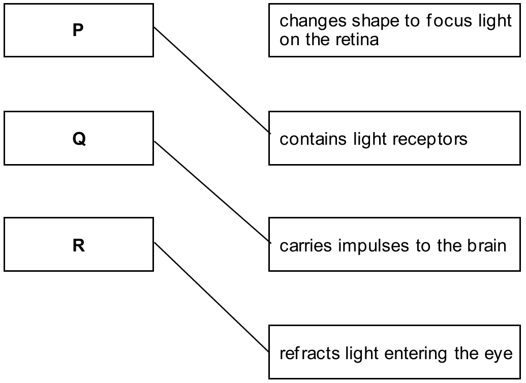
• P (Retina): contains light receptors.
• Q (Optic Nerve): carries impulses to the brain.
• R (Cornea): refracts light.
Part (a): Sense organs contain groups of specialized receptor cells that detect changes in the environment, known as stimuli. For example, the ear responds to sound, while the skin responds to touch and temperature.
Part (b): This image demonstrates the pupil reflex. In dim light, the pupil dilates (widens) to maximize light capture. Although the Core syllabus focuses on changes in diameter, the mechanism (Supplement) involves the contraction of radial muscles and the relaxation of circular muscles in the iris.
Part (c):
R (Cornea): This is the transparent frontal part of the eye that refracts (bends) light as it enters.
P (Retina): The inner lining at the back of the eye containing photoreceptors (rods and cones).
Q (Optic Nerve): Transmits electrical impulses generated by the receptor cells to the brain for processing. The point where this nerve exits the eye lacks receptors, creating the blind spot.
▶️ Answer/Explanation
(a)(i)
The correct conclusions to be ticked are:
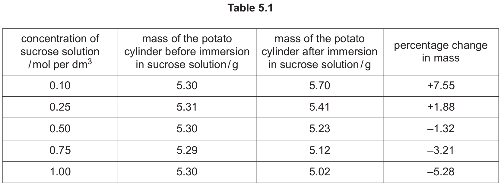
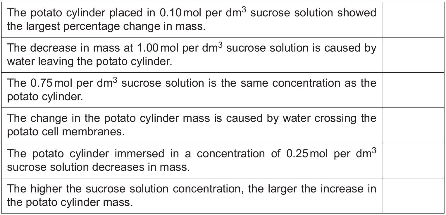
- The potato cylinder placed in \(0.10\,\text{mol per dm}^3\) sucrose solution showed the largest percentage change in mass.
Explanation: Comparing the values in the “percentage change” column, \(+7.55\%\) has the largest magnitude (absolute value) compared to the others. - The decrease in mass at \(1.00\,\text{mol per dm}^3\) sucrose solution is caused by water leaving the potato cylinder.
Explanation: A decrease in mass indicates that water has moved out of the potato cells into the surrounding hypertonic solution via osmosis. - The change in the potato cylinder mass is caused by water crossing the potato cell membranes.
Explanation: Osmosis is the movement of water molecules across a partially permeable membrane (the cell membrane) down a water potential gradient.
(a)(ii)
Osmosis.
Explanation: The experiment demonstrates the movement of water across a partially permeable membrane due to differences in solute concentration (sucrose).
(a)(iii)
Any two of the following factors:
- Temperature (Higher temperature increases kinetic energy and the rate of diffusion/osmosis).
- Surface area (Larger surface area allows more molecules to cross the membrane per unit of time).
(b)
(Cell) wall.
Explanation: The cell wall is a rigid structure made of cellulose that surrounds the cell membrane in plant cells. It resists the outward pressure (turgor pressure) exerted by the swelling vacuole and cytoplasm when water enters, preventing the cell from lysing (bursting).
(c)(i)
The two circled uses of water should be:
- as a solvent (Water is the universal solvent for metabolic reactions).
- for transport (Water is the main component of blood plasma and xylem sap, transporting substances around the body/plant).
(c)(ii)
Any two of the following components of a balanced diet (excluding water):
- Carbohydrates
- Proteins
- Fats (Lipids)
- Vitamins
- Minerals
- Fibre (Roughage)
▶️ Answer/Explanation
(a)(i)
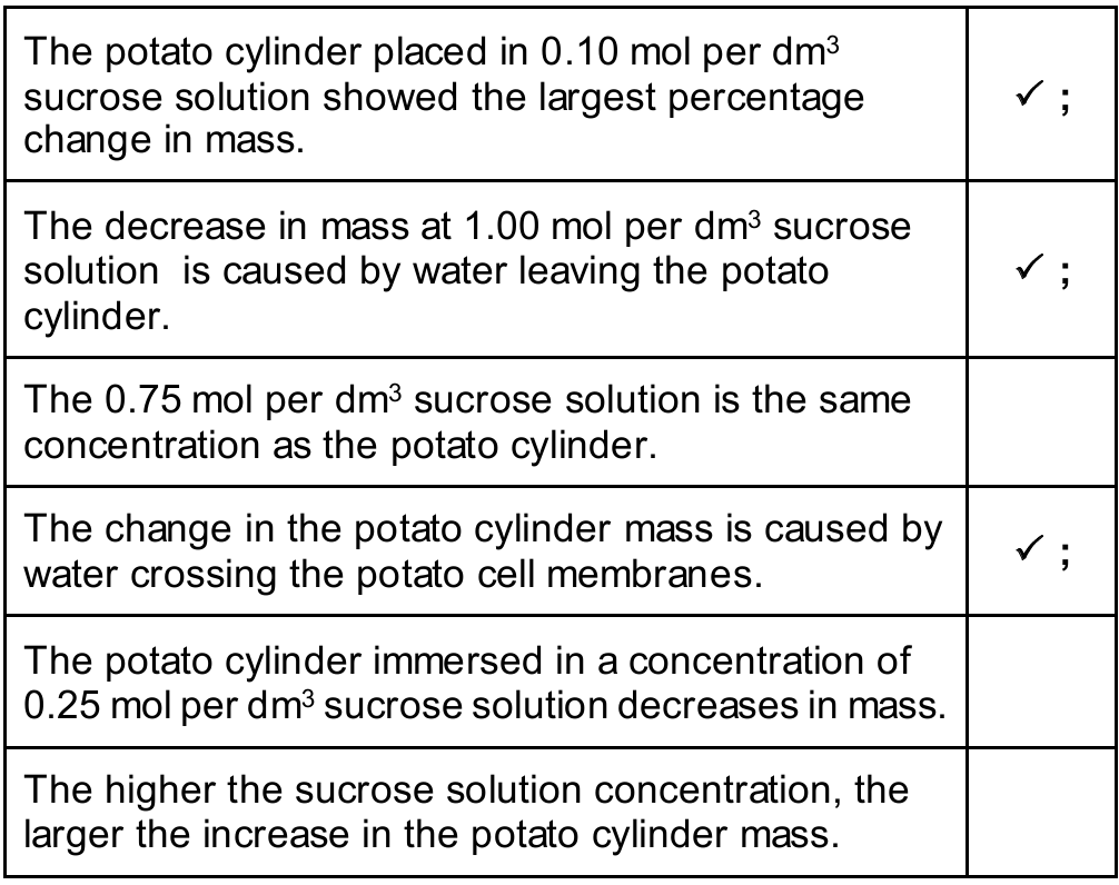
The correct identifications based on wall thickness and lumen size are:
• Row X: Vein (Thin wall, large lumen)
• Row Y: Capillary (Very thin wall, very narrow lumen)
• Row Z: Artery (Thick wall, narrow lumen)
(a)(ii)
• Name: Valves
• Function: To ensure the one-way flow of blood (prevent backflow). Veins carry blood at low pressure, often against gravity, so valves are necessary to keep blood moving towards the heart.
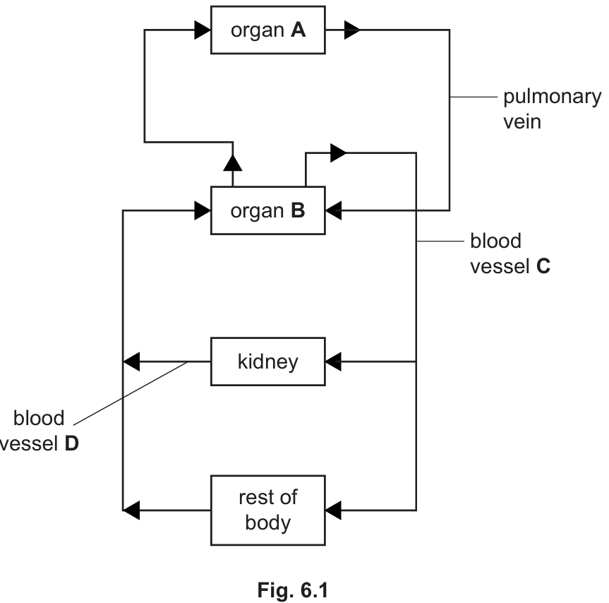
(b)
• organ A: Lungs (Blood flows from the heart to the lungs via the pulmonary artery and returns via the pulmonary vein).
• organ B: Heart (The central pump of the circulatory system).
• blood vessel C: Aorta (The main artery carrying oxygenated blood from the heart to the rest of the body).
• blood vessel D: Renal vein (The vessel carrying deoxygenated blood away from the kidney back towards the heart).
(c)
Any two of the following:
• Urea
• Excess water
• Excess ions (or salts)
(d)
Any two valid organ systems other than circulatory or digestive, such as:
• Nervous system
• Respiratory system (or gas exchange system)
• Reproductive system
• Excretory system
• Endocrine system
▶️ Answer/Explanation
(a)(i)
(5 \text{ mm})
(a)(ii)
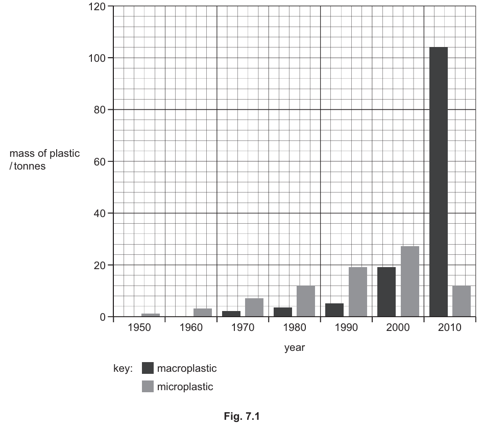
Based on Fig. 7.1, the changes can be described as follows:
- Microplastics: The mass increased steadily from 1950, reaching a peak in 2000, before decreasing in 2010.
- Macroplastics: There was little to no mass recorded until 1970; it then increased slowly until 2000, followed by a sharp/rapid increase between 2000 and 2010.
- Comparison: Microplastics had a higher mass than macroplastics from 1950 to 2000, but in 2010, the mass of macroplastics exceeded that of microplastics.
(b)
Two ways plastics harm animals:
- Ingestion: Animals eat plastic mistaking it for food, which can block the digestive system, cause internal injuries, or lead to starvation due to a false sense of fullness.
- Entanglement: Animals can get trapped in plastic debris (e.g., fishing nets, six-pack rings), which restricts movement, causing drowning, injury, or inability to feed/escape predators.
(c)
One other source of pollution:
- Untreated sewage.
- Excess chemical fertiliser (leading to eutrophication).
- Insecticides/Herbicides (agricultural runoff).
- Oil spills (marine pollution).
Part (a)(i): Unit Conversion
The syllabus requires candidates to convert between units, including cm and mm. Since (1 \text{ cm} = 10 \text{ mm}), the calculation is (0.5 \times 10 = 5 \text{ mm}).
Part (a)(ii): Data Interpretation
This question assesses the ability to interpret bar charts and describe trends (AO2: Handling information). Candidates must note the rise and fall of microplastics versus the late surge in macroplastics.
Part (b): Effects of Plastics
The syllabus explicitly includes the description of the “effects of non-biodegradable plastics, in both aquatic and terrestrial ecosystems.” In the ocean, this primarily manifests as physical harm through ingestion and entanglement.
Part (c): Aquatic Pollution
The syllabus lists several other aquatic pollutants. Key examples include “untreated sewage and excess fertiliser” and “freshwater and marine pollution” associated with habitat destruction.
▶️ Answer/Explanation
(a) Other Characteristics:
Any two from: Movement, Respiration, Sensitivity, Growth, Excretion, or Nutrition. (Often remembered by the acronym MRS GREN).
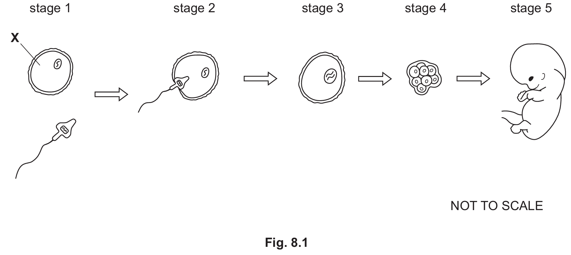
(b) Stages of Reproduction:
(i) Egg cell (or Ovum/Oocyte). Cell X is the female gamete.
(ii) Fertilisation. This is the fusion of the sperm nucleus with the egg nucleus.
(iii) Zygote. This is the diploid cell formed immediately after fertilisation.
(iv) Stage $4$. The zygote divides to form a ball of cells (embryo) which travels to the uterus and implants into the lining.
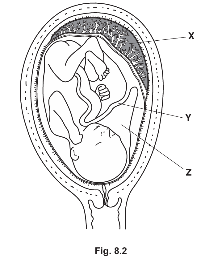
(c) Pregnancy Structures:
(i) X – Placenta; Y – Umbilical cord.
(ii) Part Z is the amniotic fluid. Its main functions are to protect the fetus against mechanical shock (bumps/knocks), maintain a constant temperature, and prevent the fetus from drying out.
(a) Characteristics of Life:
All living organisms share seven distinct characteristics defined by the syllabus (Topic 1.1): Movement, Respiration, Sensitivity, Growth, Reproduction, Excretion, and Nutrition.
(b) Human Reproduction:
Sexual reproduction involves the fusion of two haploid gametes (sperm and egg) to form a diploid zygote (Topic 16.4). The process shown in Fig 8.1 begins with the sperm approaching the egg cell (Stage $1$). Stage $2$ leads to fertilisation, where nuclei fuse. The resulting single cell (Stage $3$, the zygote) undergoes mitosis to form a ball of cells (Stage $4$, often called a blastocyst/morula), which implants in the uterus wall to develop into an embryo and then a fetus.
(c) Pregnancy:
During development (Topic 16.4), the fetus is supported by several structures:
X (Placenta): Anchors the fetus to the uterus and facilitates the exchange of nutrients, gases (oxygen/carbon dioxide), and waste between the mother’s blood and the fetus’s blood.
Y (Umbilical Cord): Contains blood vessels that connect the fetus to the placenta.
Z (Amniotic Fluid): Contained within the amniotic sac, this fluid cushions the fetus from external physical impact and allows for movement.