
▶️ Answer/Explanation
(a)(i) Identification of structures:
A: Trachea
B: Bronchus (or Bronchi)
C: Bronchiole(s)
Explanation: The trachea (windpipe) is the main tube with cartilage rings (A) leading from the mouth/nose. It splits into two bronchi (B) which enter the lungs, and further subdivides into smaller tubes called bronchioles (C).
(a)(ii) Function of cartilage:
The function is to keep the airway open or prevent the collapse of the airway.
Explanation: The C-shaped rings of cartilage provide structural support so that the trachea does not collapse inward when the pressure drops during inspiration.
(a)(iii) Mechanism of inspiration:
During inspiration (breathing in):
- The diaphragm contracts and flattens (moves downwards).
- The external intercostal muscles contract (while internal intercostal muscles relax), causing the ribs to move up and out.
- This action increases the volume of the thorax (chest cavity).
- Consequently, the pressure decreases inside the thorax/lungs.
- Air flows into the lungs to equalise the pressure difference.
(b)(i) Protection of airways:
Two common cell types are:
- Goblet cells: They secrete mucus. This sticky mucus traps dust, particles, and pathogens (bacteria/viruses).
- Ciliated cells: They have hair-like structures called cilia. These beat/move to sweep the mucus (containing trapped pathogens) up and out of the airways towards the throat to be swallowed.
Note: Lymphocytes (produce antibodies) and Phagocytes (engulf pathogens) are also acceptable answers.
(b)(ii) Effect on exercise:
Thicker mucus creates a blockage or narrowing of the airways. This leads to:
- Reduced surface area for gas exchange or reduced rate of diffusion.
- Less oxygen ($O_2$) reaches the blood and muscle cells (and less $CO_2$ is removed).
- Muscle cells perform less aerobic respiration and must rely more on anaerobic respiration.
- This results in less energy/ATP being released/produced for muscle contraction, causing fatigue more quickly.
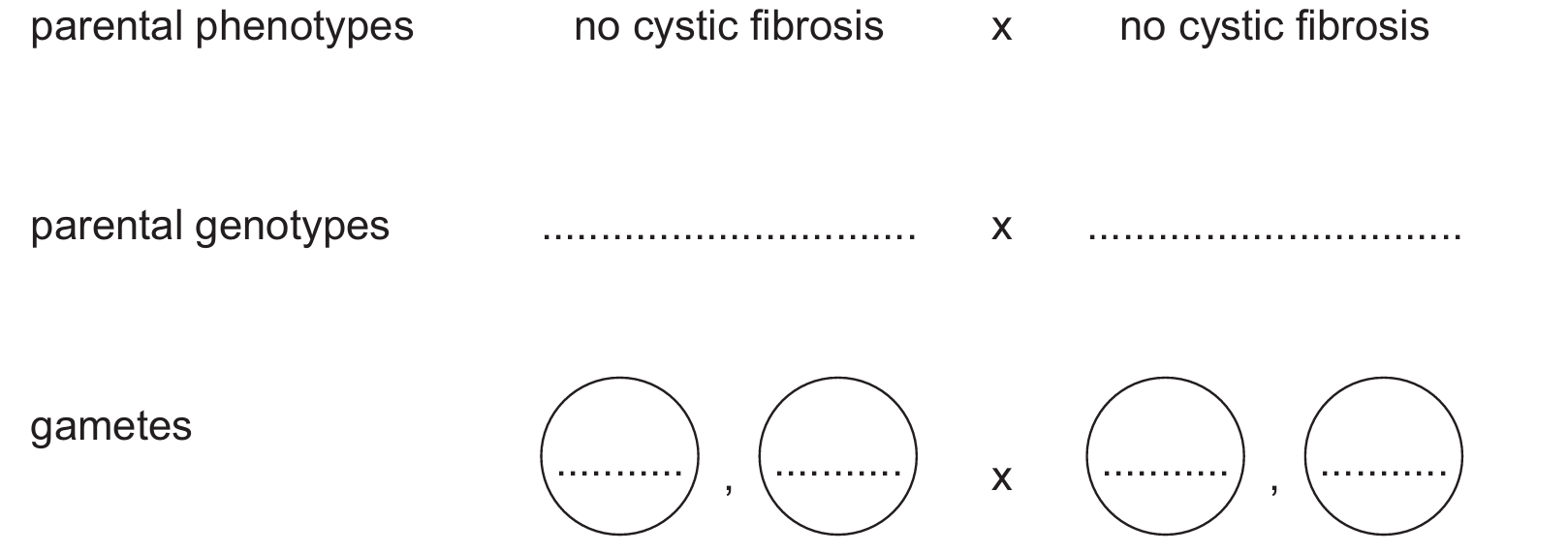
(c) Genetic Diagram:
Since both parents are heterozygous, they carry one dominant and one recessive allele.
Parental genotypes: $Ff$ x $Ff$
Gametes: ($F$), ($f$) x ($F$), ($f$)
Offspring genotypes: $FF$, $Ff$, $Ff$, $ff$
Offspring phenotypes: No cystic fibrosis, No cystic fibrosis, No cystic fibrosis, Cystic fibrosis
Probability: $0.25$ or $25\%$ or $1:3$ or $\frac{1}{4}$
Explanation: The cross yields one homozygous dominant ($FF$), two heterozygous carriers ($Ff$), and one homozygous recessive ($ff$). Since cystic fibrosis is recessive, only the $ff$ genotype presents the condition.
▶️ Answer/Explanation
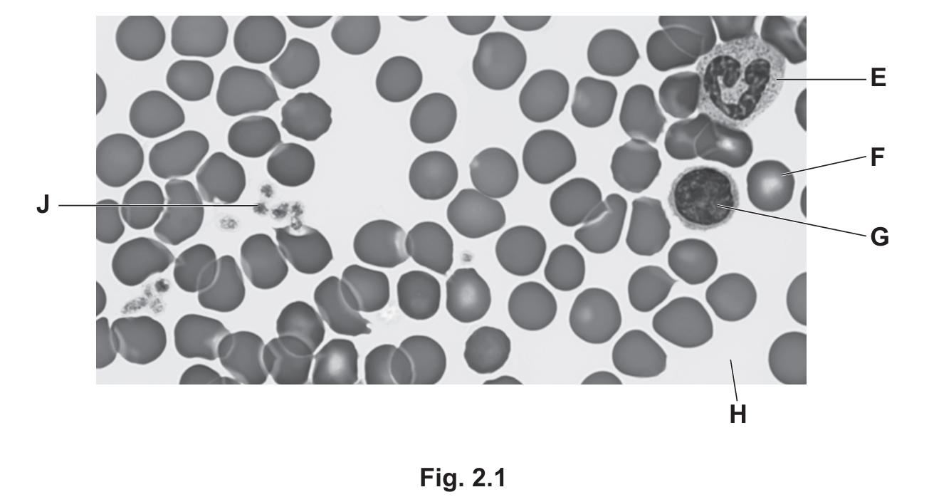
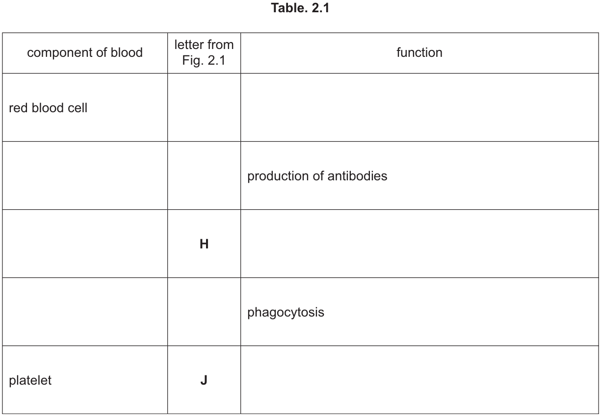
(a)
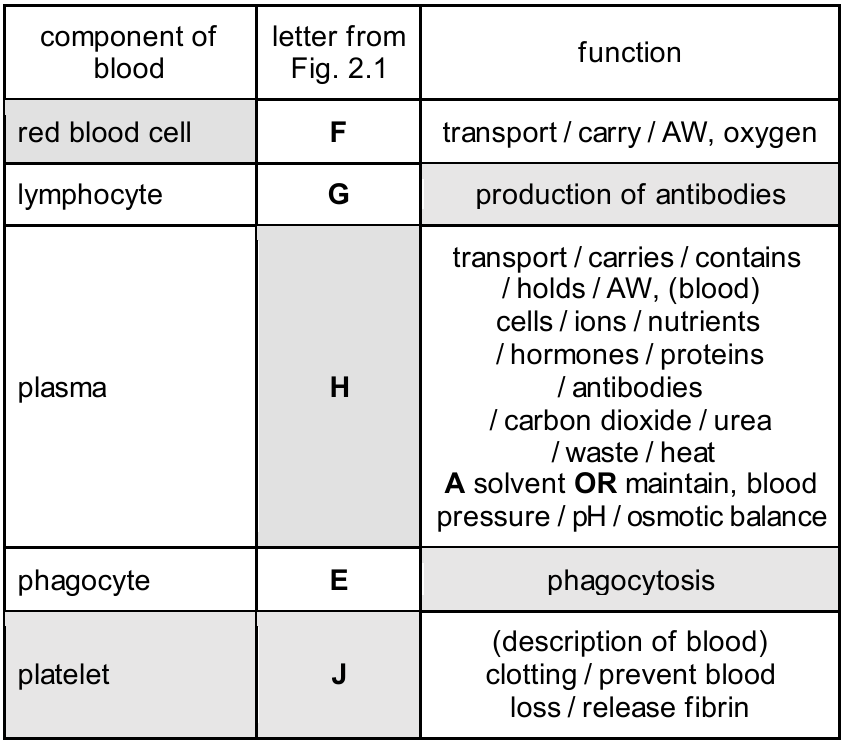
The completed table is as follows:
- Red blood cell:
- Letter: F (These are the numerous small, biconcave cells lacking a nucleus).
- Function: Transport of oxygen (mediated by haemoglobin).
- Lymphocyte:
- Component: lymphocyte (Identified by the large, round nucleus that fills most of the cell, labeled G).
- Letter: G.
- Function: Production of antibodies.
- Plasma:
- Component: plasma (The liquid medium surrounding the cells).
- Letter: H.
- Function: Transport of blood cells, ions, nutrients (e.g., glucose, amino acids), hormones, carbon dioxide, urea, and heat. It also acts as a solvent.
- Phagocyte:
- Component: phagocyte (Identified by the lobed or irregular-shaped nucleus, labeled E).
- Letter: E.
- Function: Phagocytosis (engulfing and digesting pathogens).
- Platelet:
- Letter: J (Small cell fragments).
- Function: Blood clotting (prevents blood loss and entry of pathogens) / release of fibrin.
(b)(i)
HIV (Human Immunodeficiency Virus).
(b)(ii)
Any two of the following:
• Contaminated surfaces / objects (fomites)
• Contaminated food or water
• Air (droplets / airborne)
• Vectors (animals that carry the pathogen, e.g., mosquitoes)
• Faeces / sewage
(c)
Antibodies are proteins produced by lymphocytes. Their role includes:
• Specificity: They have a shape complementary to specific antigens on the surface of pathogens.
• Binding: They bind/attach to these specific antigens.
• Destruction: This binding can mark the pathogen for destruction by phagocytes, cause the pathogens to clump together (agglutination), or neutralize toxins produced by the pathogen.
(d)
The key differences are:
• Source: Active immunity is produced by the body’s own immune system (production of antibodies by lymphocytes) after infection or vaccination. Passive immunity is the acquisition of ready-made antibodies from another individual (e.g., via placenta, breast milk, or injection).
• Memory: Active immunity produces memory cells, providing long-term protection. Passive immunity does not produce memory cells.
• Duration: Active immunity is long-term (or permanent). Passive immunity is short-term (antibodies are eventually broken down).
• Speed: Active immunity takes time to develop (slower response initially). Passive immunity provides immediate protection.
▶️ Answer/Explanation
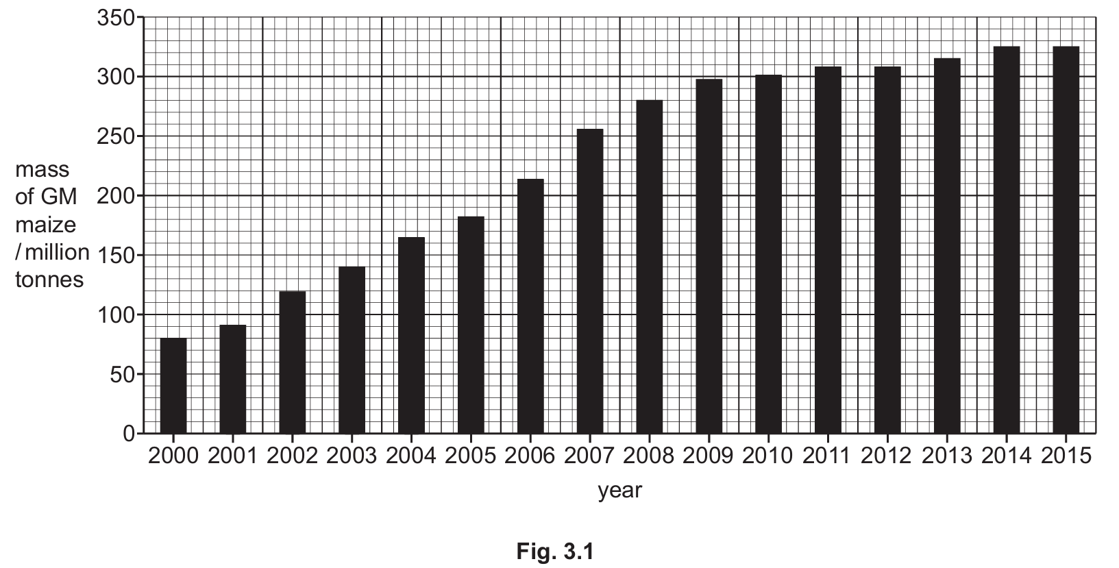
(a) 310 %
To calculate the percentage increase, you must read the values from the graph:
- Mass in \(2000 \approx 80\) million tonnes.
- Mass in \(2015 \approx 328\) (accept approx. \(330\)) million tonnes.
The formula for percentage increase is: \[ \frac{\text{Final Value} – \text{Initial Value}}{\text{Initial Value}} \times 100 \] Substituting the values: \[ \frac{328 – 80}{80} \times 100 = \frac{248}{80} \times 100 = 310\% \] The question asks for two significant figures; however, \(310\) is accepted in the mark scheme (the zero is often treated as a placeholder in integers, or the calculation results exactly in \(310\)).
(b)(i) DNA
Genes are specific sections or lengths of DNA (deoxyribonucleic acid) that code for proteins.
(b)(ii)
Scientists breed plants for multiple generations to ensure stability and viability. Key reasons include:
- To check that the gene is expressed (i.e., the plant actually produces the insecticide/toxin).
- To ensure the crop has a high enough yield and good quality.
- To ensure the gene does not cause unexpected or unwanted side effects.
- To confirm the crop can grow successfully in the intended environment.
- To produce a large enough quantity of seed to sell to farmers.
(b)(iii)
This describes the standard process of genetic engineering using a bacterial plasmid:
- Isolation: The specific gene (e.g., for the human protein) is isolated and cut from the donor organism’s DNA using restriction enzymes.
- Plasmid preparation: A bacterial plasmid (a circular ring of DNA) is cut using the same restriction enzyme.
- Sticky ends: Cutting both the donor DNA and the plasmid with the same enzyme creates complementary sticky ends.
- Insertion: The human gene is inserted into the bacterial plasmid.
- Ligation: The enzyme DNA ligase joins the gene and the plasmid together to form a recombinant plasmid.
- Transformation: The recombinant plasmid is inserted back into bacteria.
- Replication: The bacteria are placed in a fermenter to reproduce/multiply, producing the specific protein.
(c)(i)
To prevent the transfer of pollen (gene flow) to wild relatives:
- Grow the GM crops in enclosed environments like glasshouses.
- Cover the flowers to prevent pollinators (insects/wind) from accessing them.
- Remove the male parts of the flower (stamens/anthers) so no pollen is produced.
- Plant a buffer zone of a different species around the GM crop.
(c)(ii)
Advantages of GM crops (excluding insect resistance mentioned in the question):
- Herbicide resistance: Allows farmers to spray weeds without killing the crop.
- Environmental resistance: Ability to survive drought, heat, salinity (salt), or cold.
- Nutritional enhancement: Adding vitamins (e.g., Golden Rice with Vitamin A) or other nutrients.
- Improved characteristics: Better flavour, longer shelf life, or higher yield.
▶️ Answer/Explanation
(a)
An adaptive feature is an inherited feature that helps an organism to survive and reproduce (or breed/produce offspring) in its environment.
(b)
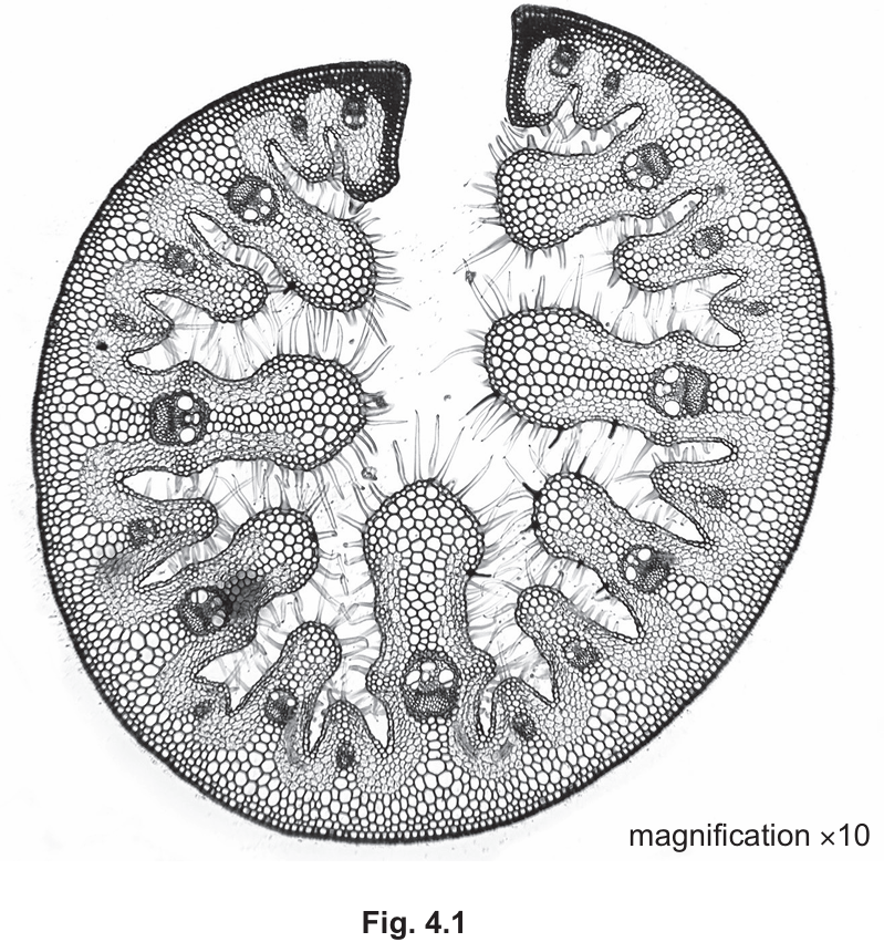
Based on the cross-section of the marram grass leaf (Fig. 4.1), the visible xerophytic features include:
- Rolled or curled leaf: The leaf is rolled inwards to reduce the surface area exposed to the drying effects of the wind and to trap moist air inside.
- Hairs: Epidermal hairs are visible lining the inside of the roll. These trap water vapour, creating a humid microclimate that reduces the water potential gradient and slows down transpiration.
- Sunken stomata: The stomata are located in pits or grooves on the inner surface (often protected by the hairs), further shielding them from air currents.
- Thick cuticle: A thick waxy layer on the outer surface prevents water loss through the epidermis.
(c) (i)
To convert micrometers (\(\mu\text{m}\)) to millimeters (mm), you divide by 1000 (since \(1 \text{ mm} = 1000 \, \mu\text{m}\)). $$ 1.276 \div 1000 = 0.001276 \text{ mm} $$ Alternatively expressed as \(1.276 \times 10^{-3} \text{ mm}\).
(c) (ii)
Cellulose. (This is the standard structural carbohydrate found in the cell walls of all plants, including xerophytes).
(c) (iii)
Adaptations of a xerophyte stem may include:
- Swollen / Succulent: The stem acts as a water storage organ (e.g., in cacti).
- Photosynthetic (Green): The stem contains chloroplasts to carry out photosynthesis, allowing the plant to reduce leaf size (or modify leaves into spines) to minimize water loss.
- Thick waxy cuticle: To reduce evaporation from the stem surface.
(d)
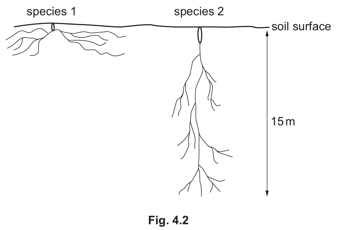
The two species show different root adaptations for water absorption in dry environments:
- Species 1 (Shallow, spreading roots): The roots are shallow and spread out over a wide area close to the soil surface.
Adaptation: This allows the plant to absorb water quickly and immediately after light rainfall before it evaporates from the surface soil. - Species 2 (Deep, vertical roots): The roots grow very deep into the soil (tap roots).
Adaptation: This allows the plant to access deep water sources or the water table, which provides a supply of water even when the surface soil is completely dry.
▶️ Answer/Explanation
(a)(i)
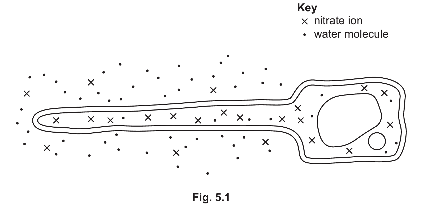
The process is active transport.
Explanation: By observing Fig. 5.1, you can see there are fewer nitrate ions ($\times$) outside the cell in the soil than inside the cell. Therefore, the ions must move from a region of low concentration to a region of high concentration (against the concentration gradient). This process requires energy (from respiration) and utilizes protein carriers located in the cell membrane.
(a)(ii)
1. Lightning
2. Nitrogen-fixing bacteria
(b)
Osmosis.
(c)
Differences:
• Nucleus: A root hair cell has a nucleus and a nuclear membrane, whereas a bacterial cell does not.
• DNA Structure: Root hair cells have linear DNA; bacteria have circular DNA and may have plasmids.
• Organelles: Root hair cells contain mitochondria and a large permanent vacuole; bacterial cells lack these membrane-bound organelles.
• Cell Wall: The root hair cell wall is made of cellulose, while the bacterial cell wall is made of a different substance (peptidoglycan).
Similarities:
• Both have a cell membrane.
• Both have cytoplasm.
• Both contain ribosomes.
• Both have a cell wall (though the composition differs).
• Both contain genetic material (DNA).
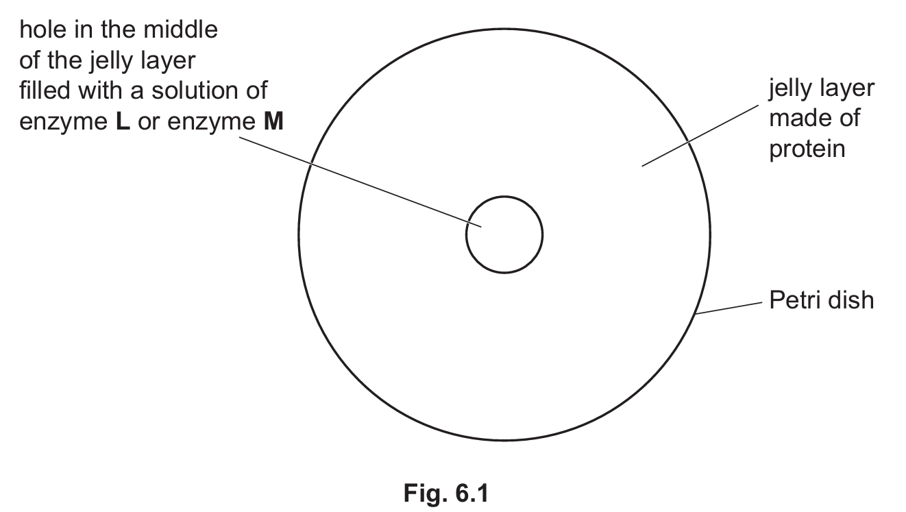
▶️ Answer/Explanation
(a)
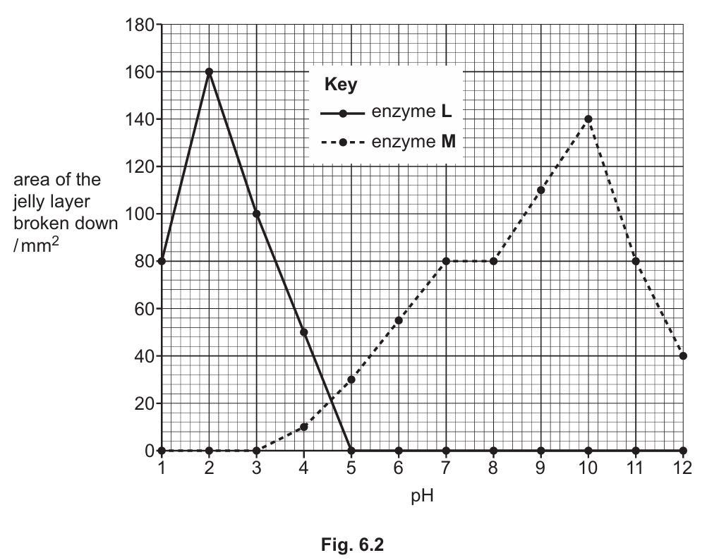
The results show that pH affects the activity of both enzymes, but they have different optimums.
- Trend: For both enzymes, activity increases to a peak and then decreases.
- Optimum pH: Enzyme \(\textbf{L}\) has an optimum pH of 2, with a maximum area digested of \(160~\text{mm}^2\). Enzyme \(\textbf{M}\) has an optimum pH of 10, with a peak activity of \(140~\text{mm}^2\).
- Activity Range: Enzyme \(\textbf{M}\) is active over a wider range (pH 3 to 12) compared to Enzyme \(\textbf{L}\) (pH 1 to 5). Enzyme \(\textbf{L}\) shows the steepest increase in activity between pH 1 and 2.
- Data Point: At pH 5, Enzyme \(\textbf{L}\) stops working (0 activity), whereas Enzyme \(\textbf{M}\) is active with an area of \(30~\text{mm}^2\).
(b)
At pH 2, the activity of enzyme \(\textbf{M}\) is zero (no jelly broken down). This is because enzyme \(\textbf{M}\) is adapted to alkaline conditions (optimum pH 10). The highly acidic environment at pH 2 causes the enzyme to become denatured. The shape of its active site changes so that it is no longer complementary to the substrate (the protein in the jelly), preventing the formation of enzyme-substrate complexes.
(c)
Name of enzyme L: Pepsin
Location: Stomach
(Explanation: Pepsin is a protease that functions in the acidic environment of the stomach, matching the optimum pH of 2 shown for enzyme L.)