▶️ Answer/Explanation
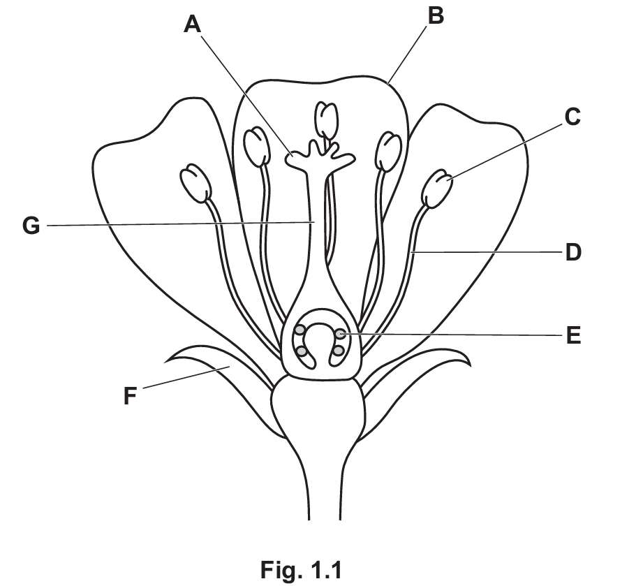
(a)(i)
Explanation: The stigma (A) is the sticky part that receives pollen. Petals (B) are colorful to attract pollinators. Sepals (F) are the protective outer covering of the flower bud.
(a)(ii) filament and anther
Explanation: The stamen, the male reproductive part of a flower, consists of the filament (stalk) that supports the anther (pollen-producing part).
(b)(i)
1. Self-pollination occurs when pollen is transferred from the anther to the stigma of the same flower or plant.
2. The pollen grain germinates and grows a pollen tube down through the style.
3. The pollen tube carries the male nucleus to the ovule through the micropyle.
4. Enzymes are released to help the pollen tube penetrate the ovule.
5. Fertilization occurs when the male nucleus fuses with the female nucleus in the ovule, forming a diploid zygote.
Explanation: Self-pollination begins with pollen transfer within the same plant, followed by pollen tube growth through the style to reach the ovule. The male nucleus travels down this tube to fuse with the female nucleus, completing fertilization inside the ovary.
(b)(ii)
Advantages of self-pollination:
1. Guarantees pollination even in isolated plants
2. Preserves successful genetic combinations
3. Requires less energy than attracting pollinators
4. Ensures reproduction when pollinators are scarce
Disadvantages of self-pollination:
1. Reduces genetic variation in offspring
2. Increases risk of harmful recessive traits appearing
3. Limits adaptability to environmental changes
4. May lead to weaker offspring over generations
Explanation: While self-pollination ensures reproduction, it limits genetic diversity, making populations more vulnerable to diseases and environmental changes. Cross-pollination promotes genetic variation but depends on external factors like pollinators.

▶️ Answer/Explanation
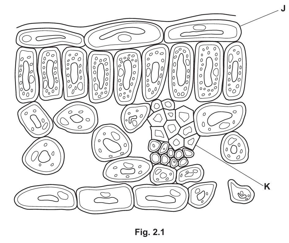
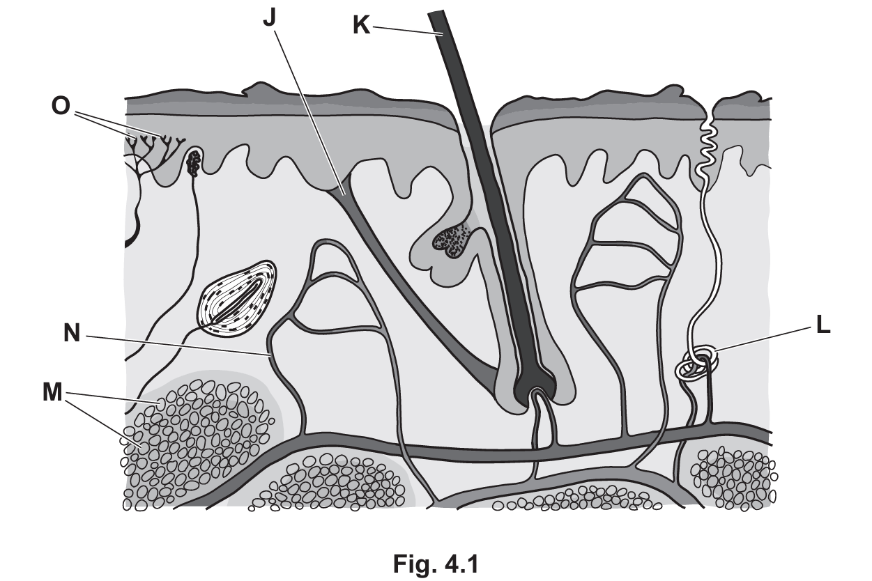
(a)
J – (Upper) epidermis cell:
The upper epidermis is transparent (translucent) and thin, which allows maximum light penetration to reach the palisade mesophyll cells underneath where most photosynthesis occurs. The lack of chloroplasts in these cells ensures no light is absorbed before reaching the photosynthetic cells.
K – Xylem vessel:
The xylem is adapted with its hollow, continuous tube structure (no end walls) and lignin-strengthened walls to efficiently transport water and mineral ions to the leaf. This constant water supply is crucial for photosynthesis. The structural support from lignin also helps maintain leaf shape for optimal light absorption.
(b)
Table 2.1 shows that increasing the carbon dioxide concentration caused more starch to be produced in the leaves. This shows that, at a normal carbon dioxide concentration, carbon dioxide is a limiting factor for photosynthesis.
During photosynthesis, 6 molecules of carbon dioxide are required to make one molecule of glucose.
The greater quantity of starch stored in the leaves grown in a high carbon dioxide concentration means, when needed, more sucrose can be produced for transport in the phloem, so the leaves act as a source.
The greater number of chloroplasts per cell in the leaves grown in the higher carbon dioxide concentration means that more energy can be absorbed from sunlight and transferred to chemical energy.
The transpiration rate is lower when the carbon dioxide concentration is higher. This means reduced loss of water vapor from the leaves.
Magnesium ion concentration is lower in these leaves because they have used the magnesium ions to make chlorophyll.
Detailed Explanation:
The experiment demonstrates several key concepts about photosynthesis and plant responses to CO2 concentration:
- The increased starch production shows CO2 is normally limiting – when more is available, photosynthesis increases.
- The chemical equation for photosynthesis (6CO2 + 6H2O → C6H12O6 + 6O2) explains why 6 CO2 molecules are needed per glucose.
- Leaves become sources when they produce excess carbohydrates that can be transported to other plant parts.
- More chloroplasts develop to handle the increased photosynthetic activity, absorbing more light energy.
- Stomata don’t need to open as wide when CO2 is plentiful, reducing water loss through transpiration.
- Magnesium is a key component of chlorophyll molecules, so more chlorophyll production uses up available magnesium ions.
▶️ Answer/Explanation
(a) homeostasis; negative feedback
Explanation: The human body maintains a constant internal temperature through homeostasis, which is the process of maintaining stable internal conditions despite changes in the external environment. When temperature deviates from the set point, negative feedback mechanisms work to return it to normal. For example, if body temperature rises, mechanisms like sweating are triggered to cool the body down, while if temperature drops, shivering generates heat to warm the body up.
(b)(i)
L – sweat gland
O – receptor(s)
J – (hair) erector muscle
Explanation: These structures are key components of the skin’s thermoregulatory system. Sweat glands produce sweat for cooling, receptors detect temperature changes, and hair erector muscles control hair position to trap insulating air when contracted.
(b)(ii)
When external temperature decreases:
- Temperature receptors in the skin detect the cold stimulus
- Nerve impulses are sent to the hypothalamus in the brain
- Hair erector muscles contract, making hairs stand up to trap insulating air
- Blood vessels near the skin surface constrict (vasoconstriction) to reduce heat loss
- Sweat glands reduce or stop sweat production to conserve heat
- Shivering may occur as muscles contract rapidly to generate heat
- Fatty tissue under the skin acts as insulation
Explanation: This coordinated response helps maintain core body temperature by reducing heat loss and increasing heat production when exposed to cold environments. The hypothalamus acts as the body’s thermostat, coordinating these responses through the nervous and endocrine systems.
(c)(i) pancreas
Explanation: Glucagon is secreted by the alpha cells in the islets of Langerhans within the pancreas, which is both an exocrine and endocrine gland located behind the stomach.
(c)(ii) Glucagon stimulates the breakdown of glycogen to glucose in the liver, increasing blood glucose concentration.
Explanation: When blood glucose levels drop too low, glucagon is released. It acts primarily on liver cells, triggering the conversion of stored glycogen into glucose through glycogenolysis. This glucose is then released into the bloodstream, raising blood sugar levels back to normal. Glucagon works in opposition to insulin to maintain glucose homeostasis.
▶️ Answer/Explanation
(a) Cell membrane and cytoplasm (or ribosomes or DNA).
Explanation: Both animal and bacterial cells share these fundamental structures. The cell membrane controls what enters and exits the cell, while the cytoplasm is the jelly-like substance where metabolic reactions occur. Ribosomes are present in both for protein synthesis, and DNA carries genetic information in both cell types.
(b) Any three from: rapid reproduction rate, reproduce asexually, small size, simple growth requirements, ability to make complex molecules, few ethical concerns, presence of plasmids, same genetic code as other organisms.
Explanation: Bacteria are extremely useful in biotechnology due to several key characteristics. Their rapid reproduction allows quick production of desired products. Asexual reproduction ensures genetic consistency. Their small size means they can be grown in large quantities in small spaces. They have simple nutritional needs, often just requiring basic nutrients. Many bacteria naturally produce complex molecules like enzymes. There are fewer ethical concerns compared to using animal or human cells. Plasmids make genetic modification easier, and their universal genetic code means genes from other organisms can be expressed in bacteria.
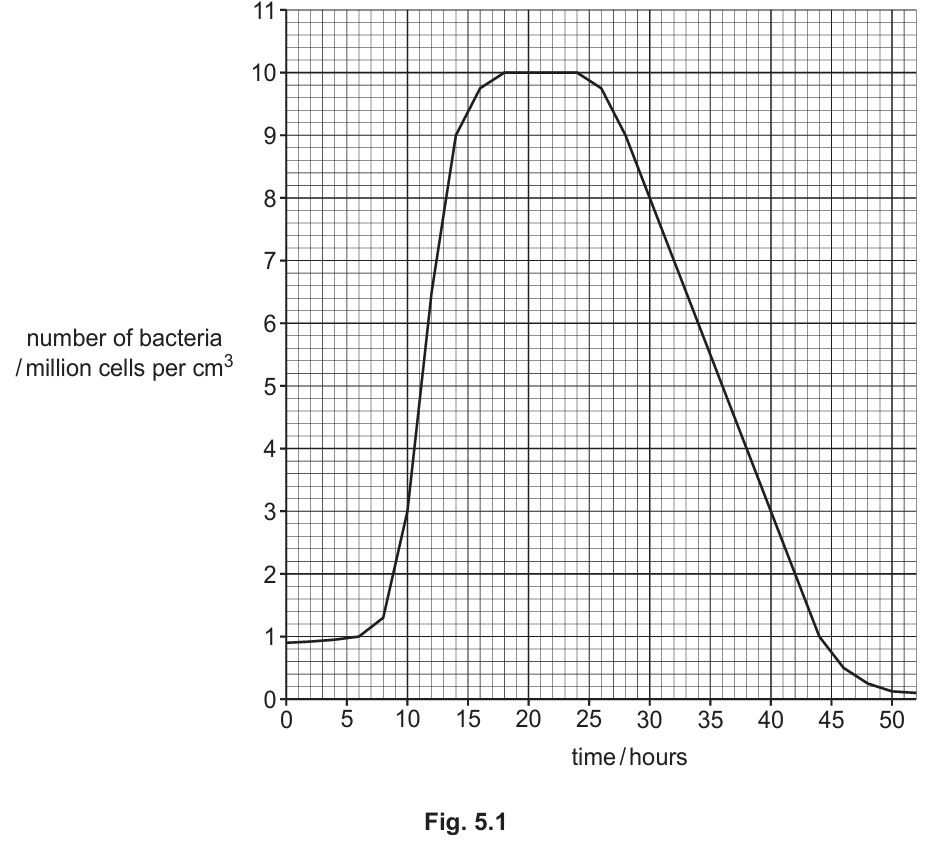
(c)(i) X placed between 0-8 hours.
Explanation: The lag phase is the initial period where bacteria are adjusting to their environment, synthesizing enzymes and preparing for growth, before population numbers begin to increase significantly.
(c)(ii) Y placed between 18-24 hours.
Explanation: The stationary phase occurs when birth rate equals death rate, resulting in no net population growth. This typically happens when resources become limited.
(c)(iii) 12 hours.
Explanation: At 24 hours the population is at its peak (about 10 million cells/cm³). By 36 hours it has halved to about 5 million cells/cm³, so the halving time is 12 hours.
(c)(iv) The population enters death phase where death rate exceeds birth rate, decreasing due to limited resources, increased competition, waste buildup, or other unfavorable conditions.
Explanation: After 24 hours, the bacterial population begins to decline. This death phase occurs because essential nutrients become depleted, waste products accumulate to toxic levels, and competition for remaining resources intensifies. The environment becomes increasingly hostile, causing more cells to die than are being produced through reproduction.
(c)(v) To maximize enzyme activity while preventing denaturation.
Explanation: Enzymes function best at their optimum temperature. At this temperature, they have sufficient kinetic energy for frequent collisions with substrates, forming many enzyme-substrate complexes. The active site maintains its correct shape for substrate binding. Temperatures above the optimum would cause denaturation (permanent shape change), while lower temperatures would reduce reaction rates.
(c)(vi) To make proteins/enzymes/nucleic acids.
Explanation: Amino acids are the building blocks of proteins, which bacteria need for growth, enzyme production, and cellular structures. Providing amino acids in the growth medium allows for efficient protein synthesis without requiring the bacteria to produce all amino acids from scratch, thus supporting faster growth and higher yields of desired products.
▶️ Answer/Explanation
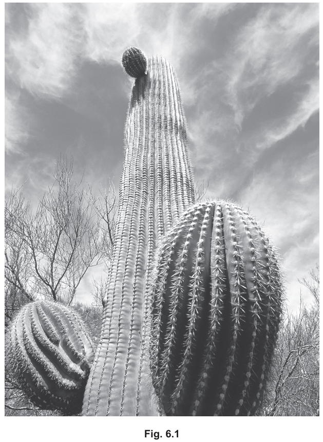
(a) Adaptation refers to the process resulting from natural selection where populations become better suited to their environment over many generations.
Explanation: Adaptations are characteristics that help organisms survive and reproduce in their specific environment. These traits develop over long periods through natural selection, where individuals with beneficial traits are more likely to survive and pass on their genes. In the case of xerophytes, their adaptations help them conserve water in arid environments.
(b)
Feature 1: Spines/needles/thorns
Explanation: These structures reduce the surface area of the plant, minimizing water loss through transpiration. They also serve as a defense mechanism against herbivores that might otherwise consume the plant’s precious water reserves.
Feature 2: Fleshy/thick/swollen stem
Explanation: The enlarged stem acts as a water storage organ, allowing the cactus to store large amounts of water during rare rainfall events and slowly utilize it during prolonged dry periods.
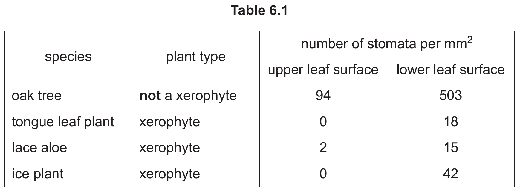
(c)(i) 33,600 stomata
Explanation: Calculation: 8 cm² = 800 mm² (since 1 cm² = 100 mm²). The ice plant has 42 stomata per mm² on its lower surface, so total stomata = 800 × 42 = 33,600.
(c)(ii) Xerophytes generally have fewer stomata than non-xerophytes to reduce water loss through transpiration. The stomata are often concentrated on the lower leaf surface which is cooler and more shaded, further minimizing water loss. The data shows that xerophytes like the tongue leaf plant and ice plant may have no stomata on their upper surfaces at all.
Detailed Explanation: The table demonstrates how xerophytes have evolved to minimize water loss while still allowing for gas exchange. The oak tree (non-xerophyte) has many more stomata (503 per mm² on lower surface) compared to the xerophytes (15-42 per mm²). Some xerophytes like the tongue leaf plant have no stomata on their upper surface at all (0 per mm²). This adaptation reduces transpiration while still allowing some gas exchange through the fewer stomata on the cooler, shaded lower surface.
(d) Reasons to conserve xerophytic ecosystems include:
- Maintaining biodiversity and genetic diversity
- Preventing extinction of unique species
- Ensuring stability of food chains and nutrient cycling
- Protecting vulnerable ecosystems that may have important ecological functions
Detailed Explanation: Xerophytic ecosystems, while appearing harsh, support unique biodiversity that may have undiscovered benefits for medicine, science, or agriculture. They often contain species found nowhere else. Conservation helps maintain ecological balance, protects potential resources, and preserves these unique environments for future generations. Additionally, many xerophytic plants are important carbon sinks and help stabilize soils in fragile environments.