▶️ Answer/Explanation
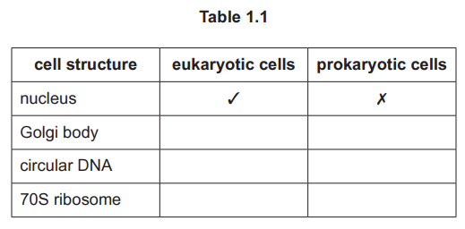
(a)
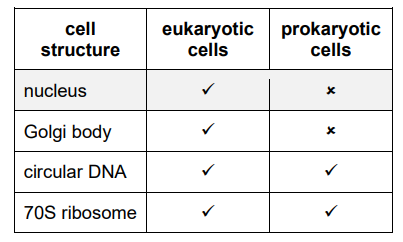
Explanation: The table is completed by identifying which structures are present in eukaryotic cells (✓ for nucleus, mitochondria, Golgi apparatus, etc.) and prokaryotic cells (✓ for cell wall, plasmids, ribosomes, etc.). Structures like the nucleus are exclusive to eukaryotes (✗ in prokaryotes), while ribosomes are present in both (✓ in both).
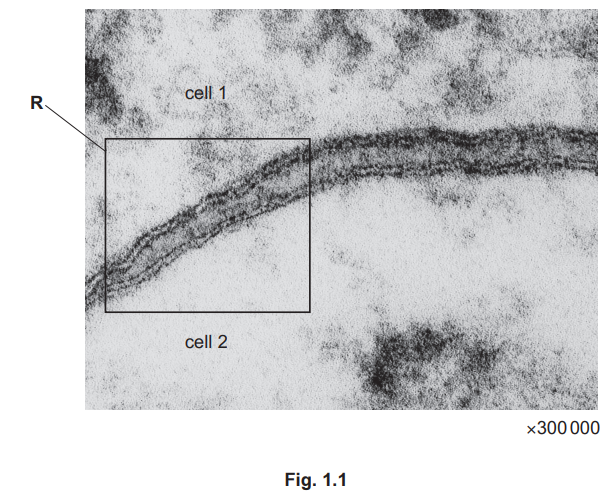
(b)
Explanation: The diagram should show the four dark lines representing the phospholipid bilayers of the two adjacent cell membranes. The dark lines are the phosphate heads, the spaces between them are the fatty acid tails (hydrophobic core), and the central space is the intercellular space (interstitial fluid).
(c)(i)
Explanation: In the \(G_1\) phase, the cell grows and synthesizes RNA, proteins, and organelles. In the \(S\) phase, DNA replication occurs, doubling the DNA content and forming sister chromatids.
(c)(ii)
Explanation: Increased mitogen concentrations can lead to uncontrolled cell division, as more cells are stimulated to progress from \(G_1\) to \(S\) phase. This may result in excessive mitosis and potential tumor formation.
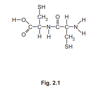
▶️ Answer/Explanation
(a)
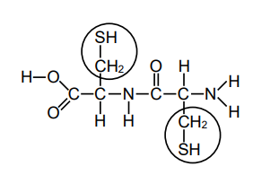
Explanation: The R-group in cysteine is the side chain containing sulfur (—CH2SH). Circling either of the R-groups in the molecule is correct as they are identical in structure.
(b)(i) Disulfide bonds.
Explanation: The covalent bonds formed between the sulfur-containing R-groups of cysteine residues are called disulfide bonds (—S—S—). These bonds stabilize the structure of mucin strands.
(b)(ii) Exocytosis.
Explanation: Mucin strands are transported out of goblet cells via exocytosis. This process involves vesicles containing mucins fusing with the cell membrane to release their contents outside the cell. It requires ATP and is facilitated by the cytoskeleton.
(c)
Explanation: Thicker mucus hinders the movement of cilia, preventing effective clearance of pathogens. This leads to accumulation of harmful microorganisms, increasing the risk of infections and damaging the gas exchange surfaces.
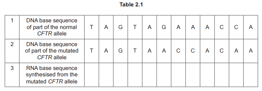
(d)(i) Deletion.
Explanation: The mutation is a deletion, as one base (T) is missing in the mutated sequence compared to the normal sequence.
(d)(ii) Template strand.
Explanation: The DNA strand used for RNA synthesis is called the template strand (or antisense strand). RNA polymerase reads this strand to produce complementary mRNA.
(d)(iii) AUC AUU GGU GUU.
Explanation: The RNA sequence is derived by transcribing the mutated DNA strand (TAG TAA CCA CAA), replacing T with U, resulting in AUC AUU GGU GUU.
(d)(iv)
Explanation: The difference arises because: (1) Only exons code for amino acids, while introns are non-coding and spliced out. (2) Three DNA bases (a triplet) code for one amino acid. (3) Some sequences (e.g., promoters, terminators) regulate transcription but do not encode amino acids.
▶️ Answer/Explanation
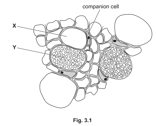
(a)(i) Cell Y shows a sieve plate/sieve pores, whereas the section misses the sieve plate in cell X. The sieve plates are at different heights in the stem, leading to their different appearances.
Explanation: Sieve tube elements have sieve plates at intervals. In a transverse section, some cells (like Y) may show these plates, while others (like X) may not, depending on where the cut is made.
(a)(ii) Companion cells use ATP to pump protons into the apoplast, creating a proton gradient. Protons then re-enter the companion cell with sucrose via cotransport, moving sucrose against its gradient. Sucrose diffuses into sieve tubes through plasmodesmata.
Explanation: This active transport mechanism ensures efficient sucrose loading into the phloem for translocation.
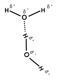
(b)(i) Hydrogen bonding occurs between the slightly positive hydrogen of one water molecule and the slightly negative oxygen of another.
Explanation: Due to water’s polar nature, oxygen attracts electrons more strongly, creating partial charges that facilitate hydrogen bonding.
(b)(ii) Hydrogen bonds create cohesion between water molecules and adhesion to xylem walls. This forms a continuous column pulled up by transpiration.
Explanation: Cohesion-tension theory relies on these bonds to maintain an unbroken water column during transport.
(b)(iii) High latent heat means evaporation removes significant heat, cooling leaves during photosynthesis and reducing enzyme denaturation.
Explanation: This property helps plants regulate temperature and minimize water loss in hot conditions.
▶️ Answer/Explanation
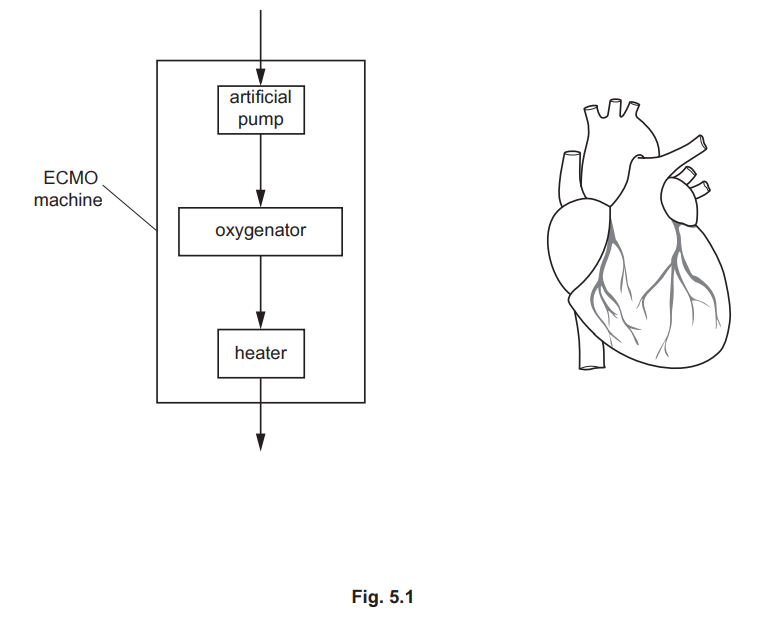
(a)
Answer: Line drawn from right atrium to the arrow into the pump and line drawn from the arrow out of the heater back to the (inferior/superior) vena cava.
Explanation: The ECMO machine is connected via two cannulas—one takes deoxygenated blood from the right atrium to the pump, and the other returns oxygenated blood from the heater to the vena cava.
(b)(i)
Answer: Alveolar wall / alveolar epithelium.
Explanation: The partially permeable membrane in the oxygenator functions similarly to the alveolar wall in the lungs, allowing gas exchange (O2 and CO2) by diffusion.
(b)(ii)
Answer:
- Oxygen diffuses from the oxygen-enriched air into the blood, and CO2 diffuses out of the blood.
- The counter-current flow maintains a steep concentration gradient, enhancing gas exchange efficiency.
- The thin membrane provides a short diffusion pathway, similar to alveoli.
Explanation: The oxygenator mimics lung function by enabling diffusion across a membrane, with opposite flow ensuring continuous gradient and efficient exchange.
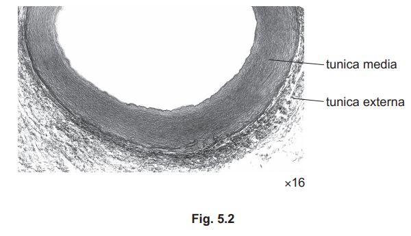
(c)
Answer: The tunica media in the aorta has more elastin than a muscular artery, allowing it to stretch during systole and recoil during diastole, maintaining blood pressure and flow.
Explanation: The aorta’s elastic structure accommodates high-pressure blood flow from the heart, preventing vessel damage and ensuring smooth circulation.
(d)(i)
Answer: Carbonic anhydrase catalyzes the conversion of CO2 and water into carbonic acid, which dissociates into hydrogencarbonate ions.
Explanation: This enzyme speeds up CO2 transport in the blood, essential for efficient respiration.
(d)(ii)
Answer: Since hydrogencarbonate ions remain bound to haemoglobin, there is no need for chloride ions to balance charges (no chloride shift).
Explanation: The physiology of C. latirostris avoids charge imbalance by retaining hydrogencarbonate ions inside red blood cells.
▶️ Answer/Explanation
(a)
Explanation: The strength of collagen in the skin arises from its structural properties. Covalent bonds between collagen molecules (specifically between R groups) provide strong intermolecular linkages. Additionally, the staggered arrangement of collagen molecules ensures there are no weak points, distributing tensile strength uniformly. In the skin, collagen fibres are arranged in layers running in different directions, enhancing resistance to forces from multiple angles.
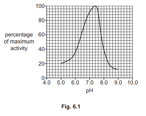
(b)
Explanation: At pH 8.0, collagenase activity decreases because the altered pH disrupts ionic and hydrogen bonds within the enzyme’s active site. This changes the shape of the active site, reducing its complementarity to the collagen substrate. As a result, fewer enzyme-substrate complexes form, leading to lower catalytic efficiency. The enzyme undergoes partial denaturation, further diminishing its activity compared to the optimum pH.