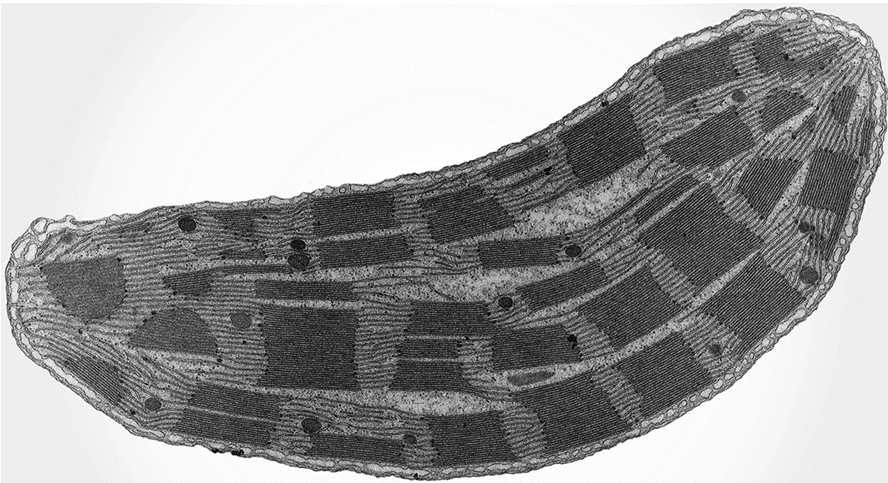
In an electron micrograph, the length of a mitochondrion is measured as 17.1 cm. The magnification of the electron micrograph is ×38 000.
What is the actual length of the mitochondrion?
A) 0.22 μm
B) 0.45 μm
C) 2.22 μm
D) 4.50 μm
▶️ Answer/Explanation
Ans: D
Actual size = Image size ÷ Magnification = 17.1 cm ÷ 38,000 = 0.00045 cm = 4.5 μm (1 cm = 10,000 μm).
There is a theory that mitochondria and chloroplasts were originally free-living prokaryotes. It is thought that millions of years ago these free-living prokaryotes were taken into larger cells by endocytosis where, instead of being digested, they became functional organelles.
Which features of mitochondria and chloroplasts support this theory?
- Mitochondria and chloroplasts are surrounded by double membranes.
- Mitochondria and chloroplasts have small, circular DNA.
- Mitochondria and chloroplasts synthesise proteins.
A) 1, 2 and 3
B) 1 and 2 only
C) 1 and 3 only
D) 2 and 3 only
▶️ Answer/Explanation
Ans: A
All three features support the endosymbiotic theory: double membranes suggest engulfing, circular DNA resembles prokaryotes, and protein synthesis shows independent function like free-living cells.
Which row correctly describes a function of each cell structure?
| lysosome | mitochondrion | smooth endoplasmic reticulum | |
|---|---|---|---|
| A | digestion of unwanted structures | abundant in sites of active transport | processing of proteins |
| B | digestion of unwanted structures | ATP synthesis | lipid production |
| C | spherical sacs containing hydrolytic enzymes | abundant in sites of active transport | lipid production |
| D | spherical sacs containing hydrolytic enzymes | ATP synthesis | processing of proteins |
▶️ Answer/Explanation
Ans: B
Lysosomes digest unwanted structures, mitochondria produce ATP, and smooth ER synthesizes lipids. Option B correctly matches all functions.
Which cell components are present in typical prokaryotic cells?
| cell wall | 80S ribosomes | |
|---|---|---|
| A | ✓ | ✓ |
| B | ✓ | X |
| C | X | X |
| D | X | ✓ |
key
✓ = present
X = not present
▶️ Answer/Explanation
Ans: B
Prokaryotes have cell walls (peptidoglycan) but 70S ribosomes, not 80S (which are eukaryotic). So cell wall present, 80S ribosomes absent.
X-ray analysis of fossilised cells found in rocks in central India dating back 1.6 billion years has revealed several features.
- The cells are up to 145 µm long.
- The cells are joined end to end to form filaments.
- The cells contain some internal cell structures.
- The cells are surrounded by a cell wall.
Which two features, when taken together, provide most support for the conclusion that the cells are plant cells?
A) 1 and 3
B) 1 and 4
C) 2 and 3
D) 2 and 4
▶️ Answer/Explanation
Ans: B
Large size (145 µm) suggests eukaryotic cells, and cell walls are characteristic of plant cells. These together best support plant cell identification.
The electron micrograph shows an organelle found in some cells of many multicellular organisms.
Which row shows structures that are expected to be present in cells that contain this organelle?
| cell wall | centrioles | plasmodesmata | |
| A | ✗ | ✓ | ✗ |
| B | ✓ | ✗ | ✗ |
| C | ✗ | ✓ | ✓ |
| D | ✓ | ✗ | ✓ |
▶️ Answer/Explanation
Ans: D
The organelle shown is likely a chloroplast, found in plant cells. Plant cells have cell walls and plasmodesmata but lack centrioles. Option D correctly shows these features.
What could take place during a hydrolysis reaction?
- A glycosidic bond is broken.
- A molecule of water is produced.
- A sucrose molecule is split into fructose and glucose.
A) 1, 2 and 3
B) 1 and 2 only
C) 1 and 3 only
D) 2 and 3 only
▶️ Answer/Explanation
Ans: C
Hydrolysis breaks bonds using water, so statement 1 is correct. Water is consumed, not produced (2 is wrong). Sucrose hydrolysis yields glucose+fructose (3 is correct). Therefore, only 1 and 3 are correct.
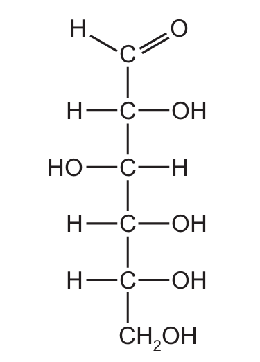
Sugars with a ring structure can also have a linear structure.
Which sugar molecules could be represented by the linear structure shown in the diagram?
A) glucose, deoxyribose and ribose
B) glucose only
C) deoxyribose and ribose only
D) deoxyribose only
▶️ Answer/Explanation
Ans: B
The diagram shows a 6-carbon linear sugar structure (hexose). Glucose is a hexose, while deoxyribose and ribose are pentoses (5-carbon sugars). Therefore, only glucose can be represented by this structure.
Which statement about triglycerides is correct?
A) Each triglyceride molecule is formed by combining three fatty acid molecules with a glycogen molecule.
B) A triglyceride molecule contains four ester bonds, each formed in a condensation reaction.
C) Triglyceride molecules form a bilayer in the cell surface membranes of cells due to hydrophobic and hydrophilic interactions.
D) The ratio of oxygen atoms to carbon atoms is lower for triglyceride molecules than for carbohydrate molecules.
▶️ Answer/Explanation
Ans: D
Triglycerides combine with glycerol, not glycogen (A is wrong). They have three ester bonds (B is wrong). Phospholipids, not triglycerides, form bilayers (C is wrong). Triglycerides have lower O:C ratio than carbohydrates (D is correct).
Hydroxyproline is synthesised by addition of an –OH group to the R-group of the amino acid proline.
Hydroxyproline is a major component of collagen and has an important role in increasing the stability of its structure.
Which statement explains why the addition of an –OH group to proline could increase the stability of collagen?
A) It strengthens hydrogen bonding between the R-groups of adjacent polypeptide chains, resulting in a tertiary structure that is more resistant to heat denaturation.
B) It increases the number of sites available for the formation of hydrogen bonds within the secondary structure of collagen, resulting in more stable alpha helices.
C) It increases the formation of hydrogen bonds between R-groups and water molecules, which help to hold the chains of the collagen triple helix together by strengthening hydrophilic interactions.
D) It strengthens the quaternary structure of collagen by providing more sites for hydrogen bonding between the R-groups of distantly separated amino acids within the same polypeptide chain.
▶️ Answer/Explanation
Ans: C
The -OH group enables hydrogen bonding between collagen strands and water molecules, stabilizing the triple helix structure. This is crucial for collagen’s structural integrity in connective tissues.
Plant cell walls are strengthened by cellulose molecules that are arranged in several layers. Within each layer, the cellulose molecules are arranged in the same direction (parallel).
Which row shows the bonds that hold adjacent cellulose molecules together within each layer and the arrangement of cellulose molecules in different layers?
| bonds that hold adjacent cellulose molecules together | arrangement of cellulose molecules in different layers | |
|---|---|---|
| A | glycosidic | in different directions |
| B | glycosidic | parallel |
| C | hydrogen | in different directions |
| D | hydrogen | parallel |
▶️ Answer/Explanation
Ans: C
Adjacent cellulose molecules are held together by hydrogen bonds between hydroxyl groups. In different layers, cellulose molecules are arranged in different directions (crossed) for added strength, making option C correct.
Galactogen is a storage polysaccharide in some animal species. It is a branched polymer that is formed from β-galactose monomers.
Which comparison of galactogen with another polysaccharide correctly summarises one similarity and one difference?
A) Glycogen and galactogen are both branched, but glycogen is not a storage polysaccharide in animals.
B) Glycogen and galactogen are both storage polysaccharides in animals, but glycogen is unbranched.
C) Cellulose and galactogen are both branched, but cellulose is a structural polysaccharide found in plants.
D) Amylopectin and galactogen are both storage polysaccharides, but amylopectin is formed from α-glucose monomers.
▶️ Answer/Explanation
Ans: D
Amylopectin (plant starch) and galactogen are both storage polysaccharides (similarity). The difference is that amylopectin is made of α-glucose while galactogen uses β-galactose monomers, making D the correct choice.
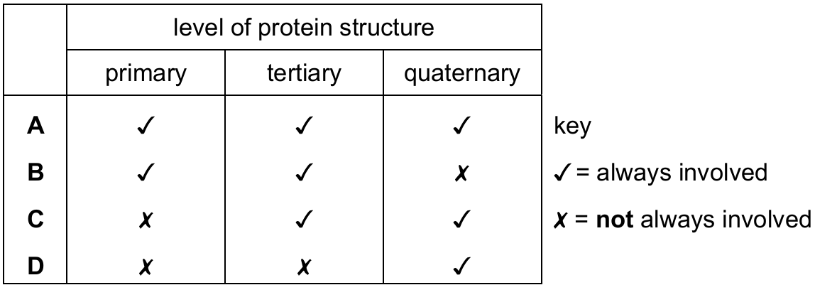
Which levels of protein structure are always involved in forming the active site of an enzyme?
▶️ Answer/Explanation
Ans: B
The active site is formed by the folding of the polypeptide chain (tertiary structure) which depends on the amino acid sequence (primary structure). Quaternary structure isn’t always involved as some enzymes are single polypeptide chains.
Catalase is an enzyme that breaks down hydrogen peroxide into water and oxygen.
Catalase was added to a solution of hydrogen peroxide and the oxygen produced was collected in a gas syringe.
The total volume of oxygen produced from the start of the reaction was recorded every 10 seconds for 1 minute.
The results are shown in the table.
| time/s | total volume of oxygen produced/cm3 |
|---|---|
| 0 | 0 |
| 10 | 22 |
| 20 | 40 |
| 30 | 50 |
| 40 | 55 |
| 50 | 57 |
| 60 | 58 |
What can be concluded from these results?
A) The reaction stopped after 60 seconds and no more oxygen was produced.
B) The highest rate of oxygen production occurred 10 seconds after the start of the reaction.
C) It took more than 20 seconds from the start of the reaction for half of the substrate to be converted to water and oxygen.
D) The mean rate of reaction between 20 and 30 seconds was twice the mean rate of reaction between 30 and 40 seconds.
▶️ Answer/Explanation
Ans: D
Between 20-30s: (50-40)/10 = 1 cm3/s. Between 30-40s: (55-50)/10 = 0.5 cm3/s. The first rate is indeed twice the second. Other options are incorrect based on the data.
Succinic dehydrogenase is an enzyme that catalyses the conversion of succinate to fumarate in aerobic respiration.
Malonate is a reversible inhibitor of succinic dehydrogenase. Malonate reduces the enzyme’s activity by binding to its active site. Malonate and succinate cannot bind to the active site at the same time.
Which statement describes the effect of malonate on the activity of succinic dehydrogenase?
A) In the presence of malonate, Vmax can still be reached if the concentration of succinate is increased.
B) Malonate has no effect on the Km.
C) In the presence of malonate, Vmax can still be reached if the concentration of fumarate is increased.
D) Malonate decreases the Km.
▶️ Answer/Explanation
Ans: A
Malonate is a competitive inhibitor – it competes with succinate for the active site. Increasing succinate concentration can overcome inhibition, allowing Vmax to be reached, though higher substrate concentrations are needed (increased apparent Km).
The diagram shows the dimensions of two blocks of agar. The diagram has been drawn to scale.
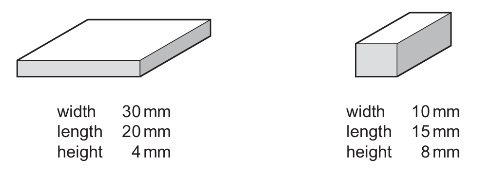
The blocks of agar were stained pink with a pH indicator. In acidic conditions, the pink pH indicator becomes colourless.
The two blocks of agar were placed in a beaker of acid at the same time. As the acid diffused into the blocks, the blocks became colourless.
What is the surface area to volume ratio of the block that became completely colourless first?
A) 0.58 : 1
B) 0.67 : 1
C) 1.50 : 1
D) 1.71 : 1
▶️ Answer/Explanation
Ans: B
Calculate SA:V for both blocks. The first block has dimensions 30×20×4 mm (SA=1840 mm², V=2400 mm³, ratio=0.77:1). The second block is 10×15×8 mm (SA=700 mm², V=1200 mm³, ratio=0.58:1). The block with higher SA:V (0.77:1) becomes colorless first, but this isn’t an option. The correct answer is the ratio of the second block (0.58:1), which matches option B.
The graph shows how the rate of facilitated diffusion of substance X across a cell surface membrane changed as the concentration of substance X increased. All conditions, except for the concentration of substance X, were kept constant. Temperature was maintained at 15°C.
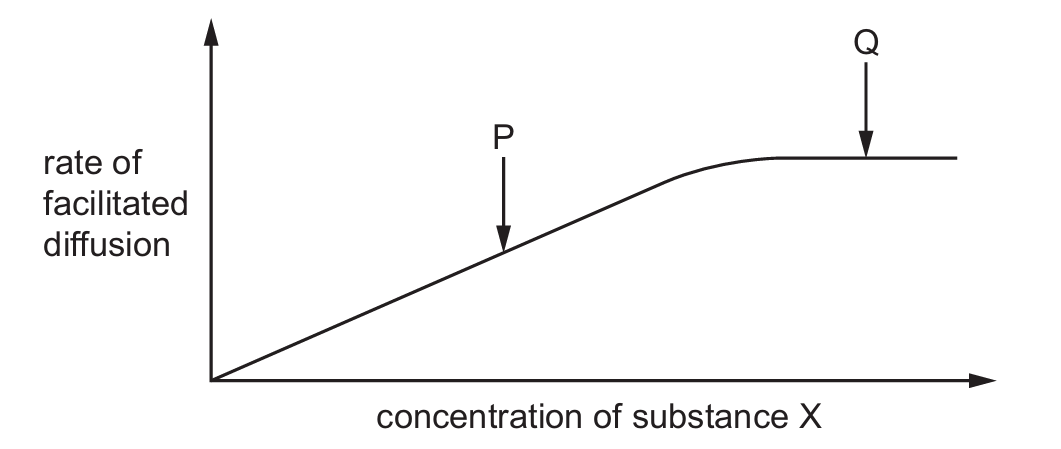
Which statement about the rate of facilitated diffusion is correct?
A) The rate of facilitated diffusion of substance X at Q will increase if the temperature is increased to 20°C.
B) The rate of facilitated diffusion of substance X at P will increase if the concentration of ATP is increased.
C) The rate of facilitated diffusion of substance X at Q will increase if the concentration of substance X is increased.
D) The rate of facilitated diffusion of substance X at P will increase if the length of time over which the rate is measured is increased.
▶️ Answer/Explanation
Ans: A
At point Q, the rate is limited by the number of carrier proteins (plateau phase). Increasing temperature (A) increases kinetic energy and diffusion rate. ATP (B) isn’t required for facilitated diffusion. At Q (C), increasing concentration won’t increase rate as carriers are saturated. Time (D) doesn’t affect the rate itself.
Which molecule forms a bilayer in the cell surface membrane of a bacterial cell?
A) fatty acid
B) peptidoglycan
C) phospholipid
D) cholesterol
▶️ Answer/Explanation
Ans: C
Phospholipids (C) form the basic bilayer structure of all cell membranes, including bacterial cells. Fatty acids (A) are components but don’t form bilayers alone. Peptidoglycan (B) is in cell walls. Cholesterol (D) stabilizes membranes but isn’t present in bacterial cell membranes.
A nucleus in a body cell of a species of fruit fly has 8 chromosomes.
How many strands of DNA are present in the nucleus at the end of interphase?
A) 8
B) 16
C) 32
D) 64
▶️ Answer/Explanation
Ans: C
At end of interphase (after S phase), DNA has replicated. 8 chromosomes become 16 chromatids (each chromosome has 2 DNA strands). Since each chromatid is one DNA molecule, total is 16 chromosomes × 2 DNA strands each = 32 DNA strands.
The photomicrograph shows plant cells in different stages of the mitotic cell cycle. Four of the cells are labelled with a number to identify them.
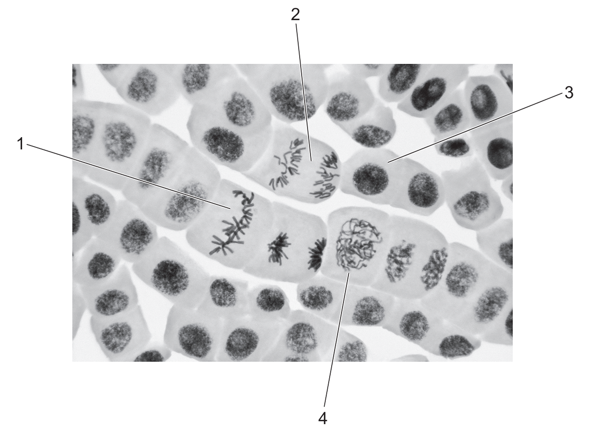
One of the numbered cells is within the main stage of mitosis in which the spindle begins to form.
A second numbered cell is within the main stage of mitosis in which the spindle fibres shorten.
Which row correctly identifies the two cel ls that are in these stages of mitosis?
| stage in which spindle begins to form | stage in which spindle fibres shorten | |
|---|---|---|
| A | 1 | 2 |
| B | 2 | 1 |
| C | 3 | 1 |
| D | 4 | 2 |
▶️ Answer/Explanation
Ans: D
The spindle begins to form in prophase (cell 4). Spindle fibers shorten during anaphase (cell 2) when chromosomes are pulled apart. Cell 1 shows telophase with new nuclei forming, and cell 3 shows metaphase with chromosomes aligned.
Which percentage of the chromosomal DNA present in a cell during G1 is present in the same cell later in the same mitotic cell cycle during prophase and during telophase?
| prophase | telophase | |
| A | 50% | 25% |
| B | 50% | 50% |
| C | 100% | 50% |
| D | 100% | 100% |
▶️ Answer/Explanation
Ans: D
During G1, DNA is unreplicated. By prophase, DNA has replicated (100%). In telophase, the DNA is still present in both daughter cells (100%), though it’s now separated into two nuclei.
The statements are about two genes and their protein products that can have a role in tumour formation.
- The protein coded for by the PTEN gene prevents cells from growing and dividing too rapidly.
- The protein coded for by the p53 gene prevents cells progressing through the mitotic cell cycle if the cells have damaged DNA.
Which combination of mutations of these two genes in an individual is most likely to result in the formation of a tumour?
| PTEN | p53 | |
| A | mutation present | mutation present |
| B | no mutation | mutation present |
| C | mutation present | no mutation |
| D | no mutation | no mutation |
▶️ Answer/Explanation
Ans: A
Both PTEN and p53 are tumor suppressor genes. Mutations in both would lead to uncontrolled cell growth (PTEN mutation) and failure to repair DNA damage (p53 mutation), making tumor formation most likely.
Telomerase is an enzyme that prevents the shortening of telomeres. It is not present in most normal cells, but is active in an estimated 85% to 95% of human tumour cells.
Which statement explains the effect of telomerase on human tumour cells?
A) Telomerase triggers a self-destruct process, known as apoptosis, ending the life of the cell.
B) Telomerase damages the chromosomes so they become genetically unstable and are unable to replicate and divide.
C) Telomerase helps human tumour cells avoid senescence, or cell death, which is usually the expected consequence of repeated cell division.
D) Telomerase enables the human tumour cells to divide more rapidly by reducing the time taken for a complete mitotic cell cycle.
▶️ Answer/Explanation
Ans: C
Telomerase maintains telomere length, allowing cancer cells to divide indefinitely by preventing the normal cellular aging process (senescence) that occurs when telomeres become too short.
Which diagram correctly represents one of the base pairs of DNA?
A) 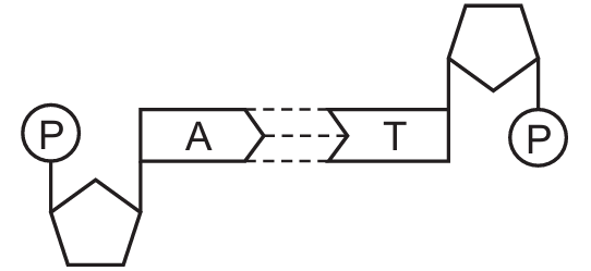
B) 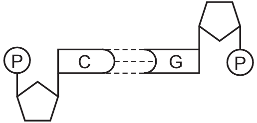
C) 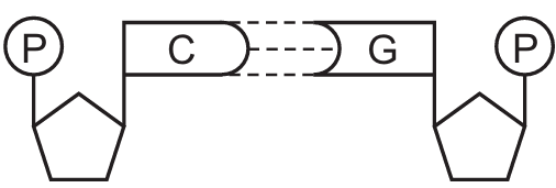
D) 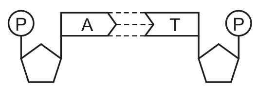
▶️ Answer/Explanation
Ans: B
The correct diagram shows C-G base pairing with three hydrogen bonds (the others show incorrect bonding patterns). In DNA, C always pairs with G via three hydrogen bonds, while A pairs with T via two hydrogen bonds.
The diagram represents the process of transcription of a gene in the nucleus of an animal cell.
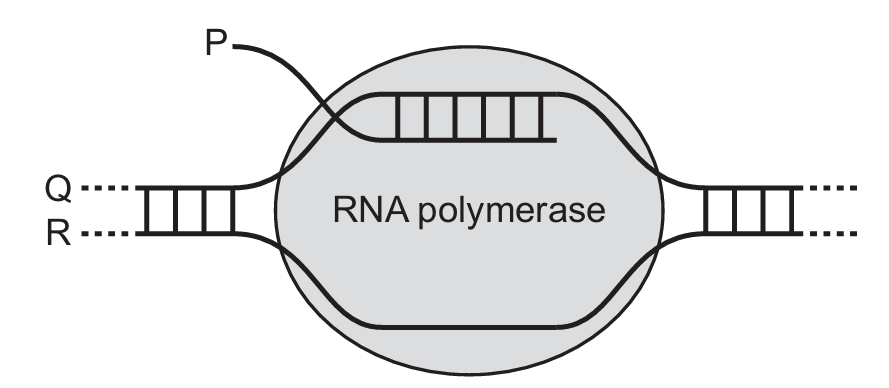
Which row correctly identifies P, Q and R?
| P | Q | R | |
| A | mRNA | transcribed strand | template strand |
| B | primary transcript | transcribed strand | template strand |
| C | mRNA | template strand | non-transcribed strand |
| D | primary transcript | template strand | non-transcribed strand |
▶️ Answer/Explanation
Ans: D
P is the primary transcript (unprocessed RNA). Q is the template strand (used to make RNA). R is the non-transcribed strand (identical to RNA sequence except T for U).
Which row correctly describes the role of growing leaves as sources or sinks for amino acids and sucrose?
| amino acids | sucrose | |
|---|---|---|
| A | leaves act as sinks only | leaves can act as sources or sinks |
| B | leaves act as sinks only | leaves act as sources only |
| C | leaves can act as sources or sinks | leaves act as sources only |
| D | leaves can act as sources or sinks | leaves can act as sources or sinks |
▶️ Answer/Explanation
Ans: D
Growing leaves can act as both sources and sinks for both amino acids and sucrose. Young leaves import both substances (acting as sinks), while mature leaves export them (acting as sources). This dual role is unique to growing leaves compared to other plant parts.
Which row correctly matches each description to cilia or root hairs?
| contain vacuoles | more than one present per cell | |
|---|---|---|
| A | root hairs | root hairs |
| B | cilia | cilia |
| C | root hairs | cilia |
| D | cilia | root hairs |
▶️ Answer/Explanation
Ans: C
Root hairs contain vacuoles as extensions of root epidermal cells, while cilia don’t. Multiple cilia are present per cell (e.g., in trachea), but each root epidermal cell typically produces just one root hair. This combination matches option C.
Which changes occur as amino acids are moved into phloem sieve tubes at a source?
| change in water potential in phloem sieve tubes | change in volume of sap in phloem sieve tubes | |
|---|---|---|
| A | decreases | decreases |
| B | decreases | increases |
| C | increases | decreases |
| D | increases | increases |
▶️ Answer/Explanation
Ans: B
When amino acids enter phloem at a source, they increase solute concentration, lowering water potential (more negative). This draws water in by osmosis, increasing sap volume. These changes create the pressure gradient needed for translocation.
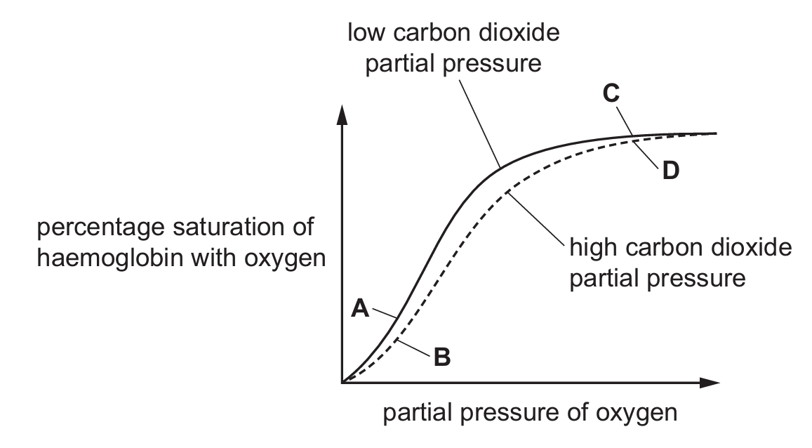
The graph shows the dissociation curves for haemoglobin at two different partial pressures of carbon dioxide.
Which labelled point on the oxygen dissociation curves will result in the highest concentration of haemoglobinic acid?
▶️ Answer/Explanation
Ans: B
Haemoglobinic acid forms when haemoglobin binds H⁺ ions. This occurs most at high CO₂ levels (Bohr effect) and when oxygen is released (low saturation). Point B represents these conditions – high CO₂ and low O₂ saturation – maximizing H⁺ binding.
Which statements about the transport of carbon dioxide in the blood are correct?
- Carbonic anhydrase catalyses the reaction of carbon dioxide and water to form carbonic acid.
- Carbonic acid dissociates into hydrogen ions and hydrogencarbonate ions.
- Some carbon dioxide combines with haemoglobin to form carbaminohaemoglobin.
- Haemoglobin readily combines with hydrogencarbonate ions to form haemoglobinic acid.
A) 1, 2 and 3
B) 1, 2 and 4
C) 1, 3 and 4
D) 2, 3 and 4
▶️ Answer/Explanation
Ans: A
Statements 1-3 are correct: CO₂ transport involves carbonic anhydrase, H⁺/HCO₃⁻ formation, and carbaminohaemoglobin. Statement 4 is incorrect – haemoglobin binds H⁺ ions (forming haemoglobinic acid), not hydrogencarbonate ions directly.
What happens as a result of the blood pressure in the left ventricle becoming higher than the blood pressure in the left atrium?
A) The left atrioventricular valve closes.
B) The left atrioventricular valve opens.
C) The semilunar valve in the aorta closes.
D) The semilunar valve in the aorta opens.
▶️ Answer/Explanation
Ans: A
When ventricular pressure exceeds atrial pressure, the atrioventricular valve closes to prevent backflow. This occurs during ventricular systole. The semilunar valve opens later when ventricular pressure exceeds aortic pressure.
The scanning electron micrograph shows the inner surface of a human trachea.
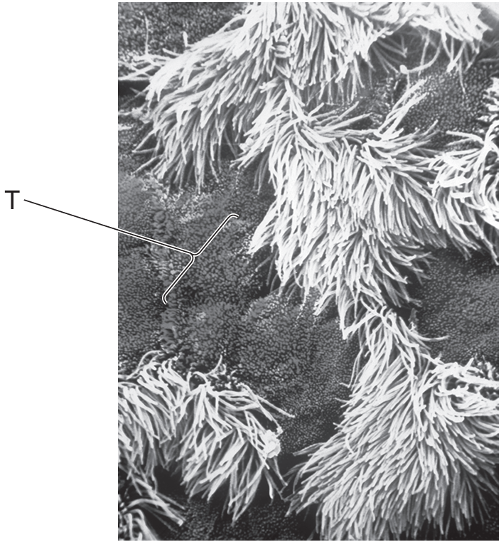
What is the identity of the part of the electron micrograph labelled T?
A) ciliated epithelial cell
B) goblet cell
C) mucus
D) squamous epithelial cell
▶️ Answer/Explanation
Ans: B
Goblet cells are specialized epithelial cells that secrete mucus. They appear as rounded structures among the ciliated cells in the tracheal lining, distinguishable by their shape and secretory function.
Which rows correctly summarise the typical distribution of cartilage and smooth muscle within different parts of the gas exchange system of humans?
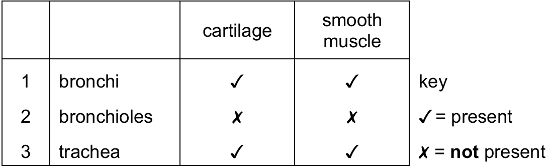
A) 1, 2 and 3
B) 1 and 2 only
C) 1 and 3 only
D) 2 and 3 only
▶️ Answer/Explanation
Ans: C
Bronchi and trachea contain both cartilage and smooth muscle, while bronchioles have smooth muscle but lack cartilage. Therefore, only rows 1 and 3 are correct (bronchi and trachea both have cartilage and smooth muscle).
A molecule of oxygen diffuses from the air in an alveolus to haemoglobin in a red blood cell.
Assuming that the molecule crosses cellular layers by passing through cells, rather than between cells, what is the minimum number of phospholipid layers that the molecule of oxygen must pass through?
A) 5
B) 6
C) 8
D) 10
▶️ Answer/Explanation
Ans: D
Oxygen must pass through: alveolar epithelium (2 layers), capillary endothelium (2 layers), and red blood cell membrane (1 layer). Each cell membrane is a bilayer (2 phospholipid layers), so total is (2+2+1)×2 = 10 layers.
Which row shows the type of pathogen that causes cholera and its mode of transmission?
| pathogen | mode of transmission | |
|---|---|---|
| A | protocist | contaminated food or water |
| B | protocist | airborne droplets |
| C | bacterium | contaminated food or water |
| D | bacterium | airborne droplets |
▶️ Answer/Explanation
Ans: C
Cholera is caused by the bacterium Vibrio cholerae, transmitted through contaminated water or food. Protists cause diseases like malaria, and airborne transmission is characteristic of diseases like tuberculosis.
Which statement about tuberculosis (TB) is not correct?
A) TB can be controlled by vaccination.
B) TB is caused by a virus spread by droplet infection.
C) HIV/AIDS increases the risk of developing TB.
D) TB may be transmitted by eating contaminated meat.
▶️ Answer/Explanation
Ans: B
TB is caused by the bacterium Mycobacterium tuberculosis, not a virus. While it spreads through droplet infection (correctly stated in option B), the causative agent is wrong. The other options are correct facts about TB.
More cases of malaria are being reported in Europe. Other diseases, formerly confined to tropical countries and transmitted in the same way as malaria, have also spread to parts of Europe. Tropical countries have higher mean temperatures and humidity than Europe.
What could explain the increase in the number of cases of these diseases in Europe?
- rising temperatures and humidity in Europe as a result of climate change
- increased travel between Europe and tropical countries
- creation of wetland areas such as marshes to increase biodiversity
A) 1, 2 and 3
B) 1 and 2 only
C) 1 and 3 only
D) 2 and 3 only
▶️ Answer/Explanation
Ans: A
All three factors contribute: (1) Climate change creates more favorable conditions for malaria vectors, (2) Increased travel introduces more cases, and (3) Wetlands provide breeding grounds for mosquitoes. The question asks for possible explanations, not definitive causes.
The diagrams show three different bonds.
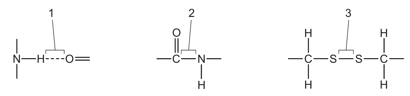
Which bonds are found in an antibody molecule?
A) 1, 2 and 3
B) 1 and 2 only
C) 1 and 3 only
D) 2 and 3 only
▶️ Answer/Explanation
Ans: A
Antibodies contain peptide bonds (1) between amino acids, disulfide bonds (2) between cysteine residues for structure, and hydrogen bonds (3) that help maintain their 3D shape. All three bond types are present in antibody molecules.
Specific monoclonal antibodies can be used to treat some forms of cancer. One example is trastuzumab, which can be used in the treatment of tumours caused by cancer.
Which statements could help to explain why monoclonal antibodies such as trastuzumab are suitable for this role?
- The variable regions of monoclonal antibodies such as trastuzumab can change, allowing the antibodies to bind to cancer cells even if their antigens mutate.
- Monoclonal antibodies such as trastuzumab bind to specific cell surface receptors on cancer cells, which prevents these receptors from receiving signals that are needed for cancer cells to grow and divide.
- The antigen-binding sites of monoclonal antibodies such as trastuzumab are complementary to antigens found on some cancer cells, and have stable tertiary structures that do not change.
A) 1, 2 and 3
B) 1 and 2 only
C) 1 and 3 only
D) 2 and 3 only
▶️ Answer/Explanation
Ans: D
Statement 1 is incorrect because monoclonal antibodies have fixed specificity – they can’t adapt to mutated antigens. Statements 2 and 3 are correct: monoclonal antibodies work by precisely binding to specific cancer cell markers (3) and can block growth signals (2).
The HIV virus causes illness by infecting and destroying T-helper cells. This leads to AIDS and an inability to produce an effective immune response.
Which components of the immune system are produced less effectively when AIDS develops?
- memory cells
- plasma cells
- antibodies
A) 1, 2 and 3
B) 1 and 2 only
C) 1 and 3 only
D) 2 and 3 only
▶️ Answer/Explanation
Ans: A
HIV attacks T-helper cells which coordinate all these immune components. Without proper T-cell help, both cellular (memory cells) and humoral (plasma cells and antibodies) immune responses are impaired. All three components listed are affected in AIDS.