▶️ Answer/Explanation
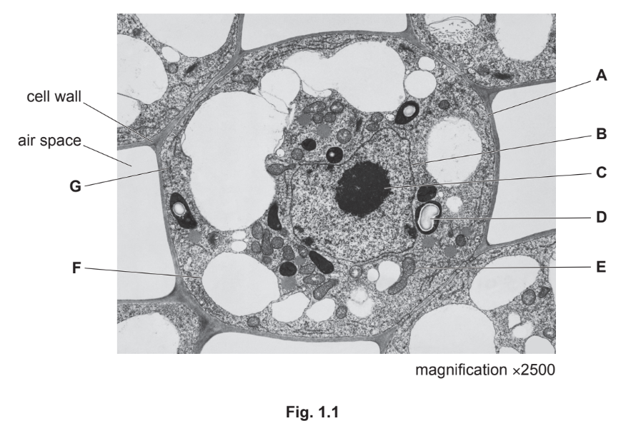
(a)(i)
Evidence: The cell lacks chloroplasts (which are present in mesophyll cells for photosynthesis) and contains many small vacuoles instead of a single large central vacuole. Additionally, the nucleus is centrally located, unlike in mesophyll cells where it is often pushed to the periphery.
(a)(ii)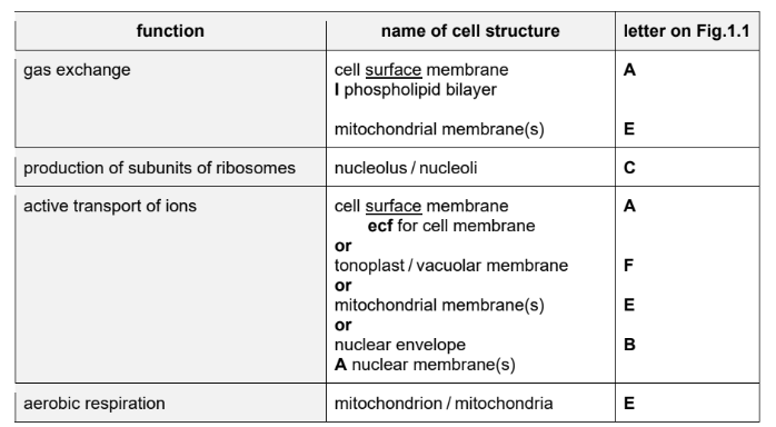
Explanation: The table is completed by identifying the correct cell structures: mitochondrion (ATP production), Golgi body (modification of proteins), rough endoplasmic reticulum (protein synthesis), and nucleus (DNA replication).
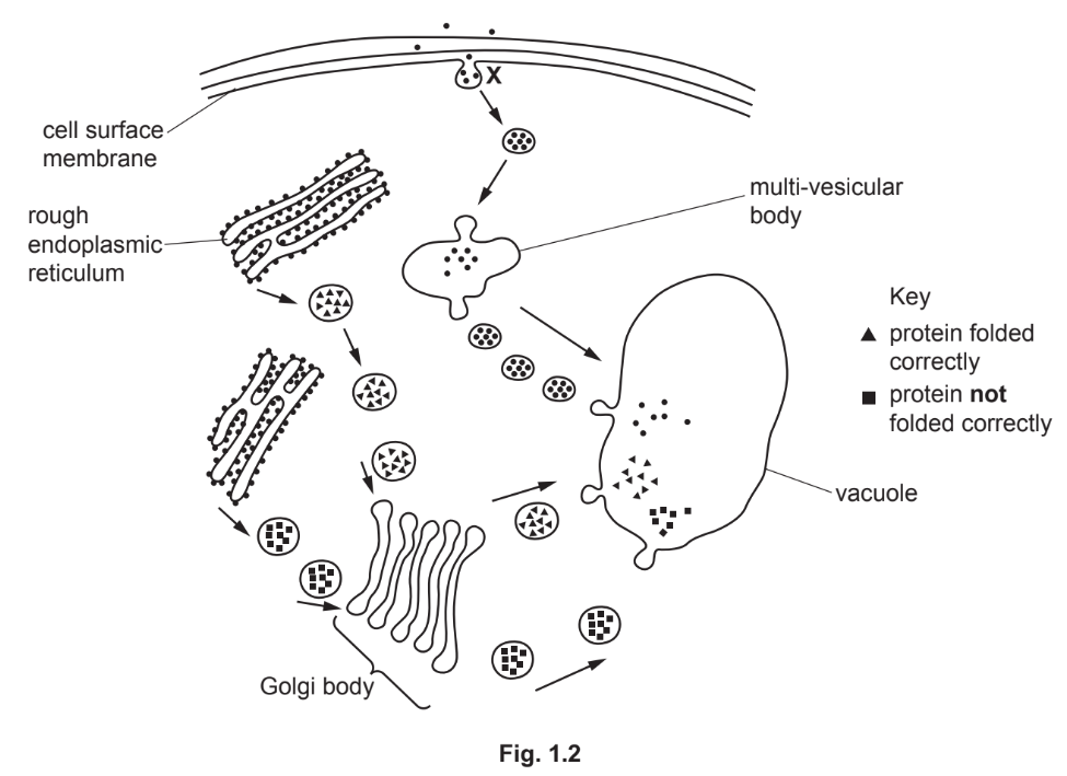
(b)(i) Endocytosis / Pinocytosis.
Explanation: The process at X is endocytosis, where the cell membrane engulfs extracellular material to form vesicles.
(b)(ii)
Explanation: Misfolded proteins are hydrolyzed in the vacuole by proteases, which break peptide bonds to form smaller peptides or amino acids. This process requires water and occurs in an acidic environment.
(c)
Explanation: Lysosomes defend against pathogens by fusing with phagocytic vesicles (forming phagolysosomes) and releasing hydrolytic enzymes (e.g., lysozyme, proteases) to digest the pathogen’s components (e.g., peptidoglycans, proteins). The breakdown products are harmless or reused by the cell.
▶️ Answer/Explanation
(a)
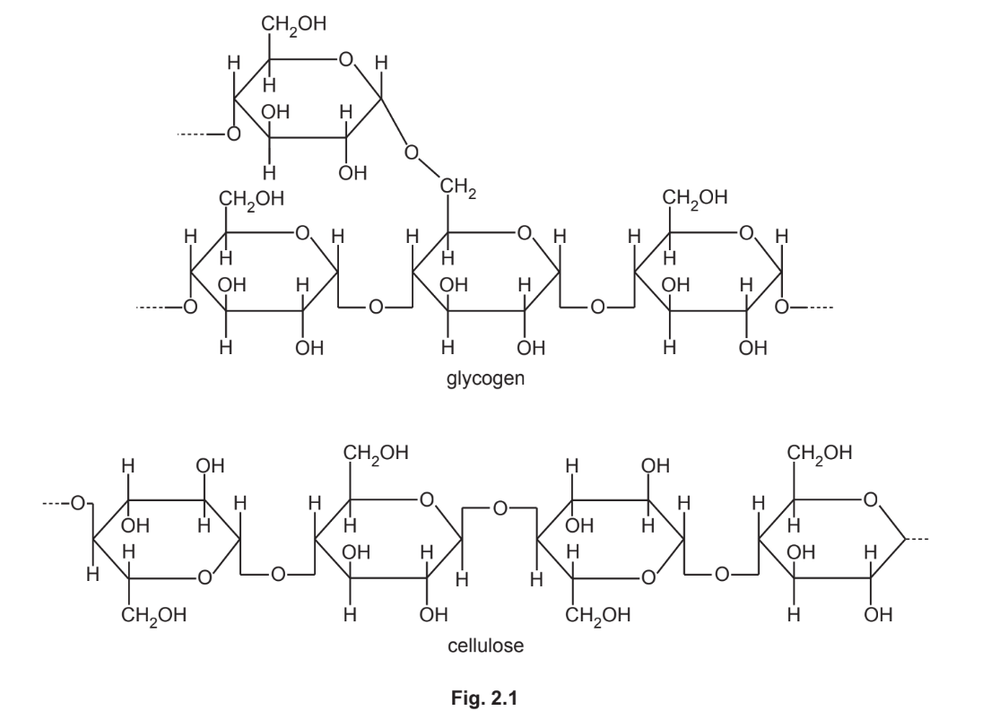
1. Glycogen is branched, whereas cellulose is unbranched.
2. Glycogen consists of α-glucose monomers, whereas cellulose consists of β-glucose monomers.
3. Glycogen has 1,4- and 1,6-glycosidic bonds, whereas cellulose only has 1,4-glycosidic bonds.
Explanation: The differences are visible in Fig. 2.1. Glycogen’s branched structure allows for rapid glucose release, while cellulose’s linear structure provides rigidity. The type of glucose monomers and glycosidic bonds also differ, affecting their biological roles.
(b)
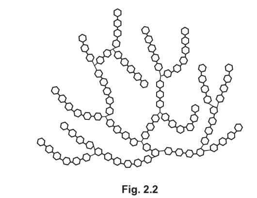
1. Glycogen serves as an energy store, releasing glucose when needed.
2. Its highly branched structure allows for quick glucose addition/removal.
3. It is compact and insoluble, preventing osmotic imbalance in cells.
Explanation: Glycogen’s structure is optimized for storage—branched for accessibility, compact for space efficiency, and insoluble to avoid disrupting cellular water potential.
(c)
1. Cellulose molecules are unbranched and form straight chains.
2. They align in parallel, forming hydrogen bonds for strength.
3. Microfibrils provide structural support to plant cell walls.
Explanation: Cellulose’s linear arrangement and hydrogen bonding create microfibrils, which resist turgor pressure and maintain cell shape, crucial for plant rigidity.
▶️ Answer/Explanation
(a) Membrane proteins are required because glucose is polar/water-soluble and cannot pass through the hydrophobic phospholipid bilayer of the cell membrane. These proteins facilitate diffusion or active transport.
Explanation: The hydrophobic core of the membrane repels polar molecules like glucose, so transporter proteins are necessary for their movement.
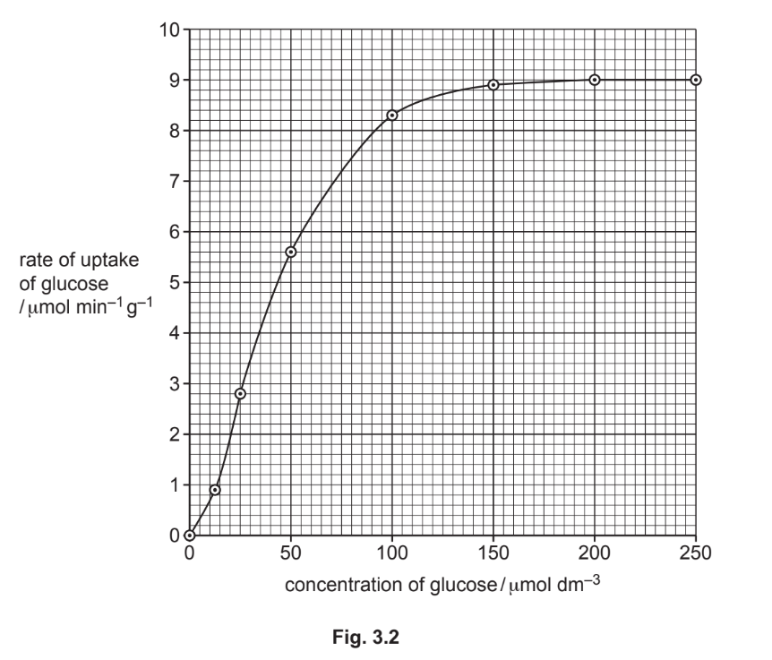
(b)(i) The graph shows the rate of glucose uptake plateaus at higher concentrations (>150 μmol dm–3), indicating that VvHT1 transporters are saturated and working at maximum capacity. This is characteristic of facilitated diffusion.
Explanation: The plateau suggests that the number of transporters (VvHT1) is the limiting factor, consistent with protein-mediated transport.
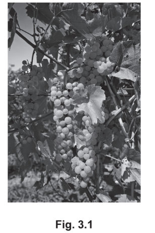
(b)(ii) Increasing hexose transporters could make grapes sweeter (more sugar uptake), improve water absorption via osmosis, enhance energy availability for growth, and increase yield/profit for growers.
Explanation: More transporters allow faster glucose accumulation, directly improving fruit quality and commercial value.
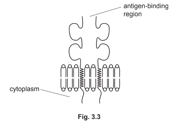
(c)(i) A non-self antigen is a foreign molecule (e.g., protein/polysaccharide) that triggers an immune response by binding to antibodies or activating lymphocytes.
Explanation: These antigens are recognized as “not belonging” to the host organism.
(c)(ii) T-lymphocytes with complementary receptors bind to the presented antigen, undergo clonal expansion, and differentiate into helper T-cells (secreting cytokines) or killer T-cells (destroying infected cells). Memory T-cells are also produced for future immunity.
Explanation: The response is highly specific to the pathogen’s antigens, ensuring targeted immunity.
▶️ Answer/Explanation
(a) A gene is a sequence of nucleotides (bases) that forms part of DNA and codes for a polypeptide (protein/enzyme).
Explanation: A gene is a functional unit of DNA that carries genetic information. It consists of a specific sequence of nucleotides that determines the sequence of amino acids in a protein.
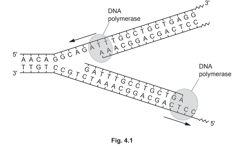
(b)(i) DNA polymerase adds complementary nucleotides to the growing DNA strand, forming phosphodiester bonds and proofreading for errors. DNA ligase joins Okazaki fragments on the lagging strand by forming phosphodiester bonds.
Explanation: DNA polymerase synthesizes new DNA strands in the 5’→3′ direction, ensuring accuracy. DNA ligase seals gaps between Okazaki fragments, completing the lagging strand.
(b)(ii) S phase (synthesis phase).
Explanation: DNA replication occurs during the S phase of interphase, where the cell’s genome is duplicated before mitosis.
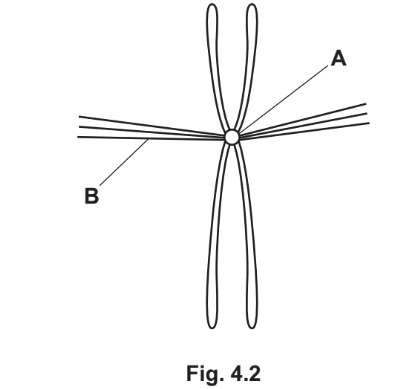
(c)(i) A – Centromere: Attaches chromatids to spindle fibres and holds sister chromatids together.
B – Spindle fibres: Align chromosomes at the equator and separate chromatids during anaphase.
Explanation: The centromere is crucial for chromosome movement, while spindle fibres ensure proper segregation of chromatids to daughter cells.
(c)(ii) In anaphase, sister chromatids separate and are pulled toward opposite poles by spindle fibres, forming V-shaped chromosomes.
Explanation: The centromere splits, allowing single chromatids to move to opposite poles, ensuring each daughter nucleus receives identical genetic material.
▶️ Answer/Explanation
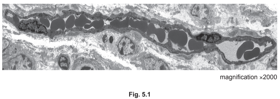
(a) any two from:
no nucleus ;
no organelles / uniform appearance / homogenous cytoplasm ;
(some are) biconcave shape / described ; A cells have different shapes
same, width / size, as the lumen of the capillary ;
A same width as capillary / diameter of cells is 6 – 7 μm
Explanation: Red blood cells (RBCs) are identified by their lack of a nucleus and organelles, uniform appearance, and biconcave shape. Their size matches the capillary lumen, confirming their identity.
(b) any three from:
1 (wall is) thin / one cell thick / 1–2 μm in thickness ;
2 ref. to, endothelial cells / endothelium ; I squamous / epithelium
3 short distance for diffusion ;
4 endothelial pores / fenestrations
5 for passage of, (named) small molecules / AW, from / to, plasma / blood / tissue fluid ;
6 AVP ; ref. to pinocytosis across endothelial cells
Explanation: The capillary wall is thin (one cell thick) to allow efficient diffusion. Endothelial cells with pores enable small molecule exchange, while the short diffusion distance ensures rapid nutrient and gas exchange.
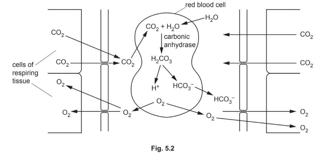
(c)(i) any four from:
1 more carbon dioxide diffuses into red blood cells ;
2 more carbonic acid is formed by carbonic anhydrase ;
3 formation of more hydrogen ions ; A H+
4 haemoglobin has a high affinity for hydrogen ions ; A H+
5 haemoglobin binds more hydrogen ions to form, haemoglobinic acid (HHb) ; AH+
6 (formation of HHb) decreases affinity of haemoglobin for oxygen ;
7 haemoglobin releases more oxygen ;
8 carbon dioxide binds to -NH2 / N terminal, of, globin / polypeptides / α chains and β chains ;
9 forms carbaminohaemoglobin ; R carboxyhaemoglobin
10 allosteric effect / change in tertiary structure / AW, in (oxy)haemoglobin (causes, release / AW, more oxygen) ;
Explanation: Increased CO2 lowers blood pH, forming more H+ ions. Haemoglobin binds H+, reducing its oxygen affinity (Bohr effect), releasing more oxygen to tissues.
(c)(ii) any two from:
1 hydrogencarbonate ions / HCO3 – , pass out of red blood cells (into the plasma to increase concentration in deoxygenated blood) ;
2 chloride ions pass into red blood cells ;
3 to replace the, negatively-charged ions / anions / HCO3-;
4 chloride shift ;
5 AVP ; chloride ions pass through, channel proteins / anion exchangers / transport proteins / by facilitated diffusion
Explanation: Chloride ions (Cl–) move into RBCs to balance the loss of HCO3– (chloride shift). This maintains electroneutrality, reducing Cl– concentration in deoxygenated blood plasma.
▶️ Answer/Explanation
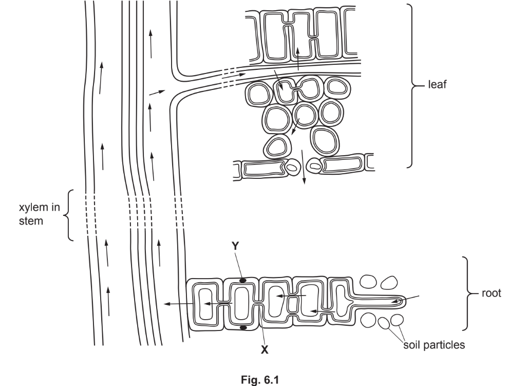
(a) plasmodesma
Explanation: Structure X is a plasmodesma, which connects plant cells and allows cytoplasmic (symplast) movement of water and nutrients.
(b) Casparian strip
Explanation: Structure Y is the Casparian strip, a waterproof layer made of suberin in the endodermis that blocks the apoplast pathway, forcing water into the symplast.
(c)
Water moves from the soil solution to the cytoplasm of root hair cells by osmosis.
Water moves from the xylem in the root to the leaf by transpiration pull (or cohesion-tension).
Water moves from mesophyll cell walls to intercellular air spaces by evaporation.
Water vapour moves from intercellular air spaces to the atmosphere outside the leaf by diffusion.
Explanation: Osmosis drives water uptake in roots, transpiration pull moves water upward, evaporation occurs in leaves, and diffusion releases water vapour into the air.