▶️ Answer/Explanation
(a)
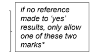
Explanation: The completed boxes show:
Box 3: Atrial systole (atria contract to push blood into ventricles).
Box 5: Ventricular systole (ventricles contract to pump blood into pulmonary artery).
Box 7: Diastole (both chambers relax, allowing blood to fill the heart).
(b) Coordination ensures:
1. Atria contract (systole) before ventricles to fully fill them.
2. Prevents simultaneous contraction, which would reduce pumping efficiency.
3. Allows complete emptying of atria and proper ventricular filling.
(c) Valve operation depends on pressure gradients:
– Tricuspid valve: Opens when atrial pressure > ventricular pressure (during atrial systole). Closes when ventricular pressure rises (ventricular systole).
– Pulmonary valve: Opens when ventricular pressure > pulmonary artery pressure (ventricular systole). Closes when ventricular pressure drops (diastole).
This ensures one-way blood flow and prevents backflow.
▶️ Answer/Explanation
2(a)
Explanation: The photomicrograph distinguishes blood vessels P (artery) and Q (vein) based on structural differences. Arteries have thicker muscular walls and a smaller lumen, while veins have valves and a larger lumen to facilitate blood flow under lower pressure.
2(b)
1. Water molecules form hydrogen bonds, creating cohesive forces.
2. Cohesion between water molecules maintains continuous water columns in xylem.
3. Adhesion of water to soil particles/clumps ensures water retention near roots.
Explanation: Hydrogen bonding enables cohesion (water-water attraction) and adhesion (water-soil attraction), stabilizing water availability for root absorption and transport.
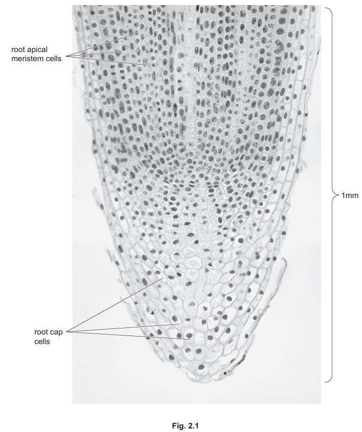
2(c)
Extracellular enzymes.
Explanation: Root cells secrete extracellular enzymes to break down organic matter in soil, releasing nutrients like minerals for absorption.
2(d)(i)
Polysaccharides are polymers of monosaccharides linked by glycosidic bonds.
Explanation: Examples include starch and cellulose, which are long chains of glucose monomers with 1,4- or 1,6-glycosidic bonds, serving as energy storage or structural components.
2(d)(ii)
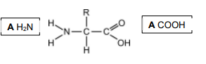
Explanation: The general structure of an amino acid includes an amino group (NH2), carboxyl group (COOH), hydrogen atom, and a variable R-group, which determines the amino acid’s properties.
2(d)(iii)
1. Mineral ions dissolve in water, lowering cellular water potential.
2. A steeper water potential gradient drives osmosis into root cells.
3. Water uptake is coupled with ion absorption for nutrient balance.
Explanation: Solutes like minerals reduce water potential in root cells, creating an osmotic gradient that draws water from the soil, facilitating nutrient uptake.
▶️ Answer/Explanation
(a) Non-self antigens are foreign molecules (e.g., proteins, glycoproteins, or polysaccharides) that:
- Stimulate an immune response by activating immune cells (e.g., lymphocytes, phagocytes).
- Bind to specific antibodies or lymphocyte receptors due to complementary shapes.
- Are found on pathogens, infected cells, or toxins.
Explanation: These features allow the immune system to distinguish “foreign” invaders from the body’s own cells.
(b) Completed Table 3.1:
Explanation: – B-lymphocytes produce antibodies (✓ in column 1). – Macrophages engulf pathogens (✓ in column 2) but do not produce antibodies (✗ in column 1). – T-killer cells destroy infected cells (✓ in column 3). – Neutrophils are phagocytic (✓ in column 2) but not antigen-presenting (✗ in column 4).
(c) A. Cytokinesis (cytoplasmic division). B. Interphase (longest phase of the cell cycle). C. DNA replication (semi-conservative synthesis of sister chromatids).
Explanation: – Cytokinesis completes cell division after mitosis. – Interphase includes G1, S (DNA synthesis), and G2 phases. – The S phase ensures genetic material is duplicated for daughter cells.
▶️ Answer/Explanation
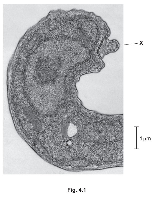
(a)(i)
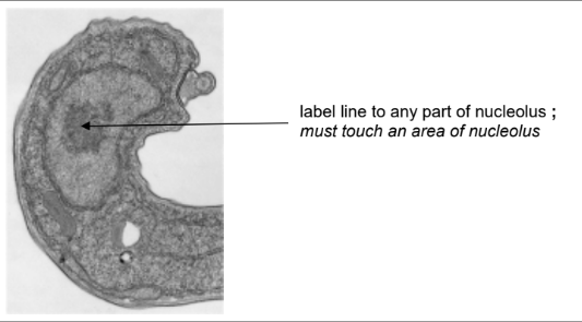
Explanation: The arrow points to the nucleolus, where rRNA and proteins assemble to form ribosomal subunits.
(a)(ii) Structure X is a flagellum, evidenced by its 9+2 microtubule arrangement and cylindrical shape, enabling motility.
Explanation: The flagellum’s microtubule structure and external position allow whip-like movements, propelling the cell.
(a)(iii) T. brucei is a eukaryote due to its nucleus, mitochondria, and membrane-bound organelles, absent in prokaryotes.
Explanation: The presence of a nuclear envelope, nucleolus, and complex organelles confirms its eukaryotic nature.
(b)(i) Pathogen.
Explanation: A pathogen is any organism that causes disease, including parasites like T. brucei.
(b)(ii) Plasmodium falciparum (or P. vivax, P. ovale, P. malariae).
Explanation: These species are known to cause malaria in humans, with P. falciparum being the most lethal.
(c)
Similarities: Both are vector-borne (tsetse flies for sleeping sickness, mosquitoes for malaria) and transmitted via blood meals.
Differences: Only female Anopheles mosquitoes transmit malaria, while both male and female tsetse flies transmit sleeping sickness.
Explanation: The key difference lies in the vector species and their feeding behaviors, though both rely on insect vectors.
▶️ Answer/Explanation
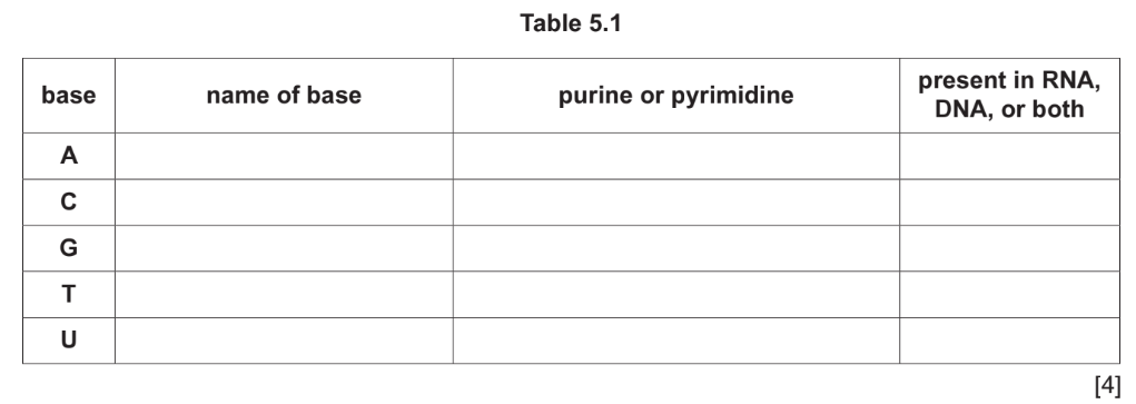
(a)
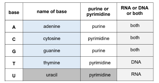
Explanation: The table is completed as follows: Adenine (A) and Guanine (G) are purines present in both RNA and DNA. Cytosine (C) is a pyrimidine present in both, while Thymine (T) is a pyrimidine found only in DNA. Uracil (U) is a pyrimidine exclusive to RNA.
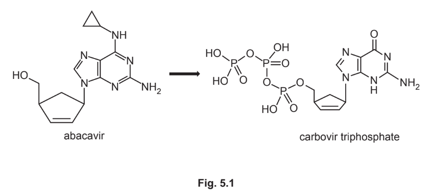
(b)(i) purine because it is a, double ring structure / has two rings ;
Explanation: Carbovir triphosphate replaces a purine nucleotide (adenine/guanine) because it has a double-ring structure, matching the purine base structure in DNA/RNA.
(b)(ii) any two from:
- carbovir triphosphate is (similar to / same as), an activated nucleotide / a nucleotide / not a nucleoside ;
- carbovir triphosphate, has (three) phosphates / is activated / is phosphorylated ;
- detail of, (DNA) polymerase action / (activated DNA) nucleotides ;
- ref. to triangle / triangular cycloalkane, no longer present / not on DNA nucleotides ;
Explanation: Conversion to carbovir triphosphate makes it resemble activated nucleotides (with three phosphates), enabling DNA polymerase to incorporate it into the growing chain. The absence of the triangular cycloalkane group further enhances compatibility.
(b)(iii) any four from:
- prevents polymerase from adding DNA nucleotide to growing chain / AW ;
- similar shape to, substrate / (activated / phosphorylated) nucleotide ;
- acts as an inhibitor ;
- fits into / binds to, active site of, enzyme / DNA polymerase ;
- (DNA polymerase) may not form phosphodiester bonds ;
- ref. to proofreading mechanism ;
Explanation: Carbovir triphosphate acts as a competitive inhibitor, binding to DNA polymerase’s active site and blocking further nucleotide addition. Its structural similarity allows incorporation but disrupts phosphodiester bond formation, halting viral DNA synthesis.
▶️ Answer/Explanation
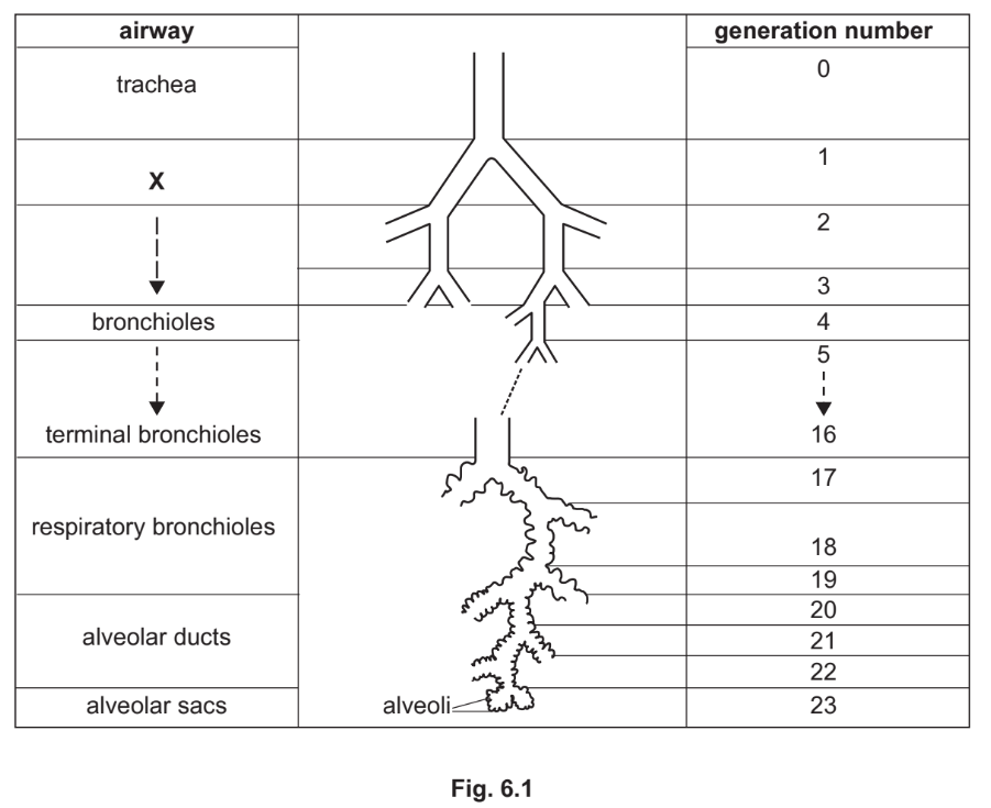
(a) Bronchi (left and right bronchus)
Explanation: Generation 0 is the trachea, which bifurcates into the primary bronchi (generation 1). These are labelled X in Fig. 6.1.
(b) Reasons for no gas exchange in generations 0–16:
- Thick walls: Multiple tissue layers (e.g., smooth muscle, cartilage) increase diffusion distance.
- Blood supply: Systemic circulation (oxygenated blood) lacks a concentration gradient for gas exchange.
- Structural role: These airways conduct air rather than facilitate diffusion (no alveoli).
Key point: Gas exchange occurs only in alveoli (generation 17+), where walls are thin and pulmonary blood flow creates gradients.
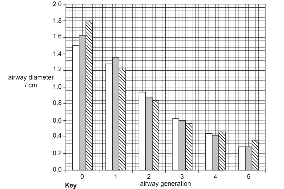
(c)(i) Inverse relationship: As airway generation increases, diameter decreases.
Explanation: The Weibel model shows a logarithmic decrease in diameter with each branching generation (e.g., trachea ≈18 mm, generation 5 ≈2 mm).
(c)(ii) HP gas MRI as an alternative:
Advantages:
- No radiation: Safe for repeated use, unlike HRCT.
- Comparable data: Both projection and multi-slice MRI follow the Weibel trend (e.g., decreasing diameter with generation).
Limitations:
- Discrepancies: MRI measurements slightly deviate from Weibel’s reference (e.g., projection MRI underestimates generation 3).
Conclusion: HP gas MRI is a viable alternative for tracking disease progression without radiation risks.