Topic: 1.1 (The microscope in cell studies)
A prokaryotic cell which is 1 μm in diameter is magnified 50 000 times in an electron micrograph.
What is the diameter of the cell in the electron micrograph?
▶️ Answer/Explanation
Ans: C
The original diameter of the prokaryotic cell is \(1 \, \mu m = 1 \times 10^{-3} \, mm\). When magnified 50,000 times, the diameter becomes:
\[ 1 \times 10^{-3} \, mm \times 50,000 = 50 \, mm = 5 \times 10^{1} \, mm \]
Thus, the correct answer is C. \(5 \times 10^{1} \, mm\).
Topic: 1.2 (Cells as the basic units of living organisms)
The diagram shows a plant cell with some labelled structures.
Which labelled structures are bound by a double membrane?
▶️ Answer/Explanation
Ans: A
In a plant cell, the nucleus (P) and mitochondria (Q) are bound by a double membrane. The nucleus has a nuclear envelope (double membrane), and mitochondria have an outer and inner membrane. Other structures like chloroplasts (if present) are also double-membraned, but based on the given options, P and Q are the correct pair.
Topic: 1.2 (Cells as the basic units of living organisms)
Which size of ribosome is found in mitochondria and typical prokaryotic cells?
▶️ Answer/Explanation
Ans: C
Mitochondria and prokaryotic cells (like bacteria) contain 70S ribosomes, which are smaller than the 80S ribosomes found in eukaryotic cytoplasm. The 70S ribosome consists of a 50S and a 30S subunit, distinguishing it from the 60S and 40S subunits of 80S ribosomes.
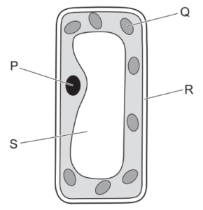
Topic: 1.2 (Cells as the basic units of living organisms)
Which row about typical prokaryotic cells and typical animal cells is correct?
▶️ Answer/Explanation
Ans: C
Prokaryotic cells lack membrane-bound organelles like mitochondria and a nucleus, while animal cells have these structures. The genetic material in prokaryotes is circular and free-floating, whereas animal cells have linear DNA enclosed in a nucleus. Thus, the correct row is C, as it accurately contrasts these features.
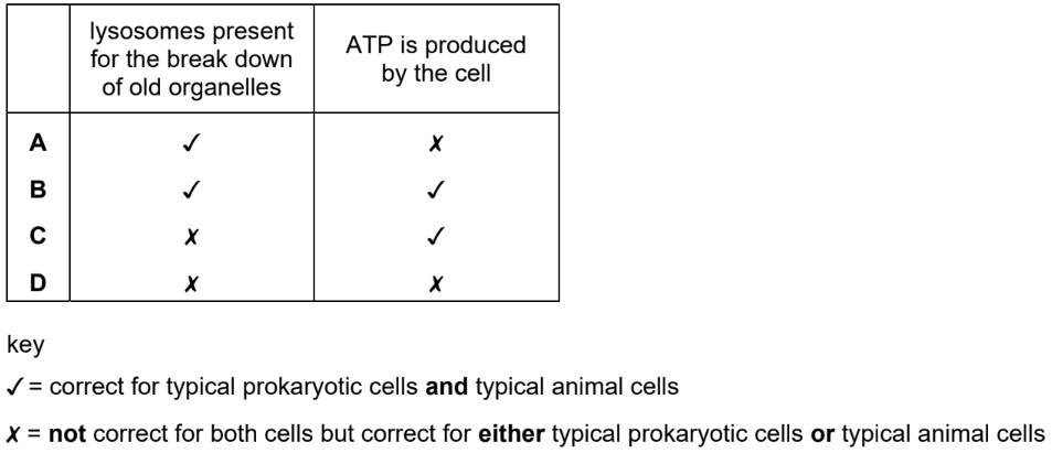
Topic: 1.2 (Cells as the basic units of living organisms)
Which row is correct for the structures present in typical plant cells and typical animal cells?
▶️ Answer/Explanation
Ans: C
Plant cells have a cell wall and chloroplasts, which animal cells lack. Animal cells may have small temporary vacuoles, but plant cells have a large permanent vacuole. The image in the question highlights these differences, confirming that row C correctly identifies the structures present in each cell type.
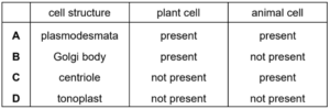
Topic: 2.2 (Carbohydrates and lipids)
Which row is correct for cellulose?
▶️ Answer/Explanation
Ans: D
Cellulose is a polysaccharide composed of β-glucose monomers, forming long straight chains with hydrogen bonds between them. This structure makes it insoluble in water but provides high tensile strength, which is crucial for plant cell walls. The correct row must reflect these properties, which corresponds to option D in the given table.
Topic: 2.3 (Proteins)
Which statements about peptide bond formation are correct?
1 The bond formation occurs between a carbon of one amino acid and a nitrogen of the next amino acid after the amino acids detach from tRNA.
2 The bond formation occurs at the ribosome while the amino acids are still attached to tRNA, and is a hydrolysis reaction.
3 The bond formation is important for growth of an organism and when the bond forms, a water molecule is removed.
▶️ Answer/Explanation
Ans: D
Statement 1 is incorrect because peptide bond formation occurs while amino acids are still attached to tRNA, not after detachment. Statement 2 is incorrect because peptide bond formation is a condensation reaction (releasing water), not hydrolysis. Statement 3 is correct—peptide bonds are essential for growth (protein synthesis), and their formation involves the removal of a water molecule. Thus, only Statement 3 is correct, making D the answer.
Topic: 2.2 (Carbohydrates and lipids)
The diagram shows naturally occurring D-glucose and a form of glucose that can be synthesised in the laboratory, known as L-glucose.
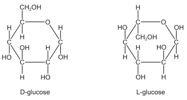
The enzyme glucose oxidase catalyses the oxidation of D-glucose. The enzyme cannot catalyse the oxidation of L-glucose.
Which statement about L-glucose explains this?
▶️ Answer/Explanation
Ans: A
Enzymes are highly specific to their substrates due to their active site structure. L-glucose is the enantiomer (mirror image) of D-glucose, meaning it has a different spatial arrangement. Since glucose oxidase is adapted to bind D-glucose, the altered shape of L-glucose prevents it from fitting into the enzyme’s active site, explaining why it cannot be oxidized.
Topic: 2.1 (Testing for biological molecules)
Tests for biological molecules were carried out on three solutions. Each solution contained only one type of biological molecule. The observations were as follows.
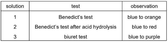
Which solutions would contain either sucrose or amylase?
▶️ Answer/Explanation
Ans: C
Sucrose is a non-reducing sugar, so it gives a negative result with Benedict’s test (Solution 2). Amylase is a protein, giving a positive result with Biuret test (Solution 3). Solution 1 contains a reducing sugar (positive Benedict’s test), which is neither sucrose nor amylase. Thus, only Solutions 2 and 3 could contain sucrose or amylase, making option C correct.
Topic: 3.2 (Factors that affect enzyme action)
Which row describes the expected effect on Vmax and Km when a competitive reversible inhibitor is added to an enzyme-catalysed reaction?
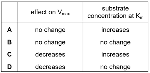
▶️ Answer/Explanation
Ans: A
A competitive inhibitor competes with the substrate for the active site of the enzyme, increasing the apparent \( K_m \) (substrate concentration needed for half-maximal activity) because more substrate is required to outcompete the inhibitor. However, \( V_{max} \) remains unchanged since, at high substrate concentrations, the inhibitor can be fully outcompeted, allowing the enzyme to reach its maximum reaction rate.
Topic: 3.2 (Factors that affect enzyme action)
The graph shows the effect of substrate concentration on the rates of reaction of three enzymes, X, Y, and Z.
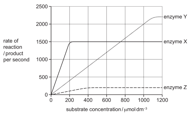
What is the correct order of affinity of these enzymes for their substrates, starting with the enzyme with the highest affinity?
▶️ Answer/Explanation
Ans: B
The enzyme with the highest affinity for its substrate reaches maximum reaction rate (Vmax) at the lowest substrate concentration. From the graph, enzyme X saturates first, followed by Z, and then Y. Thus, the order of affinity is X → Z → Y, making option B correct.
Topic: 2.3 (Proteins)
Which row correctly identifies the weak and strong bonds in the tertiary and quaternary structure of a typical protein?
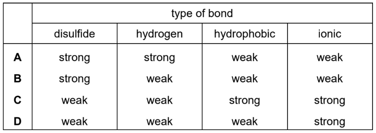
▶️ Answer/Explanation
Ans: B
In protein structures:
- Tertiary structure is stabilized by weak bonds (e.g., hydrogen bonds, ionic bonds, hydrophobic interactions, and van der Waals forces) and strong bonds (disulfide bridges).
- Quaternary structure involves weak interactions (e.g., hydrogen bonds, ionic bonds) between polypeptide subunits.
Option B correctly identifies these bonds, distinguishing between weak and strong interactions in both tertiary and quaternary structures.
Topic: 2.3 (Proteins)
Which row correctly describes haemoglobin?
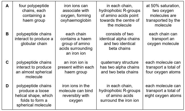
▶️ Answer/Explanation
Ans: A
Haemoglobin is a globular protein with a quaternary structure (made of four polypeptide chains). It contains a prosthetic haem group with an iron ion (Fe²⁺), which binds oxygen. The correct row must reflect these properties, and Option A matches this description accurately.
Topic: 4.2 (Movement into and out of cells)
Which process always takes place without the involvement of energy from ATP?
▶️ Answer/Explanation
Ans: D
Facilitated diffusion is a passive process where molecules move across membranes through protein channels along their concentration gradient, requiring no ATP. In contrast, active transport (A), endocytosis (B), and exocytosis (C) all require energy to move substances against gradients or transport large particles.
Topic: 4.2 (Movement into and out of cells)
The diagram shows the entry of molecule X into a cell.
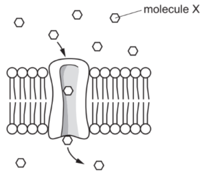
Which row shows a property of molecule X and the effect of the concentration of ATP in the cytoplasm on the rate of entry of molecule X?
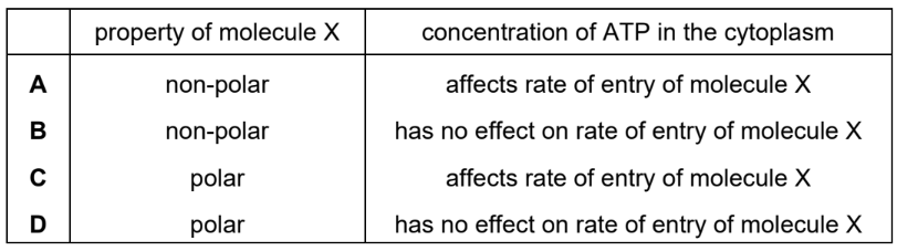
▶️ Answer/Explanation
Ans: D
The diagram suggests that molecule X enters the cell via active transport, as it moves against the concentration gradient with the help of a carrier protein. Active transport requires ATP, so the rate of entry increases with higher ATP concentration. Additionally, molecule X is large and polar, as small/nonpolar molecules diffuse freely, and ions typically use channels. Thus, the correct row is D.
Topic: 4.2 (Movement into and out of cells)
The electron micrograph shows some human blood cells.
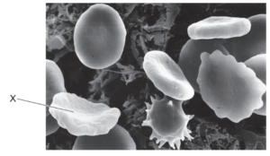
Which row correctly shows the net movement of water by osmosis and the water potential of the cytoplasm of cell X compared with the solution surrounding the cells?
▶️ Answer/Explanation
Ans: C
In the electron micrograph, cell X appears shriveled, indicating water has moved out of the cell by osmosis. This occurs when the surrounding solution has a lower water potential (higher solute concentration) than the cytoplasm. Thus, the net movement of water is out of the cell, and the water potential of the cytoplasm is higher than the external solution, matching row C.
Topic: 4.2 (Movement into and out of cells)
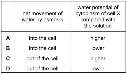
A red indicator solution was mixed with agar and the resulting solid was cut into small cylindrical blocks. The blocks were placed in an acid, which turns the indicator yellow and all other variables were kept constant. The dimensions of the blocks are shown.
block 1 height 3 mm diameter 6 mm
block 2 height 6 mm diameter 12 mm
block 3 height 8 mm diameter 16 mm
The formula for calculating the surface area of a cylinder is \(2\pi rh + 2\pi r^2\). The formula for calculating the volume of a cylinder is \(\pi r^2 h\).
Which row shows the correct surface area (SA) to volume (V) ratio for each block and the time taken for the block to turn yellow?
▶️ Answer/Explanation
Ans: C
The SA:V ratio decreases as the block size increases because volume increases faster than surface area. For diffusion (acid penetration), a higher SA:V ratio means faster color change. Calculations confirm:
- Block 1 (smallest): Highest SA:V ratio → fastest diffusion (shortest time).
- Block 3 (largest): Lowest SA:V ratio → slowest diffusion (longest time).
Only row C matches this trend, with SA:V ratios ordered as Block 1 > Block 2 > Block 3 and corresponding times increasing proportionally.
Topic: 5.1 (Replication and division of nuclei and cells)
Which metabolic processes will be very active in a cell that has just completed cytokinesis?
1. ATP formation
2. DNA replication
3. protein synthesis
▶️ Answer/Explanation
Ans: B
After cytokinesis, the cell enters the G1 phase of the cell cycle, where it grows and prepares for DNA replication. ATP formation (1) is highly active to fuel cellular activities, and protein synthesis (3) increases to support growth. DNA replication (2) occurs in the S phase, not immediately after cytokinesis. Thus, only 1 and 3 are correct, making B the answer.
Topic: 5.2 (Chromosome behaviour in mitosis)
The diagram shows a typical mitotic cell cycle and the point in the cell cycle that has been reached by each of four cells, V, W, X and Y.

Which row correctly identifies the cells that match the two descriptions?

▶️ Answer/Explanation
Ans: B
The first description refers to a cell with unreplicated chromosomes, which is found in the G1 phase (Cell V). The second description refers to a cell where chromosomes are aligned at the equator, which occurs during metaphase (Cell X). Therefore, the correct pairing is V and X, corresponding to option B.
Topic: 5.2 (Chromosome behaviour in mitosis)
The graph shows the mean length of the spindle fibres during mitosis.
Which region of the graph shows when all the centromeres have detached from the spindle fibres?

▶️ Answer/Explanation
Ans: D
During mitosis, spindle fibres shorten during anaphase to separate sister chromatids. When all centromeres detach, spindle fibres disappear, leading to a sharp decline in their mean length. Region D represents this phase, where spindle fibre length drops to zero, indicating complete detachment of chromosomes.
Topic: 6.2 (Protein synthesis)
The mRNA codons ACU, ACC, ACA, and ACG all code for the same amino acid, threonine.
Which anticodons could specify an amino acid other than threonine?
1. UCA
2. ACC
3. UGU
4. UGC
▶️ Answer/Explanation
Ans: B
1. UCA (anticodon) pairs with AGU/AGC, which code for serine, not threonine.
2. ACC (anticodon) pairs with UGG, which codes for tryptophan, not threonine.
3. UGU/UGC (anticodons) pair with ACA/ACG, which code for threonine.
Thus, only 1 (UCA) and 2 (ACC) specify non-threonine amino acids, making option B correct.
Topic: 6.1 (Structure of nucleic acids and replication of DNA)
Which bond formation does DNA polymerase catalyse?
▶️ Answer/Explanation
Ans: D
DNA polymerase catalyzes the formation of phosphodiester bonds between nucleotides during DNA replication. This enzyme adds new nucleotides to the growing DNA strand, linking the 3′-OH group of one nucleotide to the 5′-phosphate group of the next, creating the sugar-phosphate backbone. Hydrogen bonds (options A and B) form between complementary bases (e.g., A-T, C-G) and are not catalyzed by DNA polymerase.
Topic: 6.2 (Protein synthesis)
In eukaryotes, the RNA molecules formed during transcription are modified by the removal of non-coding sequences. This is followed by the joining together of coding sequences to form mRNA. What are the coding sequences also called?
▶️ Answer/Explanation
Ans: B
During eukaryotic mRNA processing:
- Introns (non-coding sequences) are removed via splicing.
- Exons (coding sequences) are joined together to form mature mRNA.
Thus, the correct term for coding sequences is exons, making option B the right answer.
Topic: 7.2 (Transport mechanisms)
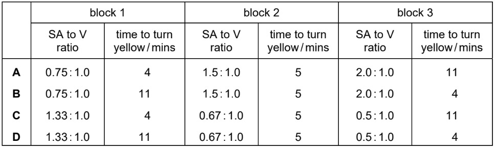
Which row correctly identifies sinks for sucrose transported by mass flow in plants?
▶️ Answer/Explanation
Ans: A
In plants, sucrose is transported via mass flow from sources</strong (e.g., leaves) to sinks (e.g., roots, fruits, growing tissues). Sinks are regions where sucrose is used for growth (e.g., meristems) or stored (e.g., tubers). Option A correctly lists roots and fruits as sinks, as they actively consume or store sucrose.
Topic: 7.1 (Structures of transport tissues)
The diagram shows a transverse section through a transport tissue in a plant.
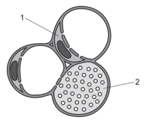
Which row correctly identifies cell 1 and cell 2?
▶️ Answer/Explanation
Ans: A
Cell 1 is a xylem vessel (identified by its large, hollow lumen and thick lignified walls for water transport). Cell 2 is a phloem sieve tube element (smaller, with companion cells nearby, responsible for sugar translocation). The image matches vascular bundle anatomy, confirming option A as correct.
Topic: 7.2 (Transport mechanisms)
Which statement correctly describes the movement of solutes in the symplast pathway?
(A) Cell surface membranes regulate the selective absorption of solutes into the symplast pathway.
(B) Plasmodesmata control the movement of solutes from the symplast pathway to the apoplast pathway.
(C) The symplast pathway transports dissolved mineral ions from the soil that cannot be transported by the apoplast pathway.
(D) The movement of solutes through plasmodesmata in the symplast pathway is prevented in the endodermis by suberin.
▶️ Answer/Explanation
Ans: A
The symplast pathway involves movement of solutes through the cytoplasm of plant cells connected by plasmodesmata. Option A is correct because solutes must first enter the cytoplasm via selective absorption through cell surface membranes. Options B, C, and D are incorrect: plasmodesmata do not regulate solute movement to the apoplast (B), the apoplast can transport mineral ions (C), and suberin blocks the apoplast (not symplast) in the endodermis (D).
Topic: 7.2 (Transport mechanisms)
Which statement helps to explain why water molecules are forced to move through xylem vessel elements as a consequence of transpiration?
▶️ Answer/Explanation
Ans: B
The movement of water through xylem vessels is primarily driven by the cohesion-tension theory. Water molecules form hydrogen bonds with each other (cohesion), creating a continuous column. As water evaporates from leaves during transpiration, tension pulls the cohesive water column upward. While adhesion (A) plays a role in water movement along walls, it is cohesion (B) that explains the bulk movement of water in xylem. Options C and D are incorrect because ionic bonds are not involved, and latent heat of vaporization does not directly drive water transport.
Topic: 8.3 (The heart)
The diagram shows the internal structure of the mammalian heart. Which letter identifies the location of the atrioventricular node?
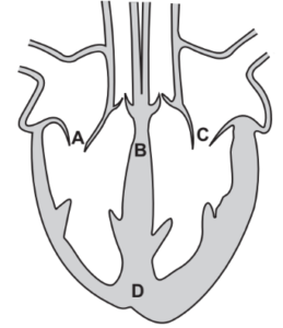
▶️ Answer/Explanation
Ans: B
The atrioventricular (AV) node is located in the lower interatrial septum, near the tricuspid valve. It delays electrical impulses from the sinoatrial (SA) node, ensuring atria contract before ventricles. In the diagram, B marks this critical region, while other labels (A, C, D) typically indicate structures like the SA node, Purkinje fibers, or ventricles.
Topic: 8.3 (The heart)
The diagram shows pressure changes in the left side of the heart during the cardiac cycle.
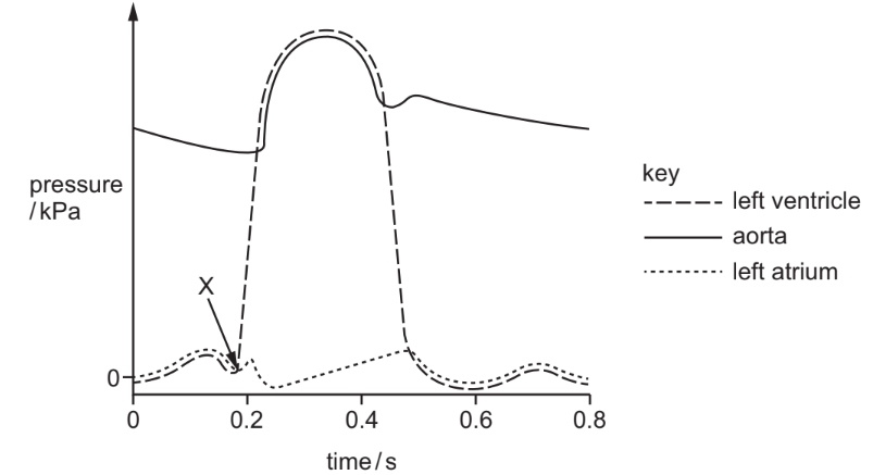
What happens in the heart at X?
(A) The atrioventricular valves close.
(B) The atrioventricular valves open.
(C) The semilunar valves close.
(D) The semilunar valves open.
▶️ Answer/Explanation
Ans: A
At point X, the left ventricular pressure rises sharply, exceeding atrial pressure. This causes the atrioventricular (AV) valves (mitral/bicuspid) to close, producing the first heart sound (“lub”). The semilunar valves (aortic/pulmonary) open later when ventricular pressure exceeds arterial pressure. Thus, the correct answer is A.
Topic: 8.1 (The circulatory system)
Which components of blood are present in tissue fluid?
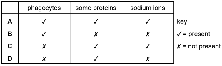
▶️ Answer/Explanation
Ans: A
Tissue fluid is formed when blood plasma is filtered out of capillaries, leaving behind large components like red blood cells and platelets. The remaining fluid contains water, small solutes (glucose, amino acids, ions), and white blood cells, which can migrate through capillary walls. Thus, the correct option is A (white blood cells and plasma proteins), as these are the only blood components present in tissue fluid.
Topic: 9.1 (The gas exchange system)
In the lungs, movement of dissolved carbon dioxide out of the capillaries occurs in one of two ways:
● by diffusion through the endothelial cells of the capillaries
● by leakage through pores in the endothelial cells of the capillaries.
What is the minimum number of times that a carbon dioxide molecule that has been transported to the lungs in a red blood cell must cross a cell surface membrane to reach an air space in an alveolus?
▶️ Answer/Explanation
Ans: B
A CO2 molecule in a red blood cell must cross: (1) the red blood cell membrane → (2) the capillary endothelial cell membrane (if diffusing through cells) → (3) the alveolar epithelium membrane to enter the alveolus. If leaking through pores, it skips the endothelial membrane but still crosses 2 membranes (RBC + alveolus). However, the question asks for the minimum crossings, which is 3 (assuming diffusion through cells).
Topic: 9.1 (The gas exchange system)
What maintains the steep concentration gradients needed for successful gas exchange in the lungs?
1. Air flow in the alveoli is in the opposite direction to blood flow in the capillaries.
2. Blood arrives in the lungs with a lower oxygen concentration and a higher carbon dioxide concentration than the air in the alveoli.
3. Blood is constantly flowing through and out of the lungs, bringing a fresh supply of red blood cells.
▶️ Answer/Explanation
Ans: D
1. False: Air and blood flow direction (countercurrent exchange) is not a mechanism in mammalian lungs (it occurs in fish gills).
2. True: Blood entering the lungs has low \( O_2 \) and high \( CO_2 \), creating a gradient for diffusion.
3. True: Continuous blood flow replaces oxygen-depleted blood, maintaining the gradient.
Thus, only 2 and 3 are correct, making option D the right choice.
Topic: 9.1 (The gas exchange system)
Where is cartilage tissue always found in the human gas exchange system?
▶️ Answer/Explanation
Ans: D
Cartilage is a rigid connective tissue that provides structural support to prevent airway collapse. In the gas exchange system, it is present in:
- Trachea: C-shaped rings of cartilage
- Bronchi: Irregular plates of cartilage
Cartilage is absent in bronchioles, which rely on smooth muscle for support. Thus, the correct answer is D (bronchi and trachea).
Topic: 9.1 (The gas exchange system)
Scientists compared the density of goblet cells in the lungs and the density of mucus in the lungs of three groups of people:
- people who do not smoke and do not have lung disease
- people who smoke tobacco but do not have lung disease
- people who smoke tobacco and have lung disease.
The results are shown in the table.
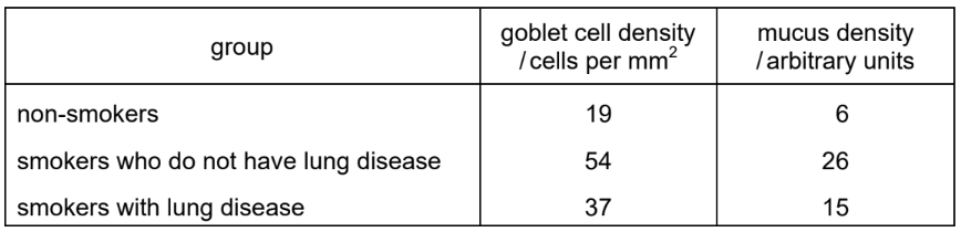
What is indicated by these data?
- There is a positive correlation (relationship) between density of goblet cells and density of mucus.
- Lung disease results in an increase in goblet cell density.
- There is an association between tobacco smoking and an increase in mucus density.
▶️ Answer/Explanation
Ans: C
Analysis of the data:
- Positive correlation between goblet cell density and mucus density:
As goblet cell density increases (non-smokers → smokers → smokers with disease), mucus density also increases. This supports statement 1. - Lung disease and goblet cell density:
The table does not isolate the effect of lung disease alone (no data for non-smokers with disease). Thus, statement 2 cannot be confirmed. - Association between smoking and mucus density:
Smokers (with/without disease) show higher mucus density than non-smokers, supporting statement 3.
Only statements 1 and 3 are valid, making C the correct choice.
Topic: 10.1 (Infectious diseases)
Which disease does Mycobacterium bovis cause?
▶️ Answer/Explanation
Ans: D
Mycobacterium bovis is a pathogenic bacterium that primarily causes tuberculosis (TB) in cattle and can be transmitted to humans. While M. tuberculosis is the main cause of human TB, M. bovis is zoonotic and accounts for a small percentage of TB cases. The other options are caused by different pathogens: cholera by Vibrio cholerae, HIV/AIDS by a virus, and malaria by Plasmodium parasites.
Topic: 10.2 (Antibiotics)
An antibiotic inhibits the formation of cross-links between the molecules that form cell walls in bacteria.
Which statements explain why bacteria are killed by the antibiotic?
1. The bacterial cell is destroyed by osmotic lysis.
2. Cellulose molecules cannot form hydrogen bonds.
3. The cell wall is no longer partially permeable.
▶️ Answer/Explanation
Ans: C
Statement 1 is correct: Without cross-links, the bacterial cell wall weakens and ruptures due to osmotic pressure (lysis). Statement 2 is incorrect because bacterial cell walls contain peptidoglycan, not cellulose. Statement 3 is irrelevant—permeability changes don’t directly cause cell death. Thus, only 1 explains the antibiotic’s effect.
Topic: 10.2 (Antibiotics)
Scientists investigated the effect of increasing concentrations of an antibiotic on the development of antibiotic resistance in bacteria.
The scientists grew four groups of bacteria and added a different concentration of antibiotic to each group. The number of resistant bacteria and the total population of bacteria were measured at intervals for 24 hours for each group.
The graphs show the results.
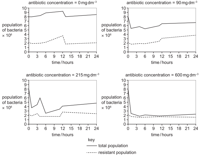
Which statements are correct conclusions that can be made from the results of this investigation?
(1) Increasing the concentration of antibiotic decreases the population of non-resistant bacteria at the end of 24 hours.
(2) The proportion of antibiotic-resistant bacteria increases with increasing concentrations of antibiotics.
(3) Increasing the concentration of antibiotic always increases the number of resistant bacteria.
(A) 1, 2 and 3
(B) 1 and 2 only
(C) 1 and 3 only
(D) 2 and 3 only
▶️ Answer/Explanation
Ans: B
Statement (1) is correct: The graphs show that higher antibiotic concentrations reduce the total bacterial population (mostly non-resistant bacteria). Statement (2) is correct: The proportion of resistant bacteria (relative to the total population) increases with antibiotic concentration due to selective pressure. Statement (3) is incorrect: While resistance proportion rises, the absolute number of resistant bacteria does not always increase (e.g., at the highest concentration, resistant counts may plateau or decline). Thus, only 1 and 2 are valid conclusions.
Topic: 11.1 (The immune system)
What is the correct sequence of events in a primary immune response?
▶️ Answer/Explanation
Ans: B
The correct sequence in a primary immune response is:
1. Antigen presentation by macrophages or dendritic cells to T-helper cells.
2. T-helper cells release cytokines, activating B-lymphocytes.
3. B-lymphocytes differentiate into plasma cells (antibody producers) and memory cells.
Option B matches this sequence. Option A skips antigen presentation, C incorrectly involves neutrophils and memory cells (which form later), and D describes a secondary (not primary) response.
Topic: 11.2 (Antibodies and vaccination)
Which statement about the properties of the antigen-binding sites in different antibody molecules is correct?
▶️ Answer/Explanation
Ans: D
The antigen-binding sites of antibodies are formed by the variable regions of both heavy and light chains (not light chains only, eliminating A). These regions have unique amino acid sequences that confer specificity for different antigens. While the hinge region (B) provides flexibility, it is not part of the binding site. Phagocyte receptors (C) bind to the constant (Fc) region, not the antigen-binding site.
Topic: 11.2 (Antibodies and vaccination)
The diagram shows a stage in monoclonal antibody production.
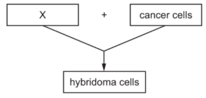
What is represented by X?
A T-lymphocytes
B B-lymphocytes
C antigens
D antibodies
▶️ Answer/Explanation
Ans: B
In monoclonal antibody production, X represents B-lymphocytes. These cells are isolated from an immunized animal (e.g., a mouse) because they produce specific antibodies against the target antigen. The B-lymphocytes are later fused with myeloma cells to form hybridomas, which continuously produce identical (monoclonal) antibodies. T-lymphocytes (A) are not involved in antibody secretion, while antigens (C) and antibodies (D) are the target and product, respectively, not the cell type.