Topic: 1.1 (The microscope in cell studies)
An eyepiece graticule can be calibrated using a stage micrometer.
What is the correct reason why an eyepiece graticule is calibrated?
▶️ Answer/Explanation
Ans: A
The eyepiece graticule is calibrated to enable accurate measurements of microscopic specimens. While it appears as a scale in the eyepiece, its divisions are arbitrary until calibrated against a stage micrometer. Once calibrated, each division corresponds to a specific real-world measurement (e.g., micrometers), allowing precise size determination of observed specimens.
Topic: 1.1 (The microscope in cell studies)
The image is an electron micrograph of a typical eukaryotic cell. What can be concluded about the eukaryotic cell from the electron micrograph?
▶️ Answer/Explanation
Ans: A
Animal cells lack a cell wall, which is a distinguishing feature compared to plant cells. The absence of a cell wall in the electron micrograph confirms it is an animal cell. Permanent vacuoles (B) and chloroplasts (C) are plant cell features, while lysosomes (D) are more abundant in animal cells but not definitive for plant cells.
Topic: 1.2 (Cells as the basic units of living organisms)
Which features are found in typical eukaryotes and also in typical bacteria?

▶️ Answer/Explanation
Ans: C
Both eukaryotes and bacteria share certain cellular features. The correct option is C because it includes cytoplasm, cell membrane, and ribosomes, which are common to both cell types. Eukaryotes have membrane-bound organelles (e.g., nucleus), while bacteria lack them, ruling out other options.
Topic: 1.2 (Cells as the basic units of living organisms)
Which type of cell will have the highest proportion of its volume taken up with cell structures bound by a single membrane?
▶️ Answer/Explanation
Ans: B
Goblet cells (option B) are specialized secretory cells that produce large amounts of mucus, which is stored in membrane-bound vesicles before secretion. These vesicles occupy a significant portion of the cell’s volume. In contrast, red blood cells (C) lack membrane-bound organelles, ciliated epithelial cells (A) have mostly double-membrane organelles, and companion cells (D) have typical plant cell structures.
Topic: 2.2 (Carbohydrates and lipids)
What causes the phosphate heads of phospholipids to become polar?
▶️ Answer/Explanation
Ans: C
The phosphate heads of phospholipids become polar because they become ionized in water, gaining a negative charge. This ionization occurs when the phosphate group loses a hydrogen ion (\(\text{H}^+\)) in aqueous environments, making it polar and hydrophilic. Options A and D are incorrect because while water interacts with the phosphate heads, it’s through ionic interactions rather than hydrogen or covalent bonds. Option B is incorrect because phosphate heads are actually water-soluble.
Topic: 2.2 (Carbohydrates and lipids)
Which statements describe features of cellulose that adapt it for its function in plant cells?
▶️ Answer/Explanation
Ans: D
Cellulose is a structural polysaccharide in plant cell walls. The correct features are:
- Statement 1 is incorrect: Cellulose forms straight chains, not a triple helix (that’s collagen).
- Statement 2 is correct: Hydrogen bonds between adjacent cellulose molecules provide strength and rigidity.
- Statement 3 is incorrect: Covalent bonds do not form between adjacent cellulose molecules; only hydrogen bonds do.
Thus, only Statement 2 is correct, making D the right answer.
Topic: 2.2 (Carbohydrates and lipids)
Which structure shows α-glucose?
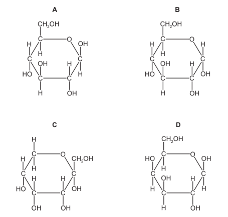
▶️ Answer/Explanation
Ans: B
In α-glucose, the hydroxyl group (-OH) on the first carbon (C1) is positioned below the plane of the ring, which is a distinguishing feature compared to β-glucose (where it is above). By analyzing the given structures, option B correctly represents this orientation, confirming it as α-glucose.
Topic: 2.2 (Carbohydrates and lipids)
What cannot occur as a result of a condensation reaction?
▶️ Answer/Explanation
Ans: A
A condensation reaction involves the joining of two molecules with the release of water (D). It forms disaccharides (B) and peptides (C), but it does not break bonds. The breaking of a glycosidic bond (A) occurs in hydrolysis, not condensation, making it the correct answer.
Topic: 2.3 (Proteins)
Which fact about the quaternary structure of proteins is correct?
▶️ Answer/Explanation
Ans: C
The quaternary structure of proteins refers to the arrangement of multiple polypeptide chains (subunits) into a functional protein complex. While it can involve interactions with metal ions (e.g., hemoglobin with heme), the primary structure (sequence of amino acids) determines how these polypeptides fold and interact. Thus, option C is correct. Options A, B, and D describe possible but not universal features of quaternary structures.
Topic: 3.1 (Mode of action of enzymes)
The diagram shows different molecules in a solution.

Which statement could explain what happens when some of the molecules are mixed together?
▶️ Answer/Explanation
Ans: C
Molecule P is likely an enzyme, and molecules R and S are substrates. Enzymes bind to their specific substrates at the active site, forming an enzyme-substrate complex (Option C). Non-competitive inhibitors (Option A) bind to allosteric sites, not the active site. Enzymes lower activation energy (Option B is incorrect). Option D is speculative as the diagram does not indicate a breakdown reaction.
Topic: 3.1 (Mode of action of enzymes)
The effect of substrate concentration on an enzyme-catalysed reaction was measured in three different conditions:
● without an inhibitor
● with a competitive inhibitor
● with a noncompetitive inhibitor.
The graph shows the results.

Which row is correct?

▶️ Answer/Explanation
Ans: A
In the graph, the competitive inhibitor increases the apparent \( K_m \) (substrate concentration needed for half-maximal velocity) but does not alter \( V_{max} \). The noncompetitive inhibitor reduces \( V_{max} \) but does not affect \( K_m \). The row matching these observations is A, as it correctly identifies the changes in \( K_m \) and \( V_{max} \) for both inhibitors.
Topic: 3.2 (Factors that affect enzyme action)
Which aspect of enzyme activity can be compared by the Michaelis-Menten constant?
▶️ Answer/Explanation
Ans: B
The Michaelis-Menten constant (\(K_m\)) represents the substrate concentration at which an enzyme achieves half of its maximum reaction rate. A lower \(K_m\) indicates higher affinity between the enzyme and its substrate, as less substrate is needed to reach half of \(V_{max}\). Thus, \(K_m\) allows comparison of affinities of different enzymes for their substrates (Option B). It does not measure activation energy (A), changing affinities at different concentrations (C), or temperature effects on \(V_{max}\) (D).
Topic: 3.2 (Factors that affect enzyme action)
The number of substrate molecules one enzyme molecule can convert to product in a second is called the turnover number. This number is obtained when all conditions are optimum for the specific enzyme-catalysed reaction.

How many times faster at converting substrate to product is catalase compared to phosphatase?
▶️ Answer/Explanation
Ans: C
From the data provided (assuming the image shows turnover numbers), catalase has a turnover number of 40,000,000 per second, while phosphatase has a turnover number of 13,860 per second. To find how many times faster catalase is compared to phosphatase, we divide the turnover number of catalase by that of phosphatase:
\[ \text{Ratio} = \frac{40,000,000}{13,860} \approx 2884 \]
Thus, catalase is approximately 2884 times faster than phosphatase, making option (C) correct.
Topic: 4.2 (Movement into and out of cells)
A red indicator solution was mixed with agar, and the resulting solid was cut into small cuboid blocks. The blocks were placed in an acid which turns the indicator yellow, and all other variables were kept constant. The dimensions of the three blocks used are shown.
block 1: 3 mm × 3 mm × 3 mm
block 2: 8 mm × 8 mm × 8 mm
block 3: 11 mm × 11 mm × 11 mm
Which row shows the correct surface area (SA) to volume (V) ratio for each block, and the time taken for the block to turn yellow?

▶️ Answer/Explanation
Ans: C
The SA:V ratio for a cube is calculated as \( \frac{6x^2}{x^3} = \frac{6}{x} \), where \( x \) is the side length. Thus:
- Block 1 (3 mm): SA:V = \( \frac{6}{3} = 2 \) (highest ratio, fastest diffusion → shortest time).
- Block 2 (8 mm): SA:V = \( \frac{6}{8} = 0.75 \).
- SA:V = \( \frac{6}{11} \approx 0.55 \) (lowest ratio, slowest diffusion → longest time).
Option C correctly matches these ratios with the observed times: smaller blocks (higher SA:V) turn yellow faster.
Topic: 4.2 (Movement into and out of cells)
The statements describe some events in the process of exocytosis of glycoprotein molecules.
1. Membrane of the Golgi body folds around glycoprotein molecules.
2. Vesicle binds to and fuses with the cell surface membrane.
3. Vesicle attached to microtubules moves through the cytoplasm.
4. Secretory vesicle forms.
What is the correct order of events for exocytosis?
▶️ Answer/Explanation
Ans: B
The correct sequence of exocytosis is: 1 → 4 → 3 → 2. First, the Golgi membrane encloses glycoproteins (1), forming a secretory vesicle (4). The vesicle then moves via microtubules (3) before fusing with the plasma membrane (2) for secretion. This matches option B as the correct order.
Topic: 4.2 (Movement into and out of cells)
Four cylinders that were identical in size, A, B, C and D, were cut from potatoes that had been stored for different lengths of time. The cylinders were weighed, immersed in 10% salt solution for 45 minutes and then reweighed. The percentage change in mass was then calculated. Which cylinder had a water potential similar to the 10% salt solution?

▶️ Answer/Explanation
Ans: C
The cylinder with a water potential similar to the 10% salt solution will show no net change in mass after immersion. This is because when the water potential inside the potato cells matches the external solution, there is no net movement of water (osmosis). From the graph, cylinder C shows a 0% change in mass, indicating equilibrium. Cylinders A and B lose mass (water leaves the cells), while D gains mass (water enters the cells), confirming C as the correct answer.
Topic: 5.1 (Replication and division of nuclei and cells)
The contents of a daughter cell are compared to the parent cell after one cell cycle. Which row is correct?

▶️ Answer/Explanation
Ans: D
After one complete cell cycle (which includes interphase and mitosis), the daughter cell should be genetically identical to the parent cell. Key points:
- DNA content: The daughter cell must have the same amount of DNA as the parent cell (since DNA is replicated in S phase and equally distributed during mitosis).
- Chromosome number: The daughter cell must maintain the same chromosome number (diploid or haploid) as the parent cell.
- Genetic variation: No new genetic variation is introduced in mitosis (unlike meiosis).
Thus, the correct row is D, where the daughter cell is genetically identical to the parent cell.
Topic: 5.1 (Replication and division of nuclei and cells)
A high-power photomicrograph shows a cell in a stage of mitosis. The chromosomes are visible and lined up along the cell equator but there is no nuclear envelope. Which stage of mitosis is shown by the photomicrograph?
▶️ Answer/Explanation
Ans: B (Metaphase)
In metaphase, the chromosomes are fully condensed and aligned at the cell’s equator (metaphase plate), and the nuclear envelope has already disintegrated (which occurs in late prophase). Since the photomicrograph shows these exact features—visible chromosomes at the equator and no nuclear membrane—the correct stage is metaphase.
Topic: 5.1 (Replication and division of nuclei and cells)
Which statements about the cell cycle are correct?
1. The cell cycle includes interphase and mitosis.
2. DNA replication takes place in interphase.
3. A cell can remain in interphase for several months.
▶️ Answer/Explanation
Ans: A
1. Correct: The cell cycle consists of interphase (growth and DNA replication) and mitosis (nuclear division).
2. Correct: DNA replication occurs during the S-phase of interphase.
3. Correct: Some cells (e.g., neurons, muscle cells) remain in interphase (G0 phase) indefinitely or for extended periods.
Thus, all three statements (1, 2, and 3) are correct, making option A the right answer.
Topic: 5.2 (Chromosome behaviour in mitosis)
A scientist stains the chromosomes of a plant cell with a fluorescent dye to observe the telomeres. This cell has 38 chromosomes.
How many telomeres will the scientist observe in one of the nuclei during telophase of mitosis?
▶️ Answer/Explanation
Ans: B
During telophase of mitosis, each chromosome consists of two sister chromatids, each with its own pair of telomeres. Since the cell has 38 chromosomes, the total number of telomeres observed is calculated as:
\[ 38 \text{ chromosomes} \times 2 \text{ chromatids/chromosome} \times 2 \text{ telomeres/chromatid} = 152 \text{ telomeres} \]
However, in one nucleus (after cytokinesis), only half of these telomeres are present (since the cell divides into two daughter cells). Thus:
\[ 152 \div 2 = 76 \text{ telomeres} \]
Therefore, the correct answer is B (76).
Topic: 6.1 (Structure of nucleic acids and replication of DNA)
During the semi-conservative replication of DNA, the double helix is unwound by an enzyme. Which diagram shows how the strands are copied?

▶️ Answer/Explanation
Ans: D
In semi-conservative replication, each new DNA molecule consists of one original (parental) strand and one newly synthesized strand. Diagram D correctly depicts this process, where the unwound parental strands serve as templates for complementary base pairing, ensuring genetic continuity. The other diagrams (A, B, C) either show incorrect strand separation or replication mechanisms (e.g., conservative or dispersive replication).
Topic: 6.2 (Protein synthesis)
A transcription error results in the deletion of one nucleotide from the middle of a primary transcript. mRNA forms from the primary transcript.
Which statement describes one possible effect of this deletion on the protein translated from this mRNA?
▶️ Answer/Explanation
Ans: D
A single nucleotide deletion in the primary transcript causes a frameshift mutation in the mRNA. This shifts the reading frame, altering all downstream codons and resulting in a completely different amino acid sequence from the point of deletion. While some mutations may only affect a single amino acid, a frameshift disrupts the entire downstream protein structure, making option D the correct choice.
Topic: 6.2 (Protein synthesis)
Which statements correctly describe the process of translation?
(1) The nucleotide sequence on an mRNA molecule is used to produce a specific amino acid chain.
(2) A section of DNA is copied into an mRNA molecule by RNA polymerase.
(3) A polypeptide is produced because anticodons on tRNA molecules attach to mRNA codons through peptide bonds.
▶️ Answer/Explanation
Ans: C
Statement 1 is correct: Translation converts the mRNA nucleotide sequence into an amino acid chain (polypeptide) via ribosomes. Statement 2 describes transcription (DNA → mRNA), not translation. Statement 3 is incorrect because tRNA anticodons bind to mRNA codons via hydrogen bonds (not peptide bonds); peptide bonds form between amino acids during polypeptide assembly. Thus, only statement 1 is accurate for translation.
Topic: 6.2 (Protein synthesis)
A molecule of mRNA was used in translation. Part of its sequence is shown.
GAU CUG UAA CGG
There were no introns present in the section of DNA that was transcribed to make this mRNA.
What is the sequence of the non-transcribed DNA strand for this section?
▶️ Answer/Explanation
Ans: C
The non-transcribed DNA strand (coding strand) has the same sequence as the mRNA, except thymine (T) replaces uracil (U). Given the mRNA sequence GAU CUG UAA CGG, the coding DNA strand is:
\[ \text{mRNA: } \text{GAU CUG UAA CGG} \\ \text{Non-transcribed DNA: } \text{GAT CTG TAA CGG} \]
Option (C) matches this sequence, while (A) is the transcribed (template) strand, (B) incorrectly includes U, and (D) is the mRNA sequence itself.
Topic: 2.4 (Water)
Which properties of water are dependent on hydrogen bonding between water molecules?

▶️ Answer/Explanation
Ans: B
Hydrogen bonding in water is responsible for:
- High surface tension (cohesion between water molecules at the surface).
- High specific heat capacity (energy needed to break hydrogen bonds before temperature rises).
- Solvent properties (polarity from hydrogen bonding enables dissolving ionic/polar substances).
Option B correctly includes these properties. Density and freezing (not in B) are less directly dependent on hydrogen bonding.
Topic: 7.2 (Transport mechanisms)
Which substances in xylem tissue are impermeable to water and prevent the collapse of the vessels?

▶️ Answer/Explanation
Ans: D
Lignin (option D) is the correct answer as it is the waterproof polymer that strengthens xylem vessel walls. It provides structural support to prevent collapse under tension during transpiration, while being impermeable to water. The other options (cellulose, hemicellulose, pectin) are water-permeable components of plant cell walls that don’t provide the same structural rigidity.
Topic: 7.1 (Structure of transport tissues)
The electron micrograph shows a longitudinal section through phloem tissue.

Which student’s drawing of a sieve tube element is correctly drawn and labelled?

▶️ Answer/Explanation
Ans: A
The correct drawing must show these key features of sieve tube elements:
- Perforated sieve plates between cells (shown in A and D, but missing in B and C)
- No nuclei (mature sieve tubes lose their nuclei, eliminating B)
- Adjacent companion cells (present in A and C, but C lacks sieve plates)
- Thin cytoplasm lining the walls (all options show this)
Only option A accurately depicts all these structural features when compared to the electron micrograph.
Topic: 7.2 (Transport mechanisms)
Carrier proteins in the cell surface membranes of companion cells are involved in the transfer of assimilates to phloem sieve tubes. The diagram represents the use of two types of carrier protein in this process.

What are the substances labelled X and Y?

▶️ Answer/Explanation
Ans: A
In the phloem loading process, assimilates (mainly sucrose) are actively transported into sieve tubes from companion cells. The mechanism involves:
- Substance X (H+ ions): Proton pumps in the companion cell membrane actively transport H+ out, creating a proton gradient.
- Substance Y (sucrose): Sucrose is co-transported back into the companion cell along with H+ ions via a sucrose-H+ symporter.
Thus, the correct identification is X = H+ ions and Y = sucrose, corresponding to option A.
Topic: 8.3 (The heart)
The diagram shows pressure changes during two cardiac cycles. Which arrow indicates atrial systole?

▶️ Answer/Explanation
Ans: B
Atrial systole occurs when the atria contract, increasing atrial pressure and pushing blood into the ventricles. In the pressure graph:
- Arrow B points to a small rise in atrial pressure (before ventricular systole), which corresponds to atrial contraction.
- Other arrows represent ventricular events (e.g., A = late diastole, C = ventricular systole, D = ventricular relaxation).
Thus, B correctly identifies atrial systole.
Topic: 8.3 (The heart)
An irregular heartbeat may be the result of ineffective electrical stimulation of the atria. Which area of the heart could be damaged, causing this irregular heartbeat?
▶️ Answer/Explanation
Ans: D
The sinoatrial (SA) node is the natural pacemaker of the heart, initiating electrical impulses that stimulate atrial contraction. If damaged, it can lead to irregular heartbeats (arrhythmias) due to improper signaling. The atrioventricular (AV) node (A) and Purkyne tissue (C) are involved in later conduction steps, while the septum (B) is structural and unrelated to electrical stimulation. Thus, the correct answer is D.
Topic: 8.2 (Transport of oxygen and carbon dioxide)
The diagram shows the effect of three different concentrations of carbon dioxide on the oxygen dissociation curve for human haemoglobin.

Which effect does increasing carbon dioxide concentration have on haemoglobin?
▶️ Answer/Explanation
Ans: B
The Bohr effect explains how increasing \(\text{CO}_2\) concentration affects haemoglobin:
- Oxygen Uptake in Lungs: Higher \(\text{CO}_2\) lowers blood pH, reducing haemoglobin’s affinity for oxygen (curve shifts right). This makes oxygen binding less efficient in the lungs.
- Oxygen Release in Tissues: The rightward shift enhances oxygen unloading in \(\text{CO}_2\)-rich tissues (e.g., muscles), making release more efficient.
Thus, option B is correct. The diagram confirms this: higher \(\text{CO}_2\) shifts the curve rightward, showing decreased oxygen affinity (less uptake) and improved release.
Topic: 8.2 (Transport of oxygen and carbon dioxide)
Which reactions will be taking place in blood in a capillary that is next to an alveolus?
1. \( \text{Hb} + 4\text{O}_2 \to \text{HbO}_8 \) Key: Hb = haemoglobin
2. \( \text{H}_2\text{O} + \text{CO}_2 \to \text{H}_2\text{CO}_3 \)
3. \( \text{H}_2\text{CO}_3 \to \text{H}^+ + \text{HCO}_3^- \)
▶️ Answer/Explanation
Ans: B
In capillaries near alveoli, oxygen binds to haemoglobin (Reaction 1: \( \text{Hb} + 4\text{O}_2 \to \text{HbO}_8 \)) for transport. Reaction 2 (\( \text{CO}_2 \) hydration) and Reaction 3 (carbonic acid dissociation) primarily occur in tissues, where \( \text{CO}_2 \) is released, not in alveolar capillaries. Thus, only Reaction 1 is correct here.
Topic: 9.1 (The gas exchange system)
Which structure of the gas exchange system always contains cartilage?
▶️ Answer/Explanation
Ans: D
In the gas exchange system, cartilage is present in the walls of the trachea and bronchi to prevent collapse during inhalation. Alveoli (A) and capillaries (C) lack cartilage, while bronchioles (B) are too small to contain it. Thus, the bronchus (D) is the correct answer.
Topic: 9.1 (The gas exchange system)
Exchange of carbon dioxide and oxygen occurs between air in the alveoli and blood in the capillaries of the lung.
Which partial pressures of the gases will allow gaseous exchange to occur?

▶️ Answer/Explanation
Ans: B
Gaseous exchange occurs down partial pressure gradients:
- Oxygen diffuses from alveoli (higher pO₂) to blood (lower pO₂).
- Carbon dioxide diffuses from blood (higher pCO₂) to alveoli (lower pCO₂).
Option B correctly shows:
- Alveolar pO₂ (13 kPa) > Capillary pO₂ (5 kPa) → O₂ moves into blood.
- Capillary pCO₂ (6 kPa) > Alveolar pCO₂ (1 kPa) → CO₂ moves into alveoli.
Other options fail to maintain these gradients (e.g., equal pO₂/pCO₂ or reversed gradients would halt diffusion).
Topic: 9.1 (The gas exchange system)
The plan diagram shows a cross-section of a trachea. Which labelled tissue prevents the trachea from collapsing?

▶️ Answer/Explanation
Ans: A
The C-shaped rings of cartilage in the tracheal wall provide structural support, preventing collapse during inhalation when air pressure decreases. Other labelled tissues serve different functions:
- B (Ciliated epithelium): Moves mucus and trapped particles.
- C (Goblet cells): Secrete mucus to trap pathogens.
- D (Smooth muscle): Adjusts airway diameter but cannot prevent collapse.
Thus, cartilage (A) is the correct answer.
Topic: 9.1 (The gas exchange system)
Which layers of cells does an oxygen molecule diffuse through when moving from an alveolus into an alveolar capillary?

▶️ Answer/Explanation
Ans: C
When oxygen moves from an alveolus into the blood, it diffuses through:
- Alveolar epithelium (single layer of flattened cells lining the alveolus)
- Capillary endothelium (single layer of cells lining the capillary)
These two thin layers (and their shared basement membrane) form the respiratory membrane, which is optimized for rapid gas exchange. The image likely shows these layers, with option C correctly identifying both epithelial layers.
Other options may incorrectly include additional layers like mucus or multiple cell types not present in the actual diffusion pathway.
Topic: 10.2 (Antibiotics)
Bacteria may be classified according to differences in cell wall structure. The differences are shown by using the Gram stain.
The diagram shows part of a Gram-positive bacterium and part of a Gram-negative bacterium, drawn to the same scale.

The antibiotic penicillin kills bacteria by inhibiting the synthesis of the cell walls during bacterial cell growth.
Which type of bacteria will be killed by penicillin more easily and why?
▶️ Answer/Explanation
Ans: A
Gram-positive bacteria (option A) are more susceptible to penicillin because their thick peptidoglycan layer is directly exposed to the antibiotic. In Gram-negative bacteria, the outer membrane acts as a protective barrier, making the peptidoglycan layer less accessible to penicillin. The thickness of the peptidoglycan layer in Gram-positive bacteria (20-80nm) compared to Gram-negative (2-7nm) provides more target sites for penicillin’s action.
Topic: 10.1 (Infectious diseases)
Which facts relate to the disease TB or its pathogen?
1. Viruses change their antigens to a limited extent.
2. TB is caused by only one species of pathogen.
3. HIV/AIDS makes the bacterial infection worse.
4. The pathogen may be transmitted by ingestion.
5. The pathogen may be transmitted from animals.
6. Multi-drug resistance occurs.
(A) 1, 4, 5 and 6
(B) 1, 2 and 6
(C) 2, 3 and 5
(D) 3, 4, 5 and 6
▶️ Answer/Explanation
Ans: D
To determine the correct answer, let’s analyze each statement in relation to TB (Mycobacterium tuberculosis):
- False: TB is caused by bacteria, not viruses.
- False: While M. tuberculosis is the main cause, other mycobacteria (e.g., M. bovis) can also cause TB.
- True: HIV weakens the immune system, exacerbating TB infections.
- True: TB can spread via contaminated food/drink (e.g., unpasteurized milk).
- True: Zoonotic transmission occurs (e.g., M. bovis from cattle).
- True: Multi-drug-resistant TB (MDR-TB) is a significant issue.
Only statements 3, 4, 5, and 6 are correct, making D the right choice.
Topic: 11.1 (The immune response)
The events listed occur during the primary immune response to a specific pathogen.
Which row identifies a correct sequence of events?

▶️ Answer/Explanation
Ans: B
The correct sequence of events in a primary immune response is:
- Phagocytosis by macrophages (2): Pathogens are engulfed and digested.
- Antigen presentation (4): Processed antigens are displayed on the phagocyte’s surface via MHC II.
- T-helper cell activation (3): These cells recognize antigens and stimulate B-cells and cytotoxic T-cells.
- B-cell activation (1): B-lymphocytes differentiate into plasma cells (antibody producers) and memory cells.
- Antibody production (5): Plasma cells secrete pathogen-specific antibodies.
Thus, the sequence 2 → 4 → 3 → 1 → 5 (Option B) is correct.
Topic: 11.1 (The immune response)
Influenza is an infectious disease caused by a virus. It is possible to have influenza more than once.
Which statements explain why it is possible to have influenza more than once?
1. The viral antigen changes as a result of mutations.
2. The immune system may be weak and make few B-memory cells.
3. Untreated HIV infection has resulted in a low T-helper cell count.
▶️ Answer/Explanation
Ans: A (1, 2, and 3)
Explanation:
- Antigenic variation: Influenza viruses mutate rapidly (antigenic drift/shift), altering surface proteins (e.g., hemagglutinin). This evades prior immunity.
- Weak immune memory: A compromised immune system may produce insufficient B-memory cells, reducing long-term protection.
- HIV and T-helper cells: HIV destroys CD4+ T-cells, crippling adaptive immunity. Without T-helper cells, B-cells cannot generate effective memory responses.
All three statements correctly explain reinfection. Thus, A is the answer.