▶️ Answer/Explanation
(a) histones
Explanation: The proteins that are complexed with DNA to form chromatin are called histones. These are basic proteins that help in the packaging of DNA into structural units called nucleosomes. Histones play a crucial role in gene regulation and DNA replication.
(b)
Diagram should show:
- Two chromosomes, each with two chromatids, aligned along the metaphase plate (equator of the cell)
- Spindle fibers attached to the centromeres of each chromosome
- Centrioles at opposite poles of the cell (if drawing includes them)
- Labels for: chromosomes, spindle fibers, centromeres, and poles
Explanation: During metaphase, chromosomes are at their most condensed state and align at the cell’s equator. The spindle apparatus, composed of microtubules, attaches to the centromeres of each chromosome. This alignment ensures equal distribution of genetic material to daughter cells.
(c)
From start of S phase to end of interphase:
- DNA replication occurs during S phase, resulting in identical copies of each chromosome
- The replicated DNA complexes with histones to form sister chromatids
- Chromatin remains diffuse and not visible under light microscope
- Nuclear material doubles in preparation for cell division
During prophase of mitosis:
- Chromatin condenses into visible chromosomes (each consisting of two sister chromatids)
- Nuclear envelope begins to break down
- Spindle fibers begin to form from the centrosomes
- Centrosomes move to opposite poles of the cell
- Chromosomes become attached to spindle fibers via their centromeres
Explanation: These changes ensure proper segregation of genetic material. The S phase prepares the cell by duplicating DNA, while prophase organizes this DNA for division. Condensation makes chromosomes manageable for movement, while spindle formation provides the mechanism for chromosome separation.
▶️ Answer/Explanation
(a) Secondary (structure).
Explanation: The classification of keratin as α-keratin or β-keratin is based on the secondary structure of the protein. α-keratin contains many α-helices, while β-keratin contains many β-pleated sheets. These are both types of secondary structure formed by hydrogen bonding between amino acids in the polypeptide chain.
(b) Any one from:
- The area to be hydrolysed could be between the same amino acids in different proteins
- The active site has some flexibility for hydrolysing similar substrates
- Proteins have similar shapes so they can fit the active site
- The active site and substrates still have complementary shapes
Explanation: Proteases can act on different proteins because they target peptide bonds, which are common to all proteins. The active site of the enzyme may have some flexibility to accommodate different amino acid sequences, or different proteins may have similar regions where the peptide bonds are accessible to the enzyme.
(c) Any three from:
- Fibrous nature of the protein
- Insolubility in water
- High proportion of disulfide bonds between cysteine residues
- Many hydrogen bonds between polypeptides
- Many hydrophobic interactions
- Tight packing of polypeptide chains
Explanation: Keratin’s stability comes from multiple structural features. The disulfide bonds between cysteine residues create strong covalent cross-links. The hydrogen bonds and hydrophobic interactions between polypeptide chains make the structure resistant to breaking. The tight packing of fibers and insolubility make it difficult for enzymes to access the peptide bonds for hydrolysis.
(d)
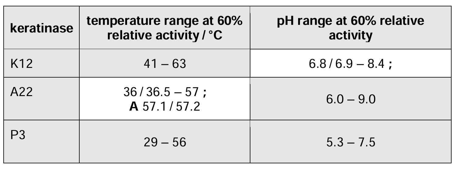
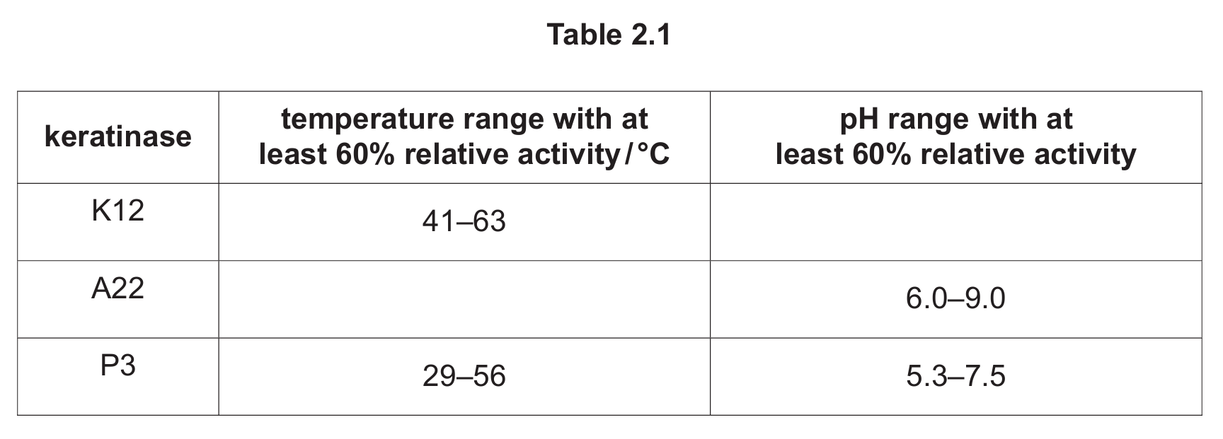
Keratinase with a relative activity of at least 60% that has:
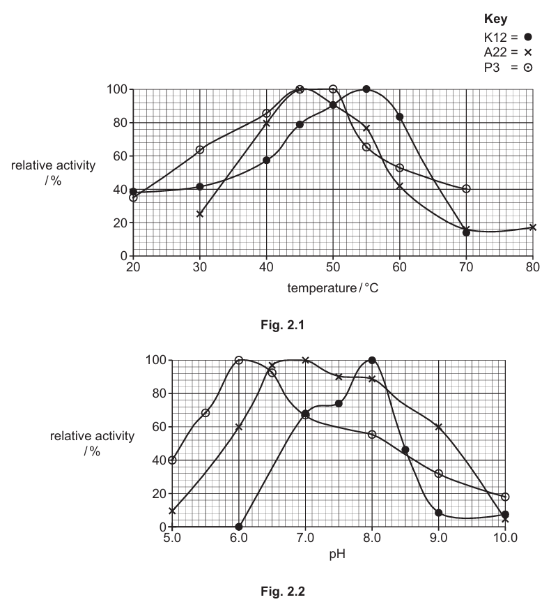
- the widest working range of temperature: P3
- the widest working range of pH: A22
Explanation: From the data, P3 has the widest temperature range (29-56°C, a span of 27°C), while A22 has the widest pH range (6.0-9.0, a span of 3 pH units). This means P3 would be more tolerant to temperature variations, while A22 would work across a broader range of pH conditions.
(e) Any three from:
Advantages:
- A22 and K12 are active at a wide range of temperatures (A22: 36-57°C, K12: 41-63°C)
- K12 shows good activity at lower temperatures (around 40% at 20°C)
- A22 has greatest activity at pH 7.5-8.0 and maintains at least 60% activity up to pH 9.0
- K12 is active between pH 7.5-8.5 with optimum at pH 8.0
Disadvantages:
- A22 has low activity at 30°C and 70-80°C
- K12 has low activity at 20°C and 30°C
- Activity decreases at higher pH values for both enzymes
Explanation: For detergent use, the enzymes need to work across various washing conditions. A22’s broad pH range makes it suitable for different detergent formulations, while K12’s temperature range allows it to work in both warm and hot washes. However, their reduced activity at certain temperatures might limit their effectiveness in some washing conditions. The alkaline tolerance is good for detergent use as most laundry detergents are alkaline.
▶️ Answer/Explanation
(a) aorta / dorsal aorta
Explanation: The hepatic artery branches directly from the aorta, which is the main artery carrying oxygenated blood from the heart to the rest of the body. The aorta gives rise to various branches that supply different organs, with the hepatic artery specifically supplying oxygen-rich blood to the liver.
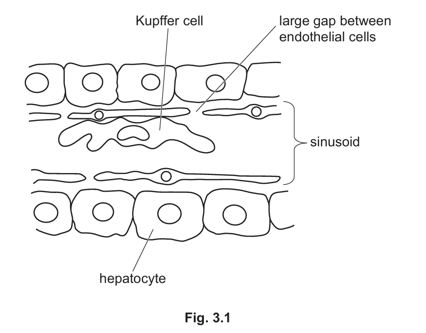
(b)(i) Higher rate of/easier exchange/movement of substances
Explanation: The large gaps between endothelial cells in liver sinusoids facilitate efficient exchange of materials between the blood and hepatocytes. This unique structure allows plasma proteins and other large molecules to pass through easily, enabling the liver to perform its vital functions in metabolism, detoxification, and protein synthesis more effectively.
(b)(ii)
- Phagocytosis occurs where the Kupffer cell engulfs the damaged red blood cell
- The red blood cell is enveloped by pseudopodia extending from the Kupffer cell
- A phagocytic vacuole (phagosome) forms around the red blood cell
- A lysosome fuses with the phagosome, releasing digestive enzymes
- The enzymes break down the red blood cell into components like haem and globin
- These components are further processed or recycled by the cell
Explanation: Kupffer cells are specialized macrophages in the liver that play a crucial role in cleaning the blood by removing old or damaged red blood cells through phagocytosis. This process involves recognition of the damaged cells, engulfment, and enzymatic breakdown within the phagolysosome. The iron from haem can be recycled, while globin proteins are broken down into amino acids for reuse.
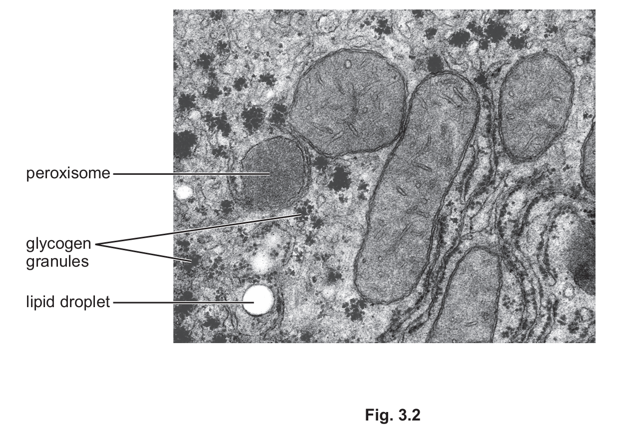
(c)(i)
- Presence of glycogen granules indicates energy storage function
- Lipid droplets visible, showing fat storage capability
- Numerous mitochondria present, suggesting high metabolic activity
- Rough endoplasmic reticulum visible, indicating protein synthesis
- Smooth endoplasmic reticulum present, involved in lipid metabolism and detoxification
Explanation: The electron micrograph provides multiple visual clues about hepatocyte functions. Glycogen granules appear as dark clusters, demonstrating the liver’s role in glucose storage. Lipid droplets are clear evidence of fat storage. The abundance of mitochondria reflects the high energy demands of liver cells for their diverse metabolic activities. The presence of both rough and smooth ER indicates the cell’s involvement in both protein synthesis and lipid metabolism/detoxification.
(c)(ii) Mitochondria have a double membrane while peroxisomes have a single membrane
Explanation: This structural difference is fundamental to their distinct functions. The double membrane of mitochondria, with its folded inner membrane (cristae), provides a large surface area for ATP production during cellular respiration. The single membrane of peroxisomes is sufficient for their role in oxidative reactions and detoxification processes.
(c)(iii)
- Mitochondria contain their own DNA and ribosomes (70S) for protein synthesis
- They have the complete machinery for transcription and translation
- Peroxisomes lack genetic material and protein-synthesizing machinery
- All peroxisomal enzymes are encoded by nuclear DNA and imported from the cytosol
Explanation: This difference stems from the endosymbiotic origin of mitochondria, which were once free-living bacteria that retained some of their original genetic machinery. Mitochondria can produce some of their own proteins, particularly those involved in oxidative phosphorylation. Peroxisomes, in contrast, are not derived from endosymbionts and rely entirely on the cell’s nuclear genome for their enzyme complement, which must be imported after synthesis in the cytosol.
▶️ Answer/Explanation
(a)
1. Blood arriving at the alveoli is deoxygenated (has low partial pressure of oxygen) while the alveoli contain oxygen-rich air, creating a steep concentration gradient for oxygen diffusion.
2. The continuous flow of blood removes oxygenated blood and brings in more deoxygenated blood, maintaining the concentration gradient by constantly replacing blood that has picked up oxygen.
Explanation: The efficient exchange of gases relies on maintaining steep concentration gradients. The blood flow system ensures that oxygen-poor blood is always arriving at the alveoli while oxygen-rich blood is constantly being carried away, preventing equilibrium from being reached and thus maintaining the diffusion gradient.
(b)
1. Elastic fibers allow alveoli to expand during inhalation by stretching, accommodating the incoming air.
2. They prevent overstretching and potential damage to alveoli during deep inhalation.
3. During exhalation, the elastic fibers recoil, helping to push air out of the lungs and return the alveoli to their resting size.
Explanation: The elastic properties of alveolar walls are crucial for the ventilation process. During inhalation, the fibers stretch to allow lung expansion, while their recoil during exhalation helps passively expel air. This elastic behavior maintains the structure and function of alveoli through repeated breathing cycles.
(c)
1. ABCA3 is a membrane transport protein specifically designed to move phospholipids across membranes.
2. It has specific binding sites that recognize and bind surfactant phospholipids.
3. It uses energy from ATP hydrolysis to actively transport phospholipids against their concentration gradient into lamellar bodies.
4. The protein undergoes conformational changes to move phospholipids across the membrane.
Explanation: As an ATP-binding cassette transporter, ABCA3 has several specialized features. Its binding sites specifically interact with surfactant phospholipids. The ATP hydrolysis provides energy for the active transport process, moving phospholipids into the lamellar bodies where they’re stored until secretion. This active transport is necessary to concentrate the surfactant components within the storage organelles.
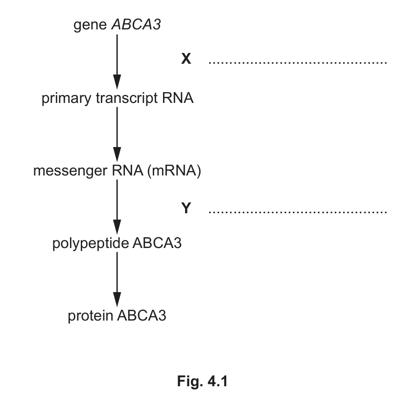
(d)(i)
X: Transcription
Y: Translation
Explanation: The process begins with transcription (X), where the DNA sequence of the ABCA3 gene is copied into a primary RNA transcript. This is then processed into mRNA which undergoes translation (Y) at ribosomes to produce the polypeptide chain that will fold into the functional ABCA3 protein.
(d)(ii)
1. The gene contains non-coding introns that are removed during RNA processing, so they don’t contribute to the final protein sequence.
2. Only the coding exons are spliced together to form the mature mRNA that gets translated.
3. Some DNA sequences are regulatory elements that control gene expression but don’t code for protein.
4. Stop codons terminate translation and don’t code for amino acids.
Explanation: Eukaryotic genes typically contain much more DNA than needed to code for their proteins. Introns are intervening sequences that are transcribed but then spliced out of the primary transcript. Additionally, there are untranslated regions and regulatory sequences that don’t contribute to the amino acid sequence. The start codon (usually coding for methionine) may also be removed post-translationally in some proteins.