▶️ Answer/Explanation
(a)
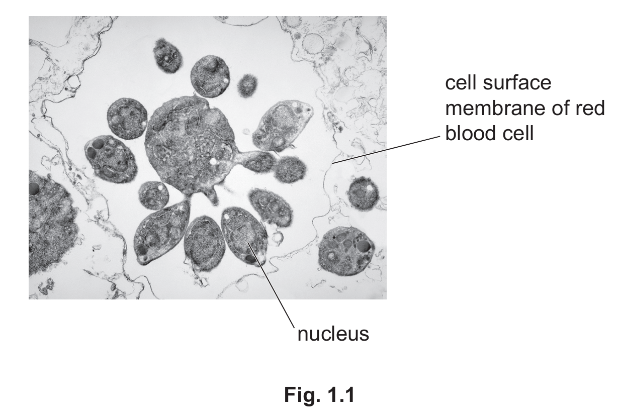
The presence of P. falciparum affects red blood cells in several ways:
- The red blood cell loses its normal biconcave shape and becomes irregular as the parasite grows inside it.
- The cell membrane may eventually burst (lysis) due to the pressure from multiple daughter cells forming inside.
- The parasite consumes hemoglobin and other cell contents, reducing the red blood cell’s ability to transport oxygen.
- The cell becomes filled with parasite daughter cells, compromising its normal function.
- The lifespan of the infected red blood cell is shortened as the parasite completes its life cycle.
(b)
The formation of daughter cells in P. falciparum involves several key steps:
- The parasite first grows inside the red blood cell, increasing its cytoplasm and synthesizing necessary biomolecules.
- The nucleus undergoes multiple rounds of DNA replication through mitosis, creating many nuclei.
- The cytoplasm then divides unequally around each nucleus through a process called budding.
- This results in many small daughter cells that are genetically identical but contain varying amounts of cytoplasm.
- The daughter cells remain temporarily within the original red blood cell membrane until they are ready to be released.
(c)
Antibodies combat malaria through several mechanisms:
- Antibodies bind specifically to antigens on the surface of P. falciparum, marking them for destruction.
- This binding can prevent the parasite from entering new red blood cells, blocking the infection cycle.
- Antibodies facilitate phagocytosis by white blood cells that recognize the antibody-coated parasites.
- Some antibodies can directly neutralize the parasite by blocking essential surface proteins.
- Antibodies may also cause agglutination (clumping) of parasites, making them easier targets for immune cells.
(d)
Successful vaccination programmes depend on multiple factors:
- Vaccine efficacy: Higher efficacy (like the 77% achieved by R21/Matrix-M™) leads to better disease control.
- Herd immunity: Vaccinating enough of the population to protect those who can’t be vaccinated.
- Accessibility: Making vaccines available to all at-risk populations, including remote areas.
- Public education: Combating misinformation and explaining vaccine benefits increases uptake.
- Logistics: Proper storage (especially for temperature-sensitive vaccines) and distribution systems.
- Duration of protection: Vaccines providing long-term immunity require fewer booster shots.
- Cost: Affordable vaccines that health systems can sustainably provide to entire populations.
- Strain coverage: Effectiveness against multiple strains of the pathogen prevents breakthrough cases.
- Safety profile: Minimal side effects increase public acceptance and compliance.
The R21/Matrix-M™ vaccine’s success in trials suggests it meets several of these criteria, particularly the crucial efficacy target set by WHO.
▶️ Answer/Explanation
(a)(i)
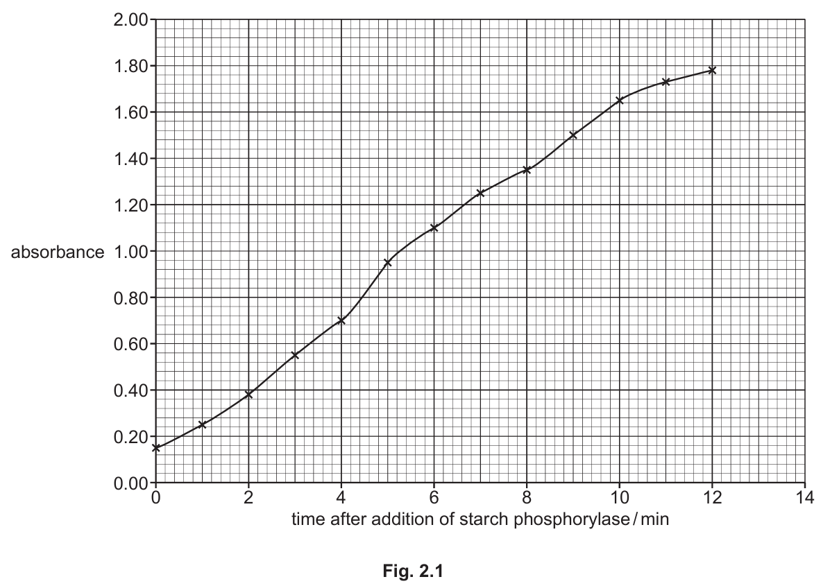
More starch/amylose is produced so iodine solution becomes darker/changes from yellow-brown to blue-black.
Explanation: The colorimeter measures absorbance, which increases as more amylose is formed. Iodine forms a blue-black complex with amylose, so as the reaction progresses and more amylose is produced, the color intensity increases, leading to higher absorbance readings.
(a)(ii)
Prediction: Absorbance remains constant/reaches plateau at about 1.80.
Explanation: All the substrate (G 1-P) would be used up/converted to amylose by this point. Once the substrate is exhausted, no more amylose can be produced, so the absorbance would stabilize at its maximum value. Alternatively, the colorimeter might reach its detection limit at about 2.00 absorbance units.
(a)(iii)
Colorimeter gives quantitative/numerical results that are not subjective.
Explanation: Unlike visual assessment which can vary between observers, the colorimeter provides objective, numerical measurements of absorbance. This allows for more precise tracking of the reaction progress and enables the creation of accurate graphs and calculations of reaction rates.
(b)
1. Substrate (G 1-P) binds to the active site of starch phosphorylase, forming an enzyme-substrate complex.
2. The end of an existing amylose molecule also binds to the active site.
3. The active site changes shape (induced fit) to accommodate both substrates.
4. The enzyme lowers the activation energy for the reaction.
5. A glycosidic (α-1,4) bond forms between the glucose molecules in a condensation reaction (water is released).
6. The phosphate ion leaves the active site.
7. The extended amylose molecule is released.
Explanation: Starch phosphorylase catalyzes the addition of glucose units to the non-reducing end of amylose molecules. The enzyme brings the glucose 1-phosphate substrate and the growing amylose chain together in its active site, facilitating the formation of glycosidic bonds through a condensation reaction. This process repeats to build the amylose polymer.
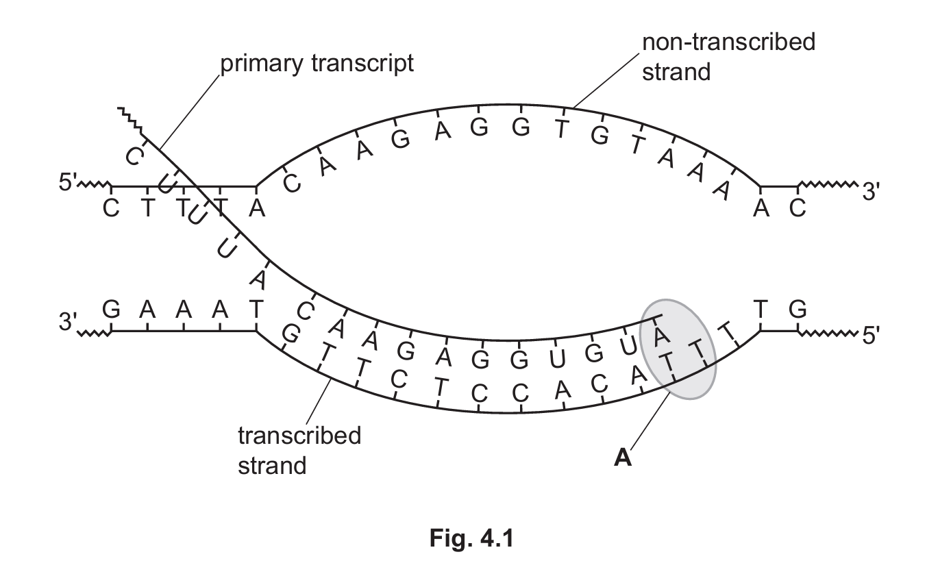
▶️ Answer/Explanation
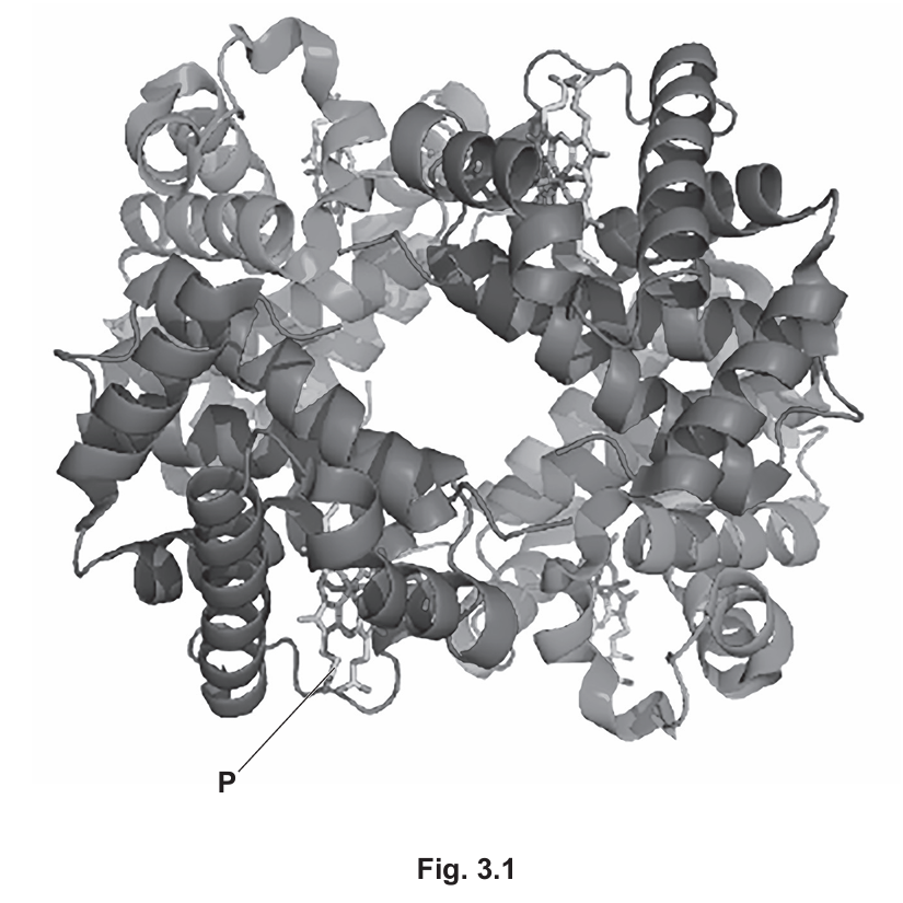
(a)(i) Alpha (α) globin and beta (β) globin polypeptides.
Explanation: Haemoglobin is a tetrameric protein composed of two alpha (α) chains and two beta (β) chains. These polypeptide chains are slightly different in their amino acid sequence but work together to form the functional haemoglobin molecule.
(a)(ii) Haem group.
Explanation: The structure labelled P is the haem group, which is a prosthetic group containing an iron ion (Fe²⁺) at its center. This iron ion is responsible for binding oxygen molecules in the lungs.
(a)(iii) Primary structure.
Explanation: The figure shows the quaternary structure (four subunits together), tertiary structure (folding of individual subunits), and secondary structure (alpha helices within subunits), but doesn’t show the primary structure, which is the specific sequence of amino acids in each polypeptide chain.
(b) Haemoglobin binds directly to carbon dioxide molecules at their terminal amine groups (-NH₂) to form carbaminohaemoglobin, transporting about 20% of CO₂ in this form. The remaining CO₂ is converted to carbonic acid.
Explanation: When carbon dioxide enters red blood cells, about 70% is converted to carbonic acid by the enzyme carbonic anhydrase. However, the remaining 20-30% binds directly to haemoglobin at the terminal amine groups of its polypeptide chains, forming carbaminohaemoglobin. This provides an alternative transport mechanism for CO₂. When blood reaches the lungs, the CO₂ is released from haemoglobin and exhaled.
(c) Alveolar capillaries in the lungs.
Explanation: Haemoglobin binds with oxygen specifically in the capillaries surrounding the alveoli in the lungs. This is where oxygen diffuses from the alveolar air spaces into the blood, binding to the iron ions in the haem groups of haemoglobin to form oxyhaemoglobin.
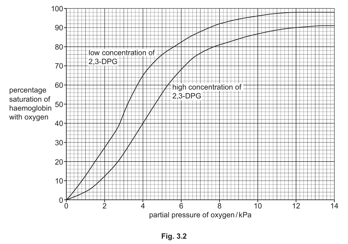
(d)(i) An increase in 2,3-DPG concentration causes the oxygen dissociation curve to shift to the right, indicating reduced oxygen affinity of haemoglobin.
Explanation: 2,3-DPG binds to haemoglobin and stabilizes its deoxygenated form, making it harder for haemoglobin to bind oxygen. This rightward shift means that at any given partial pressure of oxygen, haemoglobin will be less saturated with oxygen. The P₅₀ (partial pressure at which haemoglobin is 50% saturated) increases from about 3.2 kPa to 4.6 kPa, showing oxygen is more readily released to tissues.
(d)(ii) Blood from blood banks has reduced 2,3-DPG levels, causing haemoglobin to have higher oxygen affinity and release less oxygen to tissues during operations.
Explanation: During storage, red blood cells metabolize less and produce less 2,3-DPG. With lower 2,3-DPG, haemoglobin’s oxygen dissociation curve shifts left, increasing its oxygen affinity. This means haemoglobin holds onto oxygen more tightly and releases less to tissues during an operation, potentially causing tissue oxygen deprivation. The effect is temporary as 2,3-DPG levels are restored within 24-72 hours after transfusion.
▶️ Answer/Explanation
(a)(i)
Root as source: The root can provide assimilates like sucrose and amino acids for other parts of the plant when needed. This typically happens when the plant is growing and needs to distribute nutrients from storage areas.
Root as sink: The root acts as a storage organ, receiving assimilates from photosynthetic tissues (like leaves) to store for future use or to support its own growth. This bidirectional transport is a key feature of phloem tissue.
The direction of movement changes depending on the plant’s needs – during active growth periods, roots may be sinks receiving nutrients, while at other times they may serve as sources releasing stored nutrients.
(a)(ii)
Sucrose and amino acids are the main assimilates transported in the phloem. Other examples include certain hormones, mineral ions, and organic acids.
Sucrose is the primary transport sugar because it’s soluble, chemically stable, and contains high energy. Amino acids are transported as nitrogen-containing compounds needed for protein synthesis throughout the plant.
(b)
The movement of phloem sap occurs through the pressure flow hypothesis mechanism:
- At the source (typically leaves), sucrose is actively loaded into sieve tube elements from companion cells, either directly or through plasmodesmata.
- This increases the solute concentration in the phloem, lowering its water potential.
- Water then enters the phloem by osmosis from surrounding xylem vessels, creating a high hydrostatic pressure.
- At the sink (growing tissues or storage organs), sucrose is unloaded, decreasing the solute concentration.
- Water leaves the phloem by osmosis, reducing hydrostatic pressure at the sink.
- The pressure difference between source and sink causes mass flow of phloem sap through the sieve tubes.
This process is energy-dependent, requiring ATP for active loading and unloading of sugars, but the bulk flow itself is passive, driven by pressure gradients.
(c)
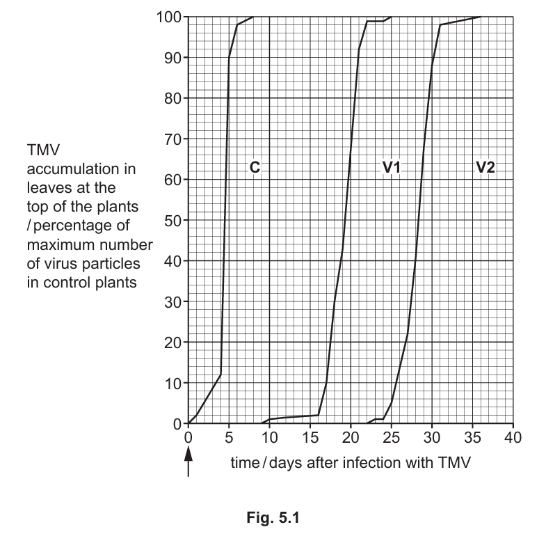
Comparison of TMV accumulation in V1, V2 versus control (C):
- The control plants (C) with normal PME levels showed rapid TMV accumulation, reaching maximum levels within about 8 days after infection.
- Variety V1 (low PME) showed delayed TMV accumulation, beginning around day 9 and reaching maximum levels by day 25.
- Variety V2 (low PME) showed even greater delay, with accumulation starting around day 22 and reaching maximum only by day 36.
- The rate of accumulation was slower in both V1 and V2 compared to the control, as seen by the less steep slopes of their curves.
- All varieties eventually reached the same maximum accumulation level, showing that low PME delays but doesn’t prevent full infection.
This suggests that PME plays a role in facilitating TMV movement through plants, and reduced PME levels significantly slow the systemic spread of the virus, though they don’t provide complete resistance.
▶️ Answer/Explanation
(a)
The ventricles have thicker walls than the atria because:
- Ventricles need to pump blood over greater distances – the left ventricle pumps blood to the entire body through systemic circulation, while the right ventricle pumps blood to the lungs through pulmonary circulation.
- The thicker muscular walls allow ventricles to generate higher pressures needed for these pumping actions. The left ventricle wall is especially thick as it must overcome the high resistance of systemic circulation.
- Atria only need to pump blood the short distance to the ventricles below them, requiring less muscular force and thus thinner walls.
This structural difference reflects their different functional demands in the circulatory system.
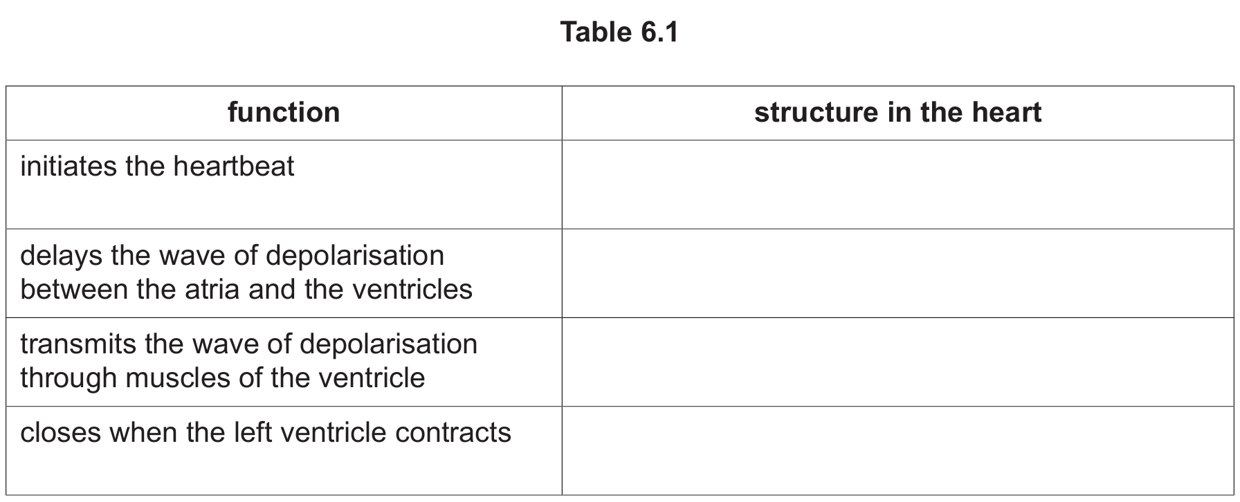
(b)
| function | structure in the heart |
|---|---|
| initiates the heartbeat | sinoatrial node (SAN) |
| delays the wave of depolarisation between the atria and the ventricles | atrioventricular node (AVN) and non-conducting tissue |
| transmits the wave of depolarisation through muscles of the ventricle | Purkyne (Purkinje) fibres |
| closes when the left ventricle contracts | left atrioventricular (bicuspid/mitral) valve |
Explanation:
- The sinoatrial node is the natural pacemaker that initiates each heartbeat through spontaneous depolarization.
- The atrioventricular node delays the electrical impulse to ensure atria contract before ventricles, with non-conducting tissue preventing direct electrical connection.
- Purkyne fibers are specialized conducting fibers that rapidly transmit the impulse through the ventricular walls for coordinated contraction.
- The left atrioventricular valve (bicuspid/mitral valve) closes when the left ventricle contracts to prevent backflow into the left atrium.
These structures work together to ensure efficient, coordinated heart function with proper timing between chambers.