▶️ Answer/Explanation
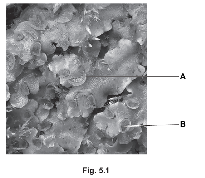
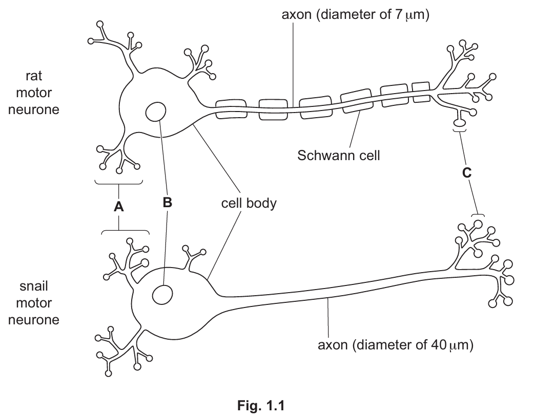
(a)(i)
A = dendrites
B = nucleus
C = synaptic knob
Explanation: The diagram shows key structures of motor neurones. A points to the branched extensions (dendrites) that receive signals. B indicates the nucleus, which contains genetic material. C marks the synaptic knob at the end of the axon where neurotransmitters are released.
(a)(ii)
The rat motor neurone has a myelin sheath/Schwann cells which allow saltatory conduction where the impulse jumps between nodes of Ranvier, making transmission faster. The snail neurone lacks this myelination.
Detailed Explanation: The rat’s motor neurone is myelinated, meaning it has Schwann cells wrapped around the axon forming an insulating layer. This allows the action potential to “jump” from one node of Ranvier to the next (saltatory conduction), significantly increasing transmission speed. Additionally, the rat’s axon has a larger diameter (40 μm vs snail’s 7 μm), which reduces resistance to ion flow. The snail’s unmyelinated neurone requires continuous propagation of the action potential along the entire axon, which is slower.
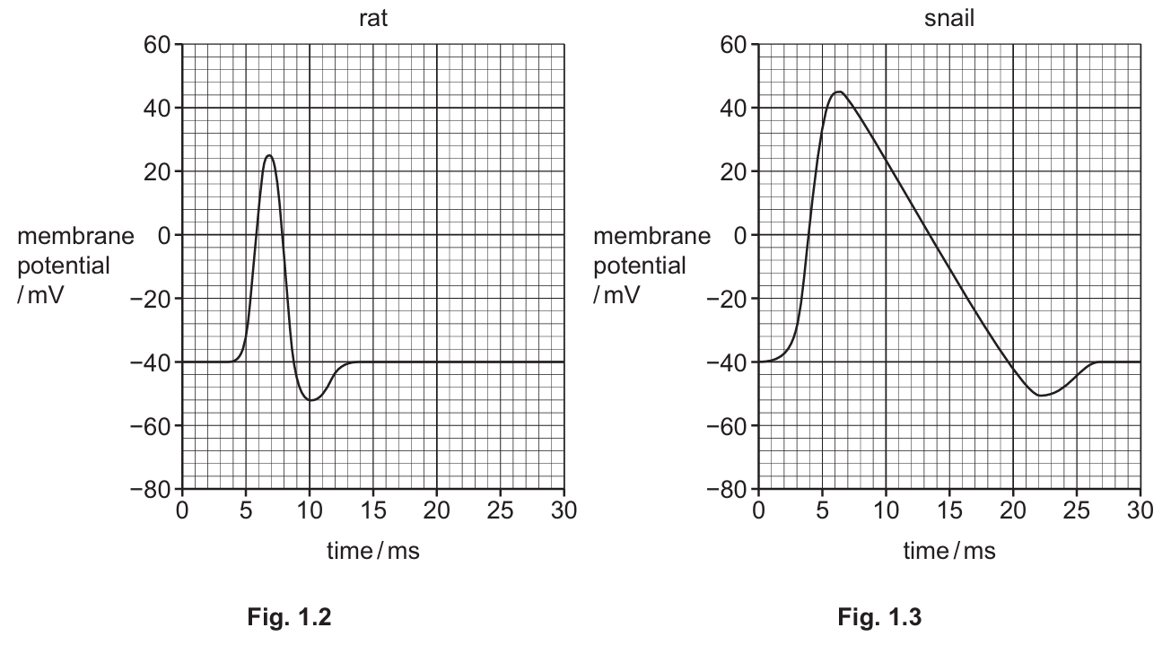
(b)
1. The snail action potential shows greater depolarization (higher peak membrane potential) compared to the rat.
2. The snail action potential takes longer to return to resting potential (slower repolarization).
3. The snail neurone has a longer refractory period before it can fire again.
Detailed Explanation: The graphs reveal significant differences in action potential characteristics between species. The snail’s action potential reaches a higher peak voltage (about +40mV vs rat’s +30mV) and has a more prolonged duration. The repolarization phase in snails is much slower, resulting in a longer absolute refractory period where the neurone cannot fire again. This slower recovery is likely due to differences in ion channel density and kinetics. The rat’s action potential is more rapid, allowing for faster neural processing, which is adaptive for its more complex nervous system requirements. The hyperpolarization (undershoot) phase is also more pronounced in the snail, suggesting differences in potassium channel activity.
▶️ Answer/Explanation
(a)
Plants open and close their stomata in a daily rhythm because:
- Stomata open during the day/light periods to allow carbon dioxide to enter for photosynthesis (light-independent stage/Calvin cycle).
- They close during the night/dark periods to reduce water loss through transpiration when photosynthesis isn’t occurring.
- This rhythm helps balance the plant’s need for CO₂ uptake with the need to conserve water.
Explanation: The daily rhythm of stomatal opening is crucial for plant survival. During daylight hours when photosynthesis is active, open stomata allow CO₂ to diffuse into leaves. However, open stomata also lead to water loss through transpiration. At night when photosynthesis stops, closing stomata conserves water while minimally affecting CO₂ uptake. This rhythm represents an evolutionary adaptation to optimize both gas exchange and water conservation.
(b)
The sequence of changes in guard cells leading to stomatal opening:
- Protons (H⁺ ions) are actively pumped out of guard cells using ATP energy.
- This creates a more negative charge inside the cells and opens potassium ion (K⁺) channels.
- K⁺ ions enter the guard cells through facilitated diffusion.
- Chloride ions (Cl⁻) also move into the cells to balance the charge.
- The increased solute concentration lowers the water potential in guard cells.
- Water enters the cells by osmosis, making them turgid.
- The guard cells’ unique structure (thicker inner walls) causes them to bend outward when turgid, opening the stoma.
Explanation: The process begins with light activation of blue-light receptors in guard cells, triggering proton pumps. As ions accumulate, the osmotic gradient drives water influx. The resulting turgor pressure changes are possible because guard cells have radial cellulose microfibrils that constrain expansion in certain directions, causing the characteristic kidney-shaped curvature that opens the pore between them.
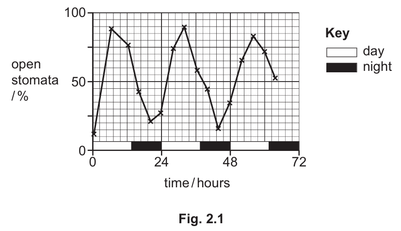
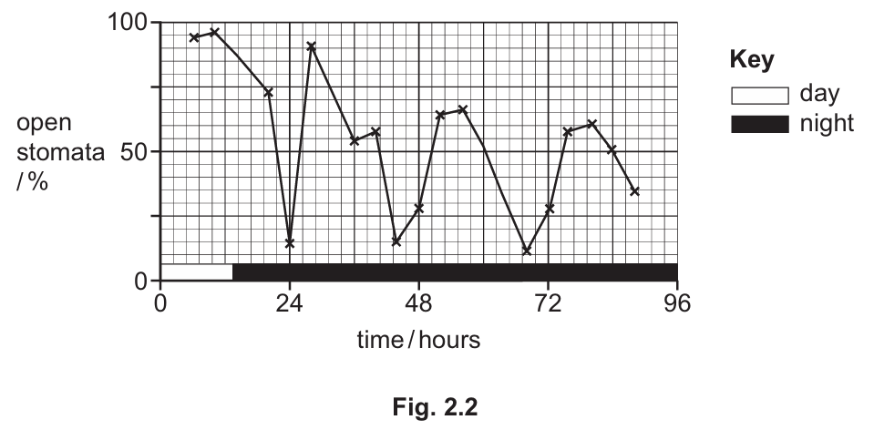
(c)
Fig. 2.2 shows:
- The environment (light) is needed to maintain the normal rhythm and achieve the same peak percentage of stomatal opening seen in Fig. 2.1.
- Genes play a role because a regular rhythm/pattern continues even in continuous darkness, though with decreasing amplitude.
- This demonstrates that stomatal rhythm is controlled by both environmental cues (light) and an endogenous circadian clock (genetic control).
Explanation: The persistence of rhythmic opening in darkness indicates an underlying biological clock (controlled by genes) that can maintain approximate 24-hour cycles without external cues. However, the decreasing amplitude shows that environmental light is required to fully sustain and synchronize the rhythm. This is characteristic of circadian rhythms in general – they’re genetically programmed but require environmental input (zeitgebers) to remain precisely timed to 24 hours. The experiment demonstrates that A. thaliana has evolved both genetic and environmental response mechanisms to optimize stomatal function.
▶️ Answer/Explanation
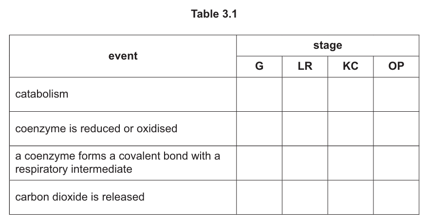
(a) 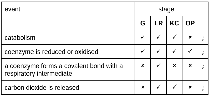
Table 3.1 completed:
Explanation:
In glycolysis (G), glucose is broken down (catabolism) and NAD+ is reduced to NADH, but no CO₂ is released. The link reaction (LR) involves pyruvate decarboxylation (CO₂ release), reduction of NAD+, and formation of acetyl-CoA (coenzyme covalent bonding). The Krebs cycle (KC) continues catabolism, reduces NAD+ and FAD, and releases CO₂. Oxidative phosphorylation (OP) involves redox reactions of coenzymes but no catabolism or CO₂ release.
(b)(i)
The device calculates RQ as the ratio of CO₂ produced to O₂ consumed (RQ = CO₂/O₂). For carbohydrates, RQ = 1 (C₆H₁₂O₆ + 6O₂ → 6CO₂ + 6H₂O). For lipids, RQ ≈ 0.7 (more O₂ needed per CO₂ produced). The air-flow meter measures O₂ inhaled, and the CO₂ sensor measures CO₂ exhaled. Higher RQ values indicate carbohydrate respiration, while lower values indicate lipid respiration.
Explanation:
The respiratory quotient differs because lipids contain more hydrogen atoms per carbon atom than carbohydrates, requiring more oxygen for complete oxidation. The device measures both gases to determine which substrate is primarily being metabolized. This is useful for understanding metabolic states like fasting (lipid-dominated) or high-carb diets.
(b)(ii)
Lipids have higher energy value (~37 kJ/g) than carbohydrates (~16 kJ/g). This is because:
- Lipids contain more C-H bonds which release energy when oxidized
- Lipids are more reduced molecules, yielding more reduced NAD/FAD
- Greater proton gradient is generated from lipid oxidation
- Lipids are anhydrous while carbohydrates are hydrated
Explanation:
The higher energy content of lipids stems from their chemical structure. Fatty acids have long hydrocarbon chains with many C-H bonds that release substantial energy when broken. Additionally, the complete oxidation of lipids generates more electron carriers (NADH and FADH₂) which drive oxidative phosphorylation to produce more ATP compared to carbohydrates.
▶️ Answer/Explanation
(a)
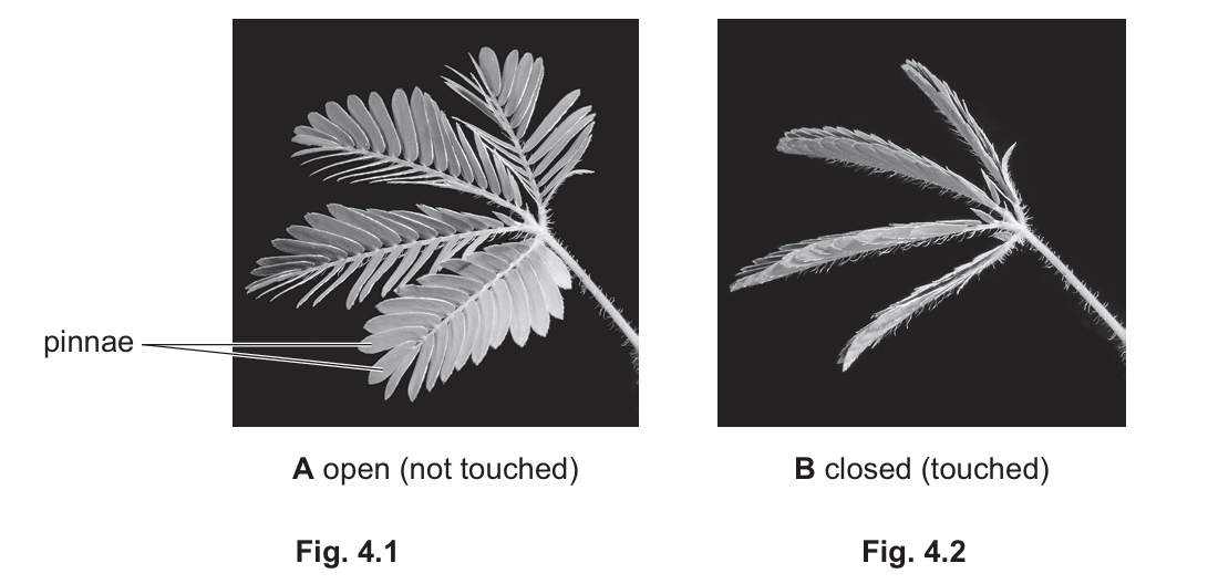
The mechanism in Mimosa pudica involves:
- Protons being pumped into extensor cells
- Chloride (Cl⁻) and potassium (K⁺) ions moving out of cells
- Water moving out of cells by osmosis
- Cells becoming flaccid and causing leaf closure
In contrast, Venus fly traps:
- Require two sensory hairs to be touched (not just one)
- Have protons leaving hinge cells (rather than entering)
- Have calcium ions (Ca²⁺) moving into cells (rather than K⁺ and Cl⁻ moving out)
- Have water moving into cells (rather than out), making them turgid
Explanation: While both plants show rapid movement responses, their cellular mechanisms are quite different. Mimosa pudica’s response is essentially a loss of turgor pressure, while Venus fly traps create turgor pressure changes in opposite directions in different cells to snap the trap shut.
(b)
- The closed leaves have less surface area exposed to light, reducing light absorption
- Fewer chloroplasts are exposed to light when the leaves are folded
- The closed position may limit gas exchange, reducing CO₂ availability
- Some stomata become covered when leaves fold, further limiting CO₂ intake
Explanation: Photosynthesis requires both light and carbon dioxide. When the leaves fold, they receive less light because the surface area facing the light source decreases. Additionally, the folding may cover some stomata, limiting CO₂ diffusion into the leaf. Both factors contribute to the significant 40% reduction in photosynthetic rate.
(c)
Similarities:
- Both occur in the thylakoid membranes of chloroplasts
- Both involve photoactivation of chlorophyll (PSI)
- Both use electron transport chains to create a proton gradient
- Both produce ATP through chemiosmosis
Differences:
- Cyclic uses only PSI while non-cyclic uses both PSI and PSII
- Non-cyclic produces NADPH (reduced NADP) while cyclic does not
- Non-cyclic involves photolysis of water and oxygen evolution while cyclic does not
- In cyclic, electrons return to PSI while in non-cyclic they end up in NADPH
- Non-cyclic produces both ATP and NADPH while cyclic produces only ATP
Explanation: These two pathways represent different strategies for light-dependent reactions. Non-cyclic photophosphorylation is the main pathway that produces both ATP and NADPH needed for the Calvin cycle, while cyclic photophosphorylation serves as a supplementary pathway that generates extra ATP when needed, without producing NADPH or oxygen.