(a) Fig. 3.1 is a photomicrograph of a transverse section through a region of the wall of the bronchus in the gas exchange system.
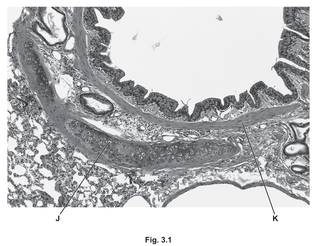
Identify the tissues J and K shown in Fig. 3.1, and suggest how the wall of a bronchiole differs from the wall of the bronchus for these two tissues.
(b) Tuberculosis (TB) is an infectious disease that affects the human gas exchange system.
The pathogen that causes TB secretes a protein that can be detected in saliva.
Early diagnosis of TB is important in reducing the transmission of the pathogen.
Scientists have developed a test strip for TB that uses monoclonal antibodies. Monoclonal antibodies are specific in their action.
This test strip contains:
- mobile monoclonal antibodies that bind to one part of the protein secreted by the pathogen
- immobilised monoclonal antibodies.
Fig. 3.2 shows a simplified diagram of the test strip.
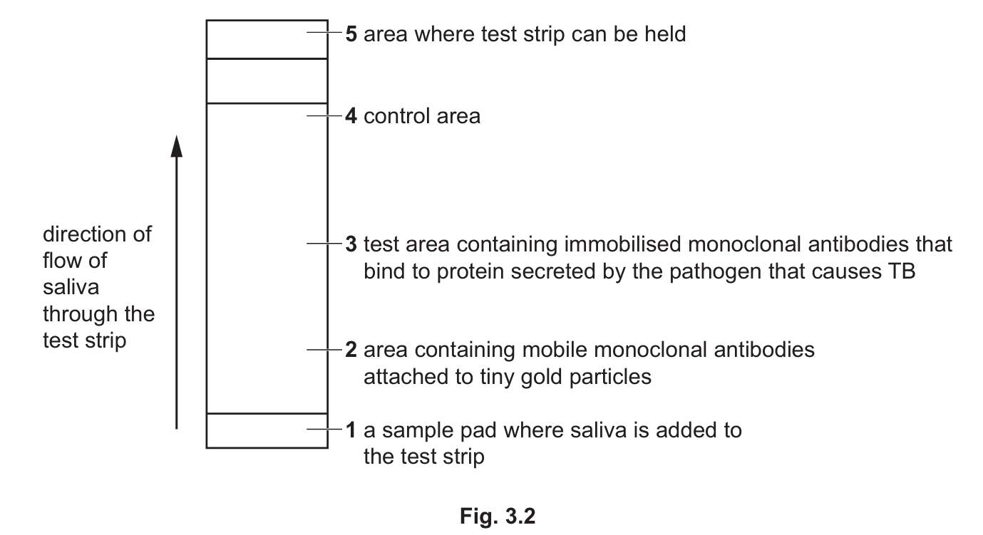
A sample of saliva is collected and put onto the sample pad in the test strip.
The saliva moves up the test strip through area 2.
The mobile monoclonal antibodies are attached to tiny gold particles. If these antibodies collect in test area 3, a gold line becomes visible on the test strip.
A gold line that becomes visible in area 4 confirms that the test strip is working and that the results are valid.
(i) State the name of the pathogen that causes TB.
(ii) Name the part of the monoclonal antibody that binds to the protein from the pathogen.
(iii) Saliva is added to a test strip to test for the presence of the protein secreted by the TB pathogen.
Fig. 3.3 is a diagram showing some of the molecules in area 3 of the test strip when a positive result for TB is obtained.
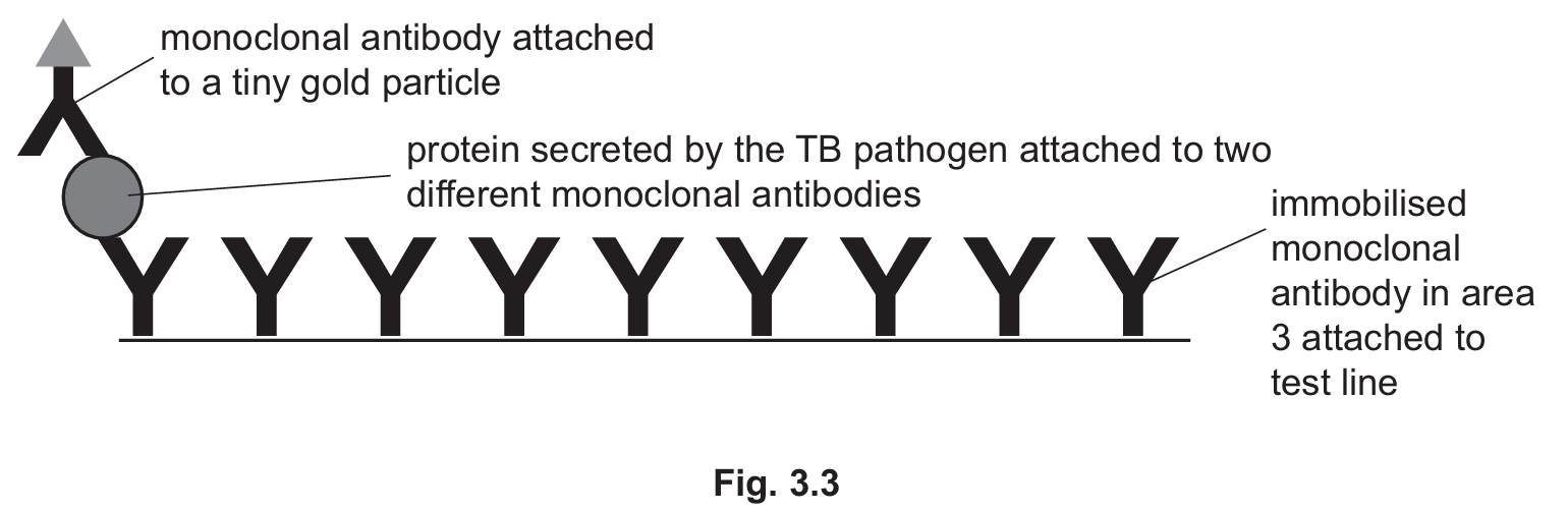
Use the information in Fig. 3.3 to suggest and explain why this test is specific for TB.
(iv) Area 4 contains different immobilised antibodies to those in area 3.
The mobile monoclonal antibodies bound to tiny gold particles will bind to these immobilised monoclonal antibodies in area 4.
If the test has functioned correctly, a gold line will be visible in area 4.
Suggest how the structure of immobilised monoclonal antibodies in area 3 differs from the structure of the immobilised monoclonal antibodies in area 4.
(c) Vaccination is another way of reducing the transmission of infectious diseases such as TB. The BCG vaccine is used to help control the spread of TB. This vaccine contains a weakened strain of the pathogen that causes TB. The BCG vaccine stimulates the development of antigen-specific memory T-lymphocytes.
Explain how memory T-lymphocytes provide protection from TB in a person who has been given a BCG vaccination.
(d) The bladder is the organ in the body used to store urine.
When cells divide uncontrollably in the bladder, a tumour develops. This can lead to bladder cancer.
The BCG vaccine has been used to treat bladder cancer.
The BCG vaccine is introduced into the bladder. The tumour cells take up the weakened pathogens in the vaccine and act as antigen-presenting cells.
(i) Name the process used by the tumour cells to take up the weakened pathogens.
(ii) Suggest how antigen presentation by tumour cells stimulates an immune response that leads to the destruction of the tumour cells.
▶️ Answer/Explanation
(a)
J: Cartilage
K: Smooth muscle
Difference: The bronchus contains cartilage in its wall which provides structural support, while bronchioles lack cartilage. Bronchioles have proportionally more smooth muscle tissue in their walls compared to bronchi, allowing for greater control over air flow.
Explanation: The bronchus is a larger airway that requires cartilage rings to prevent collapse during breathing. As airways become smaller (bronchioles), the cartilage disappears but smooth muscle becomes more prominent to regulate airflow through contraction and relaxation.
(b)(i) Mycobacterium tuberculosis / Mycobacterium bovis
Explanation: These are the bacterial species known to cause tuberculosis in humans. Mycobacterium tuberculosis is the primary causative agent, while Mycobacterium bovis is a less common cause typically associated with cattle.
(b)(ii) Antigen binding site(s)
Explanation: The variable region of the monoclonal antibody contains the antigen binding site that is specifically shaped to recognize and bind to a particular epitope on the TB protein.
(b)(iii) The test is specific for TB because the monoclonal antibodies are designed to bind only to the protein secreted by the TB pathogen. The immobilised monoclonal antibodies in the test area have binding sites that are complementary in shape to specific epitopes on the TB protein, ensuring they won’t bind to proteins from other pathogens.
Explanation: The specificity comes from the precise molecular complementarity between the antibody’s binding site and the TB protein’s epitope. This is like a lock-and-key mechanism where only the correct key (TB protein) will fit the lock (antibody binding site). The gold particles only become visible in the test line if this specific binding occurs.
(b)(iv) The immobilised monoclonal antibodies in area 3 have a different variable region/antigen binding site structure compared to those in area 4. They have different primary structures (amino acid sequences) leading to different tertiary structures and binding specificities.
Explanation: While both sets of antibodies are monoclonal, they are produced to recognize different epitopes. The antibodies in area 3 bind the TB protein, while those in area 4 are designed to bind the mobile antibodies (acting as a control). This difference in function requires different molecular structures in their binding sites.
(c) Memory T-lymphocytes provide long-term immunity by remaining in the body after vaccination. If the actual TB pathogen later enters the body, these memory cells can mount a rapid, strong secondary immune response. They quickly recognize the TB antigens, activate other immune cells, and help destroy the pathogen before it can cause disease.
Explanation: The BCG vaccine primes the immune system by exposing it to weakened TB bacteria. This stimulates the production of memory T-cells that “remember” the TB antigens. Upon subsequent exposure, these memory cells can proliferate quickly and coordinate immune defenses much faster than during the initial exposure, often preventing infection from becoming established.
(d)(i) Endocytosis
Explanation: The tumor cells engulf the weakened pathogens from the vaccine through endocytosis, a process where the cell membrane invaginates to form a vesicle containing the external material.
(d)(ii) When tumor cells present the BCG antigens on their surface, T-lymphocytes with complementary receptors recognize these foreign antigens. This activates the T-cells, which then multiply and differentiate into cytotoxic T-cells that can directly kill the tumor cells. Helper T-cells also stimulate other immune cells like macrophages to attack the tumor.
Explanation: The presentation of BCG antigens by tumor cells essentially marks them as targets for the immune system. Cytotoxic T-cells recognize these antigen-presenting tumor cells and destroy them through various mechanisms like releasing perforins and granzymes that induce apoptosis. This immune response can lead to significant tumor shrinkage.