Question [Maximum marks: 13]
Haemoglobinopathies are inherited conditions linked to the structure and function of
haemoglobin. Sickle cell anaemia is one of these conditions in which the transport and
delivery of oxygen to tissues is less than normal.
An investigation was carried out to discover the effect of sickle cell anaemia on the ability of
blood to carry oxygen. Blood samples were taken from two people:
• person L without sickle cell anaemia
• person M with sickle cell anaemia.
The percentage saturation of haemoglobin with oxygen was determined over a range of
partial pressures of oxygen.
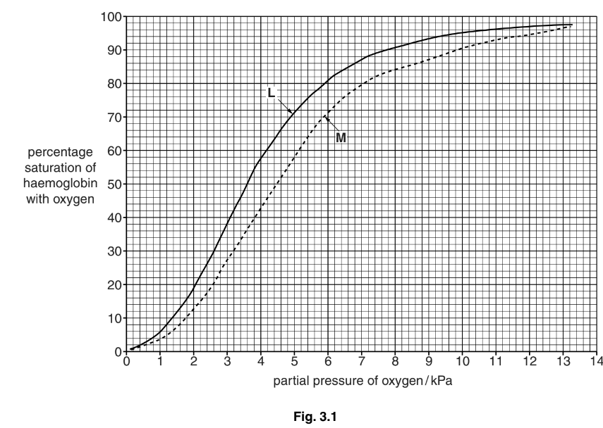
Fig. 3.1 shows oxygen haemoglobin dissociation curves for the two blood samples.

(a) P50 is the partial pressure of oxygen at which haemoglobin is 50% saturated with
oxygen. It is taken as a measurement of the affinity of haemoglobin for oxygen.
(i) State the P50 for the two blood samples, L and M.
(ii) With reference to Fig. 3.1, describe how the dissociation curve for person M differs from the dissociation curve for person L.
(b) Explain the advantage of the position of the dissociation curve for people with sickle cell
anaemia.
(c) The partial pressure of oxygen in the lungs at sea level is about 13.5 kPa. At an altitude
of 3000 metres the partial pressure of oxygen in the lungs is about 7.5 kPa.
When people move from sea level to high altitude they become adapted to the low
partial pressure of oxygen.
Describe and explain how humans become adapted to the low partial pressure of
oxygen at high altitude.
(d) Vaccination is used to control the spread of diseases, such as measles.
Explain why vaccination cannot be used to prevent sickle cell anaemia.
Answer/Explanation
Answer: 3(a) (i) no mark if no units used at all
L – 3.6 kPa ; award the mark if units only used once
M – 4.5 kPa ; A in range 4.45 to 4.55
(ii) ignore any similarities
1 to the right / lower (affinity) / qualified ; e.g. lower percentage saturation
2 at, higher / lower, partial pressures, small(er) difference in percentage saturation (than others) ; A ora 3 comparative data quote ; must refer to L and M
allow ecf from (i)
3(b) 1 at partial pressures in the tissues ; where oxygen is unloaded from Hb
2 haemoglobin is less saturated (than L) ;
3 because, haemoglobin / Hb, dissociates more readily ;
A idea of unloading oxygen more readily even if Hb not mentioned
4 to compensate for, fewer / less effective, red blood cells / Hb ;
3(c) 1 haemoglobin less well saturated (in lungs at high altitude) ;
2 data quote from Fig. 3.1 ; A 80–90% saturated at ‘about 7.5 kPa’
3 produce more red blood cells / increase in number of RBCs ;
4 more haemoglobin ;
5 idea of compensates for, smaller volume of oxygen absorbed / lower saturation (of haemoglobin) ;
also accept the following adaptations
6 increase in haematocrit / AW / decrease in plasma volume ;
A increase in RBCs per unit volume
R decrease in blood volume
7 increase in, breathing rate / tidal volume / heart rate / stroke volume ;
8 increase in, capillary density / number of mitochondria / myoglobin / respiratory enzymes, in muscle ; 9 ref. to (increased) secretion of, erythropoietin / EPO ;
10 increase in (2,3), BPG / DPG, in red blood cells ; A rightward shift in curve [max 4]
3(d) 1 not caused by (named type of) pathogen / non-infectious / non-transmissible / non-
communicable / AW ; 2 genetic / inherited / AW, disease ; A caused by a mutation / AW
A ‘passed down from parent(s)’
R idea of congenital diseases
R ‘you get it from your mother’
3 ref. to, no immune response / no antigen(s) ;
4 affects all red blood cells so vaccine would lead to their destruction ; [max 2]
Question
(a) Explain how the virus that causes measles is transmitted. [2]
(b) Antibodies against measles are produced by plasma cells during an immune response.
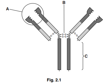
Fig. 2.1 shows a diagram of an antibody molecule.

Explain the functions of the parts labelled A, B and C.
(i) A [2]
(ii) B [1]
(iii) C [1]
[Total: 6]
Answer/Explanation
Ans:
2 (a) (infected) person, sneezes / coughs / talks / breathes out, (airborne)
droplets / aerosol/ moist air ;
ignore contact
inhaled/ inspire/ breathed in, by uninfected, person ;
ignore transplacental transmission
(b) (i) variable region
binds / attaches / combines, to antigen ;
R receptor site R ‘fit’
ref. to specificity ;
ignore complementary shape (to antigen)
R same/ similar shape
(ii) disulphide bond
ignore ref. to hinge
holds, polypeptides /heavy chains / long chains, together ;
ignore constant as description of chains
maintains, tertiary / quaternary / 3D, structure/ shape ;
R shape unqualified
(iii) constant region
binds to, receptors / cell (surface) membrane, on, phagocytes / macrophages ;
antigen, marking/ tagging, for, phagocytosis / macrophage action ; AW
A ref. to opsonisation
R agglutination
Question
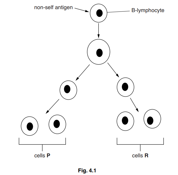
B-lymphocytes respond to the presence of a non-self antigen by dividing as shown in Fig. 4.1.

(a) (i) Explain what is meant by the term non-self antigen.[2]
(ii) Outline how B-lymphocytes recognise non-self antigens.[2]
The cells labelled P on Fig. 4.1 continue to divide to give rise to many cells that differentiate into short-lived plasma cells. The plasma cells release antibody molecules.
(b) (i) Outline how plasma cells produce antibody molecules.[4]
(ii) Describe how antibody molecules are released from the plasma cell.[2]
(c) The cells labelled R on Fig. 4.1 divide to give more cells that do not differentiate into plasma cells. These cells have an important role in the immune system.
Explain the role of these cells.[3]
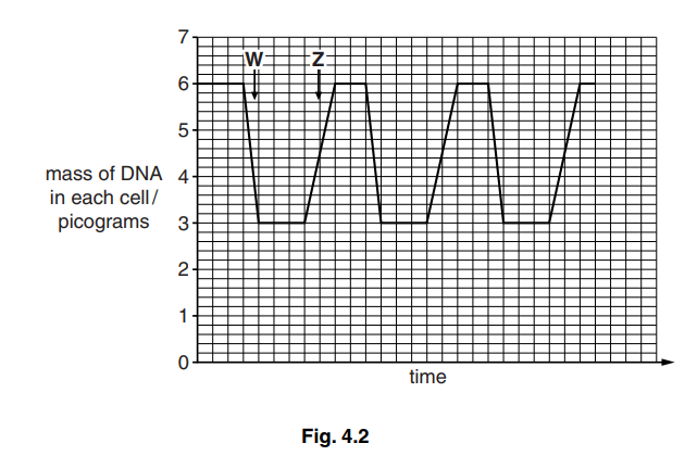
The mass of DNA in the cells shown in Fig. 4.1 was determined. The results are shown in Fig. 4.2.

(d) State what happens at W and Z to change the mass of DNA in each cell.[2]
W
Z
(e) Acute lymphoblastic leukaemia (ALL) is a cancer of B-lymphocytes. It is very rare in adults, but more common in children. A study in 2009 found that exposure to tobacco smoke in the home may put children at risk of developing ALL.
Suggest how smoking by adults in the home may put their children at risk of cancers, such as ALL.[3] [Total: 18]
Answer/Explanation
Ans:
4 (a) (i) non-self
foreign/AW ; A ref. to epitope(s) I pathogen/organism
antigen
macromolecule/(glyco)protein/ carbohydrate/ polysaccharide/ oligosaccharide ;
stimulates /AW, an immune response/production of antibodies ;
A results in formation of antigen-antibody complexes
A other described events in an immune response
(ii) antibody / immunoglobulin/ IgG, on cell surface/on cell membrane ; (act as) receptors ;
ref. to antigen-binding/AW ;
(shape) specific / complementary, to antigen ;
(b) (i) DNA/ gene transcribed/ mRNA using DNA as template/AW ;
A transcription unqualified
idea of mRNA associating with ribosome(s) ;
ref. to tRNA with specific amino acid (carried to ribosome) ;
pairing/AW of codons on mRNA with anticodons on tRNA ;
formation of peptide bonds (between adjacent amino acids) ;
antibody /protein/ polypeptide(s), enters RER/ moves to Golgi body ;
ref. to forming, secondary / tertiary structure ;
antibody /protein/ polypeptide(s), modified/processed/glycosylated/ formation
of quaternary structure/ formation of disulphide bond(s) in Golgi (body / apparatus / complex) ; I ref. to packaging
(ii) vesicles move to cell/ surface/plasma, membrane (via cytoskeleton) ;
R secreting vesicles unqualified
vesicles fuse with cell (surface) membrane/exocytosis ; R active transport
movement of vesicle/ exocytosis requires energy or ATP/ is active ;
(c) memory cells ; A form immunological memory I ‘gives immunity’
remain/ stay in circulation/ blood/lymphatic system ;
R ‘last a long time/ long lived’ unqualified
for secondary response ;
fast(er) response when exposed again to same pathogen/ same antigen ;
A fast(er) clonal selection/ fast(er) clonal expansion
A divide quickly /rapidly
A long(er) lasting response
to form plasma cells (and more memory cells) ;
more antibodies produced/higher concentration of antibodies ;
R if in context of memory cells
to prevent person feeling ill/ to prevent symptoms ;
(d) W – cytokinesis / cytoplasmic division/ cell divides into two ;
I cell division
R mitosis / telophase
Z – (semi-conservative) replication (of DNA) ;
I S phase/ interphase of cell cycle
R copying of DNA
R protein synthesis
R if replication is given in any other phase of the cell cycle
(e) 1 breathing in/ inhale smoke/ ‘second hand’ smoke/ sidestream smoke ;
A passive smoking
I exposed to smoke
2 (tobacco smoke contains) carcinogen(s) ;
3 causes mutation/ described ;
e.g. change to/alters / damages, DNA R if in wrong type of cell
4 leads to uncontrolled cell division/ mitosis / growth ;
5 forming a tumour/ mass of cells ;
6 correct ref. to (proto-)oncogenes /tumour suppressor genes ;
e.g. formation of oncogenes / mutation of tumour suppressor genes / ‘switching off’ tumour suppressing genes
mutation of correct named gene = 2 marks
e.g. mutation of tumour suppressor gene
P53 (gene) mutates = 2 marks
