▶️ Answer/Explanation
(a)(i)
1. (Excess) amino acids are deaminated/have amino group removed.
2. This occurs in the liver.
Explanation: Urea is formed as a waste product when excess amino acids are broken down in the liver through a process called deamination. The amino group (-NH2) is removed from amino acids and converted into ammonia, which is then combined with carbon dioxide to form urea in the ornithine cycle. This urea is then transported to the kidneys for excretion.
(a)(ii)
Any three from:
1. Osmoreceptors in hypothalamus (detect change/are receptors).
2. Effector(s): collecting duct/distal convoluted tubule/nephron.
3. Target cells: collecting duct (cells).
Explanation: The hypothalamus contains osmoreceptors that detect changes in blood water potential. When blood becomes too concentrated, these receptors stimulate the posterior pituitary to release ADH (antidiuretic hormone). The effectors are parts of the nephron, particularly the collecting duct and distal convoluted tubule, which respond to ADH by increasing water reabsorption. The target cells are specifically the principal cells in the collecting ducts that insert aquaporins into their membranes in response to ADH.
(b)
Any three from:
1. Ultrafiltration/forms glomerular filtrate.
2. High hydrostatic/blood pressure in glomerulus/capillaries.
3. So water/(named) solutes move (out of glomerulus/into Bowman’s capsule).
4. Through two of: fenestrae/fenestrations, basement membrane, slit pores/filtration slits/between podocytes.
Explanation: The glomerulus and Bowman’s capsule work together to perform ultrafiltration. The high hydrostatic pressure in the glomerular capillaries forces small molecules (water, glucose, amino acids, urea) through the filtration barrier consisting of fenestrated capillary endothelium, basement membrane, and podocyte filtration slits. Larger molecules like proteins and blood cells remain in the blood. This process forms the glomerular filtrate which then enters the nephron tubules.
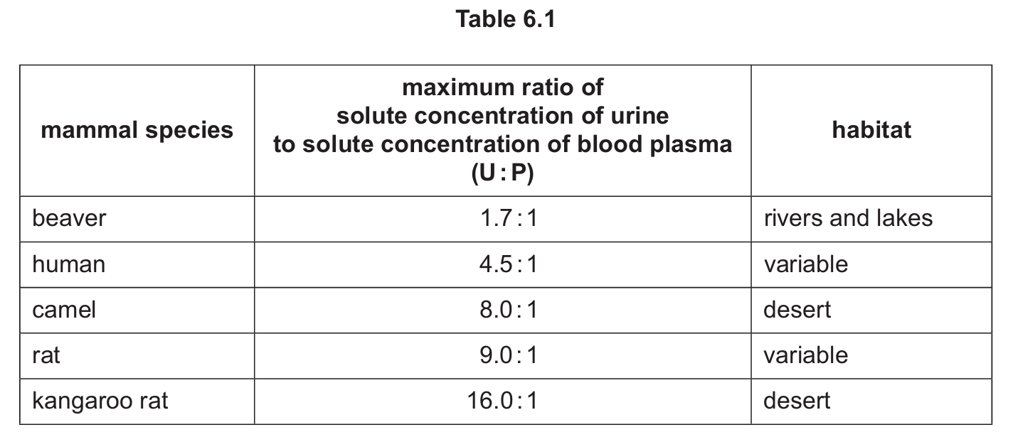
(c)
Any three from:
1. Higher U:P ratio → can tolerate water shortage.
2. Higher U:P ratio → much water reabsorbed/small volume of urine produced.
3. Higher U:P ratio → can concentrate urine/conserve water OR higher U:P ratio → (well) adapted to dry environment.
Explanation: The U:P ratio indicates how effectively a mammal can concentrate its urine. Desert species like the kangaroo rat (16:1) and camel (8:1) have much higher ratios than aquatic species like the beaver (1.7:1), showing their superior ability to conserve water. This is achieved through longer loops of Henle that create a stronger medullary concentration gradient, allowing more water reabsorption from collecting ducts when ADH is present. The high ratios in desert species demonstrate their adaptation to arid environments where water conservation is crucial for survival.