Question
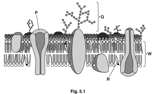
Fig. 5.1 shows a section of a cell surface membrane.

(a) State the functions of structures P, Q and R.[3]
P
Q
R
(b) Circle the width of the membrane shown as W in Fig. 5.1.
17.0 μm 1.7 μm 0.7 μm 70.0 nm 17.0 nm 7.0 nm 0.7 nm [1]
(c) Membranes, such as the cell surface membrane, are described as having a fluid mosaic structure.
Explain what is meant by the term fluid mosaic.[2]
(d) Aquaporins are membrane channel proteins in plant and animal cells. They permit the movement of water across membranes. Explain why they are necessary.[3] [Total: 9]
Answer/Explanation
Ans:
5 (a) P – moves, polar substances /hydrophilic molecules / ions, through membrane/ in or out (of cells) ;
A facilitated diffusion/ active transport/ described
Q – receptor/recognition site/ cell recognition/ binding site ;
A cell adhesion/ ‘receives’ named signal
A stabilises membrane (as forms hydrogen bonds with water)
R – regulates /AW, fluidity of, membrane/(phospholipid) bilayer ;
A contributes to hydrophobic layer/ impermeability to ions
(b) 7.0 nm ;
(c) fluid
idea of phospholipid (and protein) molecules, move about/ diffuse (within their monolayer) ;
mosaic to max 1
protein (molecules), interspersed/ scattered/not a complete layer/AW ; different/AW, proteins (molecules) ;
(d) 1 water molecules are polar ;
R hydrophilic / charged
2 idea that few polar molecules pass through the phospholipid (bilayer) ; ora for non-polar molecules
A none pass /repelled
3 core of membrane/ phospholipid tails, are hydrophobic ;
A hydrophobic core
4 channels (through aquaporins), are hydrophilic ;
A core of channel proteins /described as R-groups of amino acids
5 (aquaporins) increase permeability of membrane to water ;
6 example ;
e.g. root hairs / small intestine epithelium/ nephron
7 role of water in a cell ;
e.g. solvent /reactant/reaction medium/ turgidity or support in plant cell
ignore references to osmosis / bursting/ crenation/regulation
Question
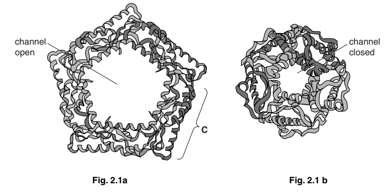
Fig. 2.1 is a diagram of the structure of a protein channel for ions in a cell surface membrane.
Fig. 2.1a shows the channel when open and Fig. 2.1b shows the same channel when closed.

(a) (i) Name the process by which ions pass across the membrane using channel proteins.[1]
(ii) Explain why a channel protein is needed for ions to pass across a cell membrane.[2]
(b) The channel protein in Fig. 2.1 is made from five identical polypeptide chains.
(i) Name the level of protein structure which is present when five polypeptide chains form the protein.[1]
(ii) The part labelled C in Fig. 2.1 is another level of protein structure.
Name this level.[1]
(c) Channel proteins are examples of transmembrane proteins. The polypeptides are held together and also interact with phospholipids in the membrane.
Suggest how the polypeptides are held together and suggest how they interact with phospholipids.[3] [Total: 8]
Answer/Explanation
Ans:
2 (a) (i) facilitated diffusion ;
(ii) ions are, charged/water-soluble ; A hydrophilic
unable to pass, through hydrophobic core/ hydrophobic (fatty acid) tails of,
phospholipid bilayer/ phospholipids(s) ;
(channel of) protein lined with amino acids with, hydrophilic/ polar, R groups /
side chains ; A hydrophilic channels
(b) (i) quaternary / 4°, (structure) ;
(ii) secondary structure ; A alpha/α, helix
(c) bonds must be named in the correct context of maintaining 4° structure and interactions with phospholipids
polypeptides held together
bonds between, R groups / side chains ;
two named bond types ; from
ionic
hydrogen
hydrophobic interactions
disulfide
van der Waal’s forces
I peptide bond
polypeptides interact with phospholipids
(regions with) hydrophilic / charged/ polar (R groups / side chains, of) amino acids
interact with, phosphate/ hydrophilic head , of phosholipid ;
(regions with) hydrophobic /non-polar (R groups / side chains, of) amino acids
interact with, fatty acid/ hydrocarbon/ hydrophobic, tails / chains ;
further detail of named bond ;
Question
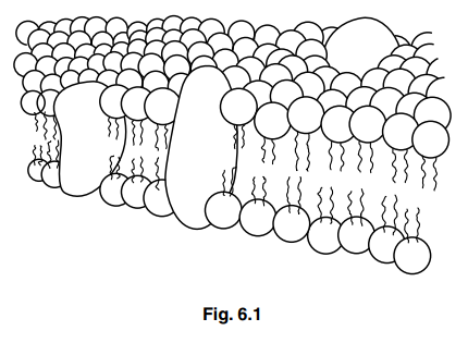
Fig. 6.1 shows an incomplete diagram of the fluid mosaic model of membrane structure. The diagram shows the cell surface membrane of a eukaryotic cell.

(a) State what is meant by the term fluid mosaic.[2]
(b) State the thickness of a cell surface membrane.[1]
(c) List four features of cell surface membranes of eukaryotic cells that are not visible in Fig. 6.1.[4]
1
2
3
4
[Total: 7]
Answer/Explanation
Ans:
6 (a) fluid
phospholipids (and proteins), move/AW ;
mosaic
proteins / glycoproteins, scattered/AW (in the phospholipid bilayer) ;
A different types of proteins
I pattern unqualified
(b) 7nm ; A any size or range within 6nm and 10nm
A 7nanometres
(c) cholesterol ;
unsaturated fatty acids ; A phospholipid tails
carbohydrate chains added to protein(s)/ glycoproteins ;
A oligosaccharides for carbohydrate chains
carbohydrate chains added to lipids /glycolipids ;
glycocalyx ;
channel protein(s)/AW ; A aquaporin(s) ;
carrier proteins /AW ;
peripheral/ extrinsic, proteins ;
attachment to, cytoskeleton/ microfilaments ;
receptor(s) ;
antigen(s) ;
AVP ;
