▶️ Answer/Explanation
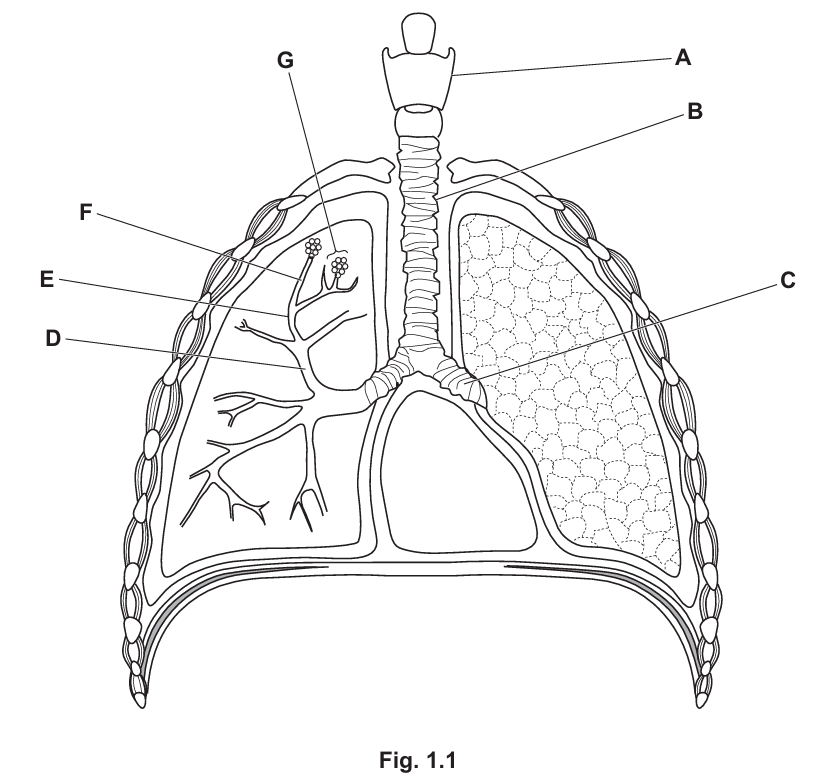
(a)(i) B and C
Explanation: In the human gas exchange system, cartilage is present in the trachea (B) and bronchi (C). These structures need cartilage to maintain their shape and prevent collapse during breathing.
(a)(ii) Any two from:
- Keeps the airways open/prevents collapse of airways
- Allows flexibility (especially important in the trachea during swallowing)
- Provides structural support to maintain airway patency
Detailed Explanation: Cartilage plays crucial roles in the respiratory system. In the trachea and bronchi, the C-shaped cartilage rings prevent these tubes from collapsing during inhalation when air pressure decreases. The incomplete rings at the back allow the esophagus to expand during swallowing. Cartilage also provides flexibility, enabling the neck to bend without obstructing airflow.
(a)(iii) 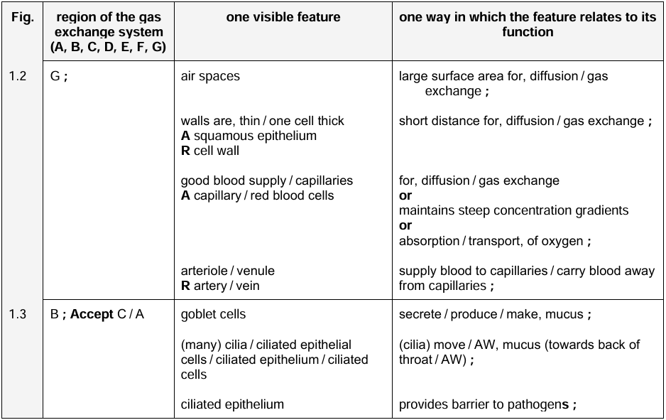
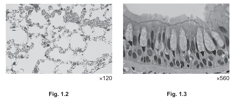
Detailed Explanation: Fig. 1.2 shows alveoli (G) with their characteristic air spaces that provide a massive surface area for efficient gas exchange. The thin walls (one cell thick) allow rapid diffusion of oxygen and carbon dioxide. Fig. 1.3 shows the trachea (B) with ciliated epithelial cells. The cilia beat in coordinated waves to move mucus and trapped particles upward, protecting the lungs from infection and debris.
(b) Any five from:
- Sinoatrial node (SAN) generates electrical impulses
- Impulses spread across atrial walls causing atrial contraction
- Tricuspid valve opens allowing blood into right ventricle
- Atrioventricular node (AVN) delays impulse briefly
- Impulse travels down Bundle of His to ventricle apex
- Right ventricle contracts, pumping blood into pulmonary artery
- Pulmonary valve prevents backflow into ventricle
Detailed Explanation: The sequence begins with the sinoatrial node (the heart’s natural pacemaker) generating electrical impulses. These impulses cause the atria to contract, pushing blood through the open tricuspid valve into the right ventricle. After a brief delay at the AV node (allowing complete ventricular filling), the impulse travels down specialized conduction pathways to the ventricle walls, causing coordinated contraction. This forces blood through the pulmonary valve into the pulmonary arteries toward the lungs. The valves ensure one-way flow, while the coordinated timing maximizes pumping efficiency.